

Black and Brown: Non-neoplastic Pigmentation of the Oral Mucosa
- PMID: 30671761
- PMCID: PMC6405786
- DOI: 10.1007/s12105-018-0980-9
Black and Brown: Non-neoplastic Pigmentation of the Oral Mucosa
Abstract
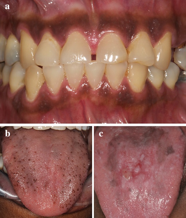
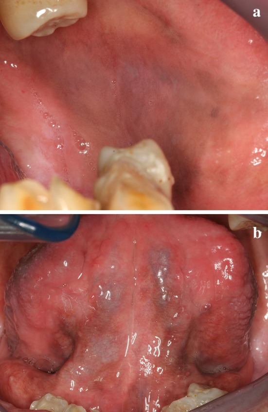
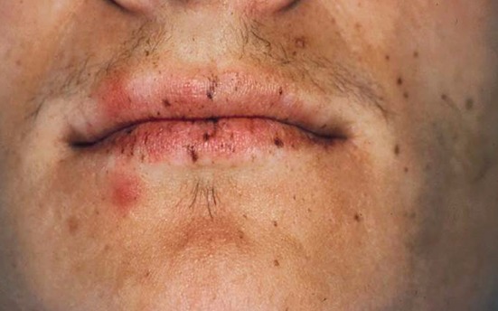
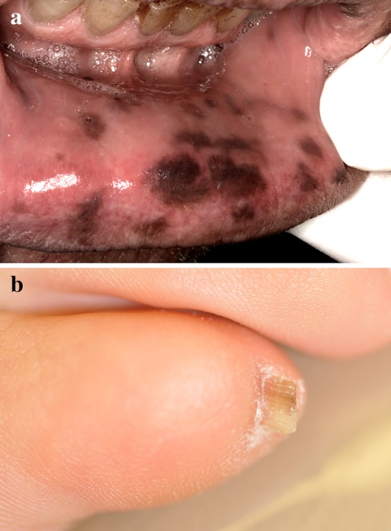
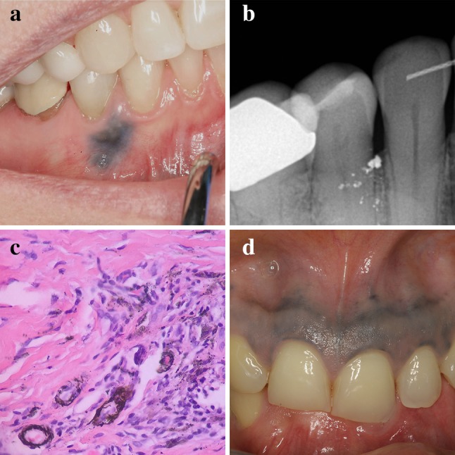
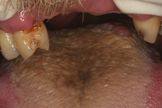
Black and brown pigmentation of the oral mucosa can occur due to a multitude of non-neoplastic causes. Endogenous or exogenous pigments may be responsible for oral discoloration which can range from innocuous to life-threatening in nature. Physiologic, reactive, and idiopathic melanin production seen in smoker's melanosis, drug-related discolorations, melanotic macule, melanoacanthoma and systemic diseases are presented. Exogenous sources of pigmentation such as amalgam tattoo and black hairy tongue are also discussed. Determining the significance of mucosal pigmented lesions may represent a diagnostic challenge for clinicians. Biopsy is indicated whenever the source of pigmentation cannot be definitively identified based on the clinical presentation.
Keywords: Biopsy; Black; Brown; Melanin; Melanotic; Oral mucosa; Pigmentation.
Conflict of interest statement
Conflict of interest
All authors declare that they have no conflict of interest.
Ethical Approval
This article does not contain any studies with human participants or animals performed by any of the authors.
Figures
Similar articles
-
Pigmented lesions of the oral mucosa: A cross-sectional study of 458 histopathological specimens.Oral Dis. 2018 Nov;24(8):1484-1491. doi: 10.1111/odi.12924. Epub 2018 Jul 10. Oral Dis. 2018. PMID: 29945290
-
Concomitant endogenous and exogenous etiology for gingival pigmentation.Dermatol Online J. 2021 Aug 15;27(8). doi: 10.5070/D327854720. Dermatol Online J. 2021. PMID: 34755968
-
Pigmented lesions of the oral cavity: an update.Dent Clin North Am. 2013 Oct;57(4):699-710. doi: 10.1016/j.cden.2013.07.006. Epub 2013 Aug 15. Dent Clin North Am. 2013. PMID: 24034073 Free PMC article. Review.
-
Oral melanotic macule--a case report.J Indian Soc Pedod Prev Dent. 2004 Jun;22(2):73-5. J Indian Soc Pedod Prev Dent. 2004. PMID: 15491090
-
Oral pigmentation in physiologic conditions, post-inflammatory affections and systemic diseases.G Ital Dermatol Venereol. 2018 Oct;153(5):666-671. doi: 10.23736/S0392-0488.17.05619-X. Epub 2017 Apr 19. G Ital Dermatol Venereol. 2018. PMID: 28421728 Review.
Cited by
-
Medication related to pigmentation of oral mucosa.Med Oral Patol Oral Cir Bucal. 2022 May 1;27(3):e230-e237. doi: 10.4317/medoral.25110. Med Oral Patol Oral Cir Bucal. 2022. PMID: 35420067 Free PMC article. Review.
-
Oral hyperpigmentation as an adverse effect of highly active antiretroviral therapy in HIV patients: A systematic review and pooled prevalence.J Clin Exp Dent. 2023 Jul 1;15(7):e561-e570. doi: 10.4317/jced.60195. eCollection 2023 Jul. J Clin Exp Dent. 2023. PMID: 37519321 Free PMC article. Review.
-
Black Hairy Tongue Observed During Esophagogastroduodenoscopy.Cureus. 2024 Nov 23;16(11):e74331. doi: 10.7759/cureus.74331. eCollection 2024 Nov. Cureus. 2024. PMID: 39720364 Free PMC article.
-
Oral manifestations in a patient with a history of asymptomatic COVID-19: Case report.Int J Infect Dis. 2020 Nov;100:154-157. doi: 10.1016/j.ijid.2020.08.071. Epub 2020 Sep 1. Int J Infect Dis. 2020. PMID: 32882435 Free PMC article.
-
Prevalence and associated factors of oral pigmented lesions among Yemeni dental patients: a large cross-sectional study.BMC Oral Health. 2025 Mar 15;25(1):391. doi: 10.1186/s12903-025-05760-6. BMC Oral Health. 2025. PMID: 40089755 Free PMC article.
References
-
- Meleti M, Vescovi P, Mooi WJ, van der Waal I. Pigmented lesions of the oral mucosa and perioral tissues: a flow-chart for the diagnosis and some recommendations for the management. Oral Surg Oral Med Oral Pathol Oral Radiol Endod. 2008;105(5):606–616. - PubMed
-
- Muller S. Melanin-associated pigmented lesions of the oral mucosa: presentation, differential diagnosis, and treatment. Dermatol Ther. 2010;23(3):220–229. - PubMed
-
- Amir E, Gorsky M, Buchner A, Sarnat H, Gat H. Physiologic pigmentation of the oral mucosa in Israeli children. Oral Surg Oral Med Oral Pathol. 1991;71(3):396–398. - PubMed
-
- Gorsky M, Buchner A, Moskona D, Aviv I. Physiologic pigmentation of the oral mucosa in Israeli Jews of different ethnic origin. Community Dent Oral Epidemiol. 1984;12(3):188–190. - PubMed
-
- Eisen D. Disorders of pigmentation in the oral cavity. Clin Dermatol. 2000;18(5):579–587. - PubMed
Publication types
MeSH terms
LinkOut - more resources
Full Text Sources
Medical
