. 2019 May;13(5):1047-1058.
doi: 10.1002/1878-0261.12457. Epub 2019 Feb 4.

Mutual regulation of MDM4 and TOP2A in cancer cell proliferation

Affiliations

Mutual regulation of MDM4 and TOP2A in cancer cell proliferation

Tao Liu et al. Mol Oncol. 2019 May.

Retraction in

Abstract

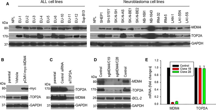
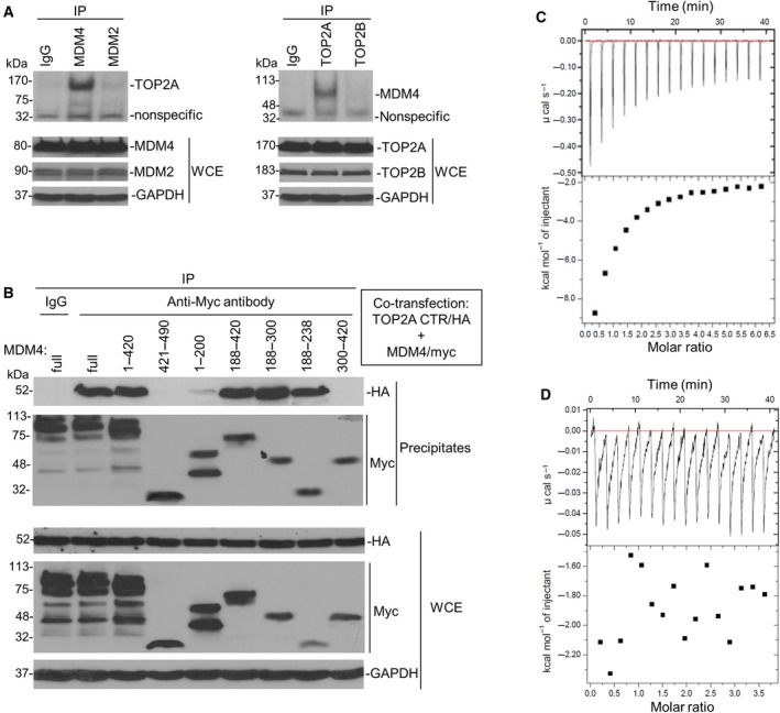
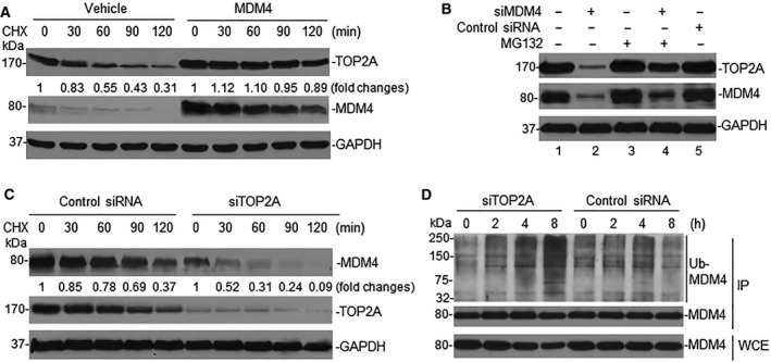
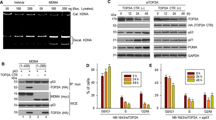
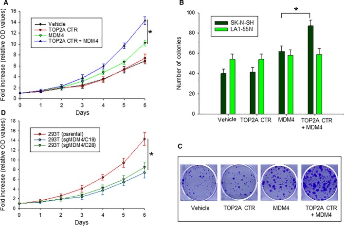
MDM4 and topoisomerase IIα (TOP2A) are overexpressed in various human cancers. MDM4 acts as an oncoprotein which promotes cancer progression by inhibiting tumor suppressor p53. As a DNA replication- and cell division-regulating enzyme, TOP2A is the main target of many anticancer therapy regimens; however, the exact role of TOP2A in cancer remains elusive. Herein, we report that MDM4 and TOP2A bind to each other and are mutually upregulated at the post-translational level, leading to TOP2A protein stabilization, inhibition of p53, and increased tumor-cell proliferation. We demonstrate that the C-terminal region (CTR) of TOP2A binds to a unique sequence (residues: 188-238) of MDM4, which contains an auto-inhibitory segment regulating the MDM4-p53 interaction. TOP2A binding in turn activates MDM4 for p53 binding, resulting in enhanced inhibition of p53 and cancer cell proliferation. Conversely, binding of the MDM4 sequence to the CTR of TOP2A stabilizes TOP2A protein, leading to increased TOP2A protein expression. These results reveal novel functions of MDM4 and TOP2A as well as their interactions in oncogenesis, suggesting that inhibition of the MDM4-TOP2A interaction may represent a novel strategy in specifically and simultaneously targeting TOP2A and MDM4 for cancer treatment.

Keywords: MDM4; TOP2A; cancer cell proliferation; p53.


Conflict of interest statement

The authors declare no conflict of interest.

Figures

Figure 1
Figure 1
Expression and mutual regulation of MDM4 and TOP2A in cancer cells. (A) MDM4 and TOP2A protein expression in ALL and neuroblastoma cell lines was detected by western blot assays. (B) TOP2A expression in LA1‐55N cells transfected with MDM4 (tagged with myc) or control (vehicle) was detected by western blot, using anti‐myc antibody for indication of transfected MDM4. (C) Western blot for expression of TOP2A and MDM4 in NB‐1643 cells transfected with siTOP2A or control siRNA. (D) Western blot results show the expression of proteins as indicated in parental 293T cells (control) and 2 clones C19 and C28, in which MDM4 was silenced using the CRISPR/Cas9 (sgMDM4). (E) The expression levels of MDM4 and TOP2A mRNA relative to GAPDH in different 293T cells as in (D) were determined by quantitative RTPCR. Data represent mean ± SD of three independent experiments.
Figure 2
Figure 2
MDM4 and TOP2A bind with each other. (A) co‐IP and western blot assays using antibodies as indicated for binding between endogenous MDM4 and TOP2A in NB‐1643 cells. WCE, whole cell extracts. (B) co‐IP and western blot for mapping the binding between transfected TOP2A CTR and different MDM4 fragments, using anti‐HA and anti‐myc antibodies, respectively. (C, D) ITC assays for binding of MDM4 (188–300) to TOP2A CTR (C) and no binding of MDM4 (421–490) and TOP2A CTR (D). The upper boxes show raw heating power over time, and the lower boxes are a fit of the integrated energy values, normalized for each injection.
Figure 3
Figure 3
Mutual regulation of MDM4 and TOP2A through protein modification. (A) LA1‐55N cells were transfected with MDM4, and turnover of TOP2A in transfected cells was detected by CHX chase assay. Numerical labels under each TOP2A band represent their relative expression levels after normalization for GAPDH, as compared with untreated (0) samples that were defined as 1 unit. (B) NB‐1643 cells transfected with siMDM4 were treated with MG132 for 4 h and TOP2A expression detected by western blot. (C) CHX chase assay for MDM4 turnover in NB‐1643 transfected with siTOP2A. (D) Denaturing protein IP (using MDM4 antibody) and western blot (using ubiquitination antibody) for detection of MDM4 ubiquitination in siTOP2A‐ and control siRNA‐transfected NB‐1643 cells.
Figure 4
Figure 4
Effect of the interaction between MDM4 and TOP2A on TOP2A catalytic activity and p53 activation. (A) LA1‐55N cells were transfected with MDM4 and TOP2A catalytic activity detected by decatenation assay. (B) LA1‐55N cells co‐transfected with p53 and plasmids containing MDM4 DNA regions corresponding to either nucleotides 1–420 or 1–200 (tagged with myc), in the presence or absence of TOP2A CTR (tagged with HA). Transfected cells were immunoprecipitated with anti‐myc antibody. The expression of p53 and TOP2A CTR (HA) in the precipitates, and expression of p53 and TOP2A as well as MDM4 (myc) in whole cell extracts (WCE), was detected by western blot. (C) NB‐1643 cells were transfected with siTOP2A in the presence and absence of TOP2A CTR for the indicated times; protein expression was detected by western blot assay. (D, E) NB‐1643 cells were transfected either with siTOP2A alone (D) or with combination of siTOP2A and sip53 (D) for the indicated time, and cell cycle distribution was detected by flow cytometry. Data represent mean of three independent experiments, bars ± SD.
Figure 5
Figure 5
Effect of TOP2A CTR on MDM4‐mediated cell proliferation and growth. (A) WST proliferation assay comparing the growth of SK‐N‐SH cells transfected with plasmids as indicated. *< 0.01. (B) colony assays of SK‐N‐SH and LA1‐55N cells transfected with plasmids as indicated, *< 0.01, as determined by two‐tailed t‐test. (C) representative colony formation of transfected SK‐N‐SK cells as in (B). (D) WST proliferation assay comparing the growth of different 293T cells (parental and sgMDM4 clones), *< 0.01. Data in (A, B, and D) represent mean ± SD of three independent experiments.

References

    1. Bista M, Petrovich M and Fersht AR (2013) MDMX contains an autoinhibitory sequence element. Proc Natl Acad Sci USA 110, 17814–17819. - PMC - PubMed
    1. Carpenter AJ and Porter AC (2004) Construction, characterization, and complementation of a conditional‐lethal DNA topoisomerase IIalpha mutant human cell line. Mol Biol Cell 15, 5700–5711. - PMC - PubMed
    1. Carrillo AM, Bouska A, Arrate MP and Eischen CM (2015) Mdmx promotes genomic instability independent of p53 and Mdm2. Oncogene 34, 846–856. - PMC - PubMed
    1. Chen L, Borcherds W, Wu S, Becker A, Schonbrunn E, Daughdrill GW and Chen J (2015) Autoinhibition of MDMX by intramolecular p53 mimicry. Proc Natl Acad Sci USA 112, 4624–4629. - PMC - PubMed
    1. Chen L, Li C, Pan Y and Chen J (2005) Regulation of p53‐MDMX interaction by casein kinase 1 alpha. Mol Cell Biol 25, 6509–6520. - PMC - PubMed

Publication types

MeSH terms