

Bisperoxovanadium Mediates Neuronal Protection through Inhibition of PTEN and Activation of PI3K/AKT-mTOR Signaling after Traumatic Spinal Injuries
- PMID: 30672370
- PMCID: PMC6727469
- DOI: 10.1089/neu.2018.6294
Bisperoxovanadium Mediates Neuronal Protection through Inhibition of PTEN and Activation of PI3K/AKT-mTOR Signaling after Traumatic Spinal Injuries
Abstract
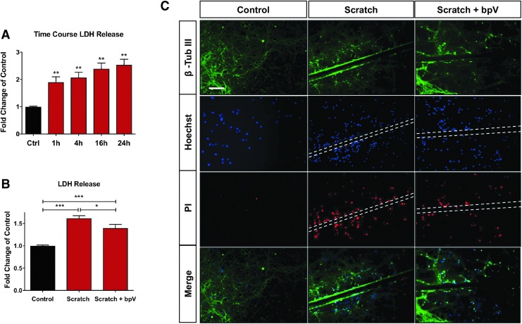
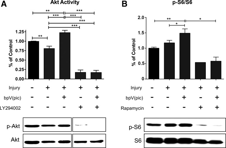
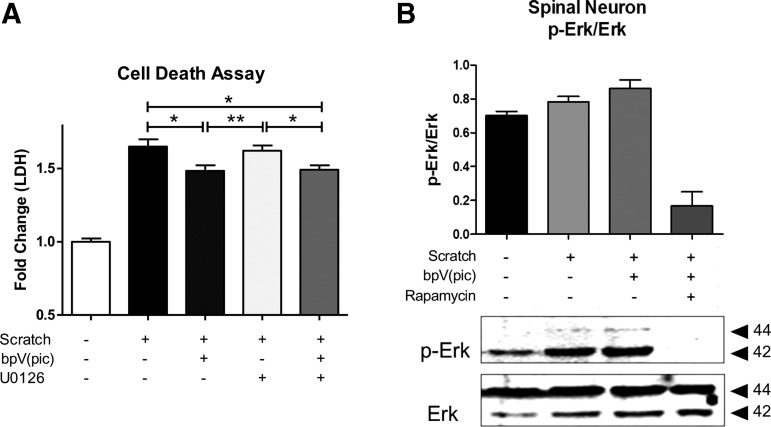
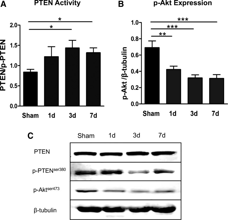
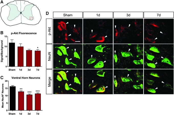
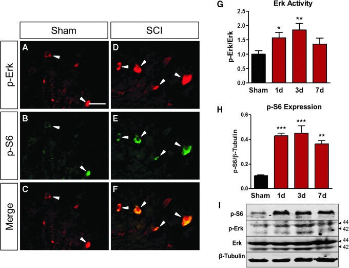
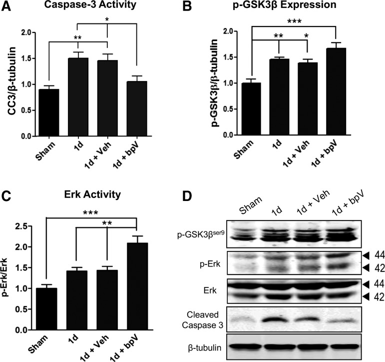
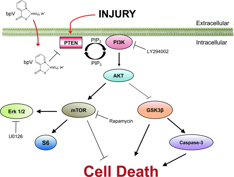
Although mechanisms involved in progression of cell death in spinal cord injury (SCI) have been studied extensively, few are clear targets for translation to clinical application. One of the best-understood mechanisms of cell survival in SCI is phosphatidylinositol-3-kinase (PI3K)/Akt and associated downstream signaling. Clear therapeutic efficacy of a phosphatase and tensin homologue (PTEN) inhibitor called bisperoxovanadium (bpV) has been shown in SCI, traumatic brain injury, stroke, and other neurological disease models in both neuroprotection and functional recovery. The present study aimed to elucidate mechanistic influences of bpV activity in neuronal survival in in vitro and in vivo models of SCI. Treatment with 100 nM bpV(pic) reduced cell death in a primary spinal neuron injury model (p < 0.05) in vitro, and upregulated both Akt and ribosomal protein S6 (pS6) activity (p < 0.05) compared with non-treated injured neurons. Pre-treatment of spinal neurons with a PI3K inhibitor, LY294002 or mammalian target of rapamycin (mTOR) inhibitor, rapamycin blocked bpV activation of Akt and ribosomal protein S6 activity, respectively. Treatment with bpV increased extracellular signal-related kinase (Erk) activity after scratch injury in vitro, and rapamycin reduced influence by bpV on Erk phosphorylation. After a cervical hemicontusive SCI, Akt phosphorylation decreased in total tissue via Western blot analysis (p < 0.01) as well as in penumbral ventral horn motor neurons throughout the first week post-injury (p < 0.05). Conversely, PTEN activity appeared to increase over this period. As observed in vitro, bpV also increased Erk activity post-SCI (p < 0.05). Our results suggest that PI3K/Akt signaling is the likely primary mechanism of bpV action in mediating neuroprotection in injured spinal neurons.
Keywords: PTEN; bisperoxovanadium; bpV; mTOR spinal cord injury; neuroprotection.
Conflict of interest statement
No competing financial interests exist.
Figures
Similar articles
-
Systemic bisperoxovanadium activates Akt/mTOR, reduces autophagy, and enhances recovery following cervical spinal cord injury.PLoS One. 2012;7(1):e30012. doi: 10.1371/journal.pone.0030012. Epub 2012 Jan 10. PLoS One. 2012. PMID: 22253859 Free PMC article.
-
PTEN inhibitor bisperoxovanadium protects oligodendrocytes and myelin and prevents neuronal atrophy in adult rats following cervical hemicontusive spinal cord injury.Neurosci Lett. 2014 Jun 24;573:64-8. doi: 10.1016/j.neulet.2014.02.039. Epub 2014 Feb 26. Neurosci Lett. 2014. PMID: 24582904 Free PMC article.
-
ERK 1/2 Activation Mediates the Neuroprotective Effect of BpV(pic) in Focal Cerebral Ischemia-Reperfusion Injury.Neurochem Res. 2018 Jul;43(7):1424-1438. doi: 10.1007/s11064-018-2558-z. Epub 2018 Jun 7. Neurochem Res. 2018. PMID: 29882124 Free PMC article.
-
PI3K/AKT Pathway and Its Mediators in Thyroid Carcinomas.Mol Diagn Ther. 2016 Feb;20(1):13-26. doi: 10.1007/s40291-015-0175-y. Mol Diagn Ther. 2016. PMID: 26597586 Review.
-
Pharmacological modulation of PI3K/PTEN/Akt/mTOR/ERK signaling pathways in ischemic injury: a mechanistic perspective.Metab Brain Dis. 2025 Feb 26;40(3):131. doi: 10.1007/s11011-025-01543-8. Metab Brain Dis. 2025. PMID: 40009091 Review.
Cited by
-
Dulaglutide Improves Gliosis and Suppresses Apoptosis/Autophagy Through the PI3K/Akt/mTOR Signaling Pathway in Vascular Dementia Rats.Neurochem Res. 2023 May;48(5):1561-1579. doi: 10.1007/s11064-022-03853-0. Epub 2022 Dec 26. Neurochem Res. 2023. PMID: 36571662
-
Genetically encoded biosensors of metabolic function for the study of neurodegeneration, a review and perspective.Neurophotonics. 2025 Jun;12(Suppl 2):S22805. doi: 10.1117/1.NPh.12.S2.S22805. Epub 2025 Sep 4. Neurophotonics. 2025. PMID: 40919264 Free PMC article. Review.
-
Acetylglutamine facilitates motor recovery and alleviates neuropathic pain after brachial plexus root avulsion in rats.J Transl Med. 2023 Aug 23;21(1):563. doi: 10.1186/s12967-023-04399-7. J Transl Med. 2023. PMID: 37612586 Free PMC article.
-
miRNA-26a-5p Accelerates Healing via Downregulation of PTEN in Fracture Patients with Traumatic Brain Injury.Mol Ther Nucleic Acids. 2019 Sep 6;17:223-234. doi: 10.1016/j.omtn.2019.06.001. Epub 2019 Jun 12. Mol Ther Nucleic Acids. 2019. PMID: 31272072 Free PMC article.
-
PTEN inhibitor bisperoxovanadium protects against noise-induced hearing loss.Neural Regen Res. 2023 Jul;18(7):1601-1606. doi: 10.4103/1673-5374.358606. Neural Regen Res. 2023. PMID: 36571368 Free PMC article.
References
-
- Ackery A., Tator C., and Krassioukov A. (2004). A global perspective on spinal cord injury epidemiology. J. Neurotrauma 21, 1355–1370 - PubMed
-
- Kirshblum S.C., Groah S.L., McKinley W.O., Gittler M.S., and Stiens S.A. (2002). Spinal cord injury medicine. 1. Etiology, classification, and acute medical management. Arch. Phys. Med. Rehabil. 83, Suppl 1, S50–S57 - PubMed
-
- Kirshblum S.C. (2002). Clinical activities of the model spinal cord injury system. J. Spinal Cord Med. 25, 339–344 - PubMed
-
- Nandoe Tewarie R.D., Hurtado A., Bartels R.H., Grotenhuis J.A., and Oudega M. (2010). A clinical perspective of spinal cord injury. NeuroRehabilitation 27, 129–139 - PubMed
Publication types
MeSH terms
Substances
Grants and funding
LinkOut - more resources
Full Text Sources
Medical
Research Materials
Miscellaneous
