. 2019 Jan 22;26(4):845-854.e6.
doi: 10.1016/j.celrep.2018.12.097.

Structural and Functional Studies of the RBPJ-SHARP Complex Reveal a Conserved Corepressor Binding Site

Affiliations

Structural and Functional Studies of the RBPJ-SHARP Complex Reveal a Conserved Corepressor Binding Site

Zhenyu Yuan et al. Cell Rep. .

Abstract

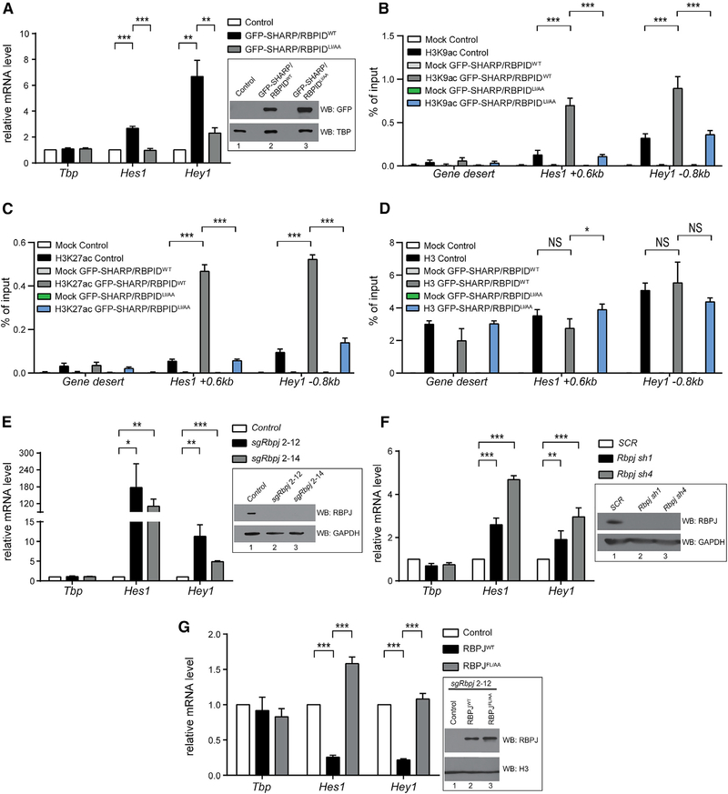
Notch is a conserved signaling pathway that is essential for metazoan development and homeostasis; dysregulated signaling underlies the pathophysiology of numerous human diseases. Receptor-ligand interactions result in gene expression changes, which are regulated by the transcription factor RBPJ. RBPJ forms a complex with the intracellular domain of the Notch receptor and the coactivator Mastermind to activate transcription, but it can also function as a repressor by interacting with corepressor proteins. Here, we determine the structure of RBPJ bound to the corepressor SHARP and DNA, revealing its mode of binding to RBPJ. We tested structure-based mutants in biophysical and biochemical-cellular assays to characterize the role of RBPJ as a repressor, clearly demonstrating that RBPJ mutants deficient for SHARP binding are incapable of repressing transcription of genes responsive to Notch signaling in cells. Altogether, our structure-function studies provide significant insights into the repressor function of RBPJ.

Keywords: CSL; MINT; Notch signaling; RBPJ; SHARP; SPEN; X-ray crystallography; isothermal titration calorimetry; signal transduction; transcriptional regulation.


Conflict of interest statement

DECLARATION OF INTERESTS

The authors declare no competing interests.

Figures

Figure 1.
Figure 1.. X-Ray Structure of the RBPJ-SHARP Corepressor Complex Bound to DNA
(A) Structure of the C. elegans RBPJ-NICD-MAM ternary complex bound to DNA (PDB: 2FO1). RBPJ is composed of three domains: the NTD (N-terminal domain), BTD (β-trefoil domain), and CTD (C-terminal domain), which are colored cyan, green, and orange, respectively. A β strand that makes hydrogen-bonding interactions with all three domains is colored magenta. The RAM and ANK domains of NICD are colored yellow and blue, respectively. MAM and DNA are colored red and light pink-blue, respectively. (B) Domain schematics of RBPJ, NICD, and MAM, colored similarly to the structure. SHARP is a multidomain transcriptional coregulator that contains N-terminal RRM (RNA recognition motif) domains, multiple NLSs (nuclear localization sequences), an RID (receptor interaction domain), an RBPID (RBPJ-interacting domain), and a C-terminal SPOC (Spen paralog and ortholog C-terminal) domain. (C) Ribbon diagram of the RBPJ-SHARP-DNA complex, with RBPJ and the DNA colored, as in (A), and SHARP is colored purple. Also shown are magnified views of the interaction of SHARP with the CTD (top) and BTD (bottom) of RBPJ. RBPJ is represented as a molecular surface, with CTD and BTD residues that contact SHARP colored orange and green, respectively. SHARP is shown in a stick representation, with carbon, oxygen, and nitrogen atoms colored purple, red, and blue, respectively. Electron density (composite omit map contoured at 1σ) corresponding to SHARP is colored gray. SHARP residues that were mutated and tested for activity are labeled. See also Figure S5. (D) Open book representation of RBPJ-SHARP interfaces. Left: CTD-SHARP interface. Right: BTD-SHARP interface. Side chains that contribute to the interface are shown. Key residues at the interface that were mutated and tested for activity are labeled and color-coded according to ΔΔG°. See also Figure S1. (E) Conformational changes in the CTD of RBPJ as a result of SHARP binding. Left: the CTD-SHARP complex with labeled secondary structural elements of the CTD. The R438-E2786 salt bridge is also shown. Center: structural overlay of RBPJ (bound) from the RBPJ-SHARP complex with an unbound structure of RBPJ (PDB: 3IAG). The RBPJ bound and unbound structures are colored orange and yellow, respectively. RBPJ residue W441, which undergoes a large conformational change in the bound structure, is shown. Right: ribbon diagram of the unbound RBPJ structure (PDB: 3IAG).
Figure 2.
Figure 2.. Comparison of Coregulator Binding Sites on RBPJ
(A) Both SHARP and NICD bind the BTD and CTD of RBPJ. Left: RBPJ-SHARP-DNA complex structure, with RBPJ represented as a gray molecular surface, SHARP as a ribbon diagram colored purple, and the DNA as CPK colored light blue and pink. The RBPJ residues that contact SHARP in the BTD and CTD are colored green and orange, respectively. Right: C. elegans RBPJ-NICD-MAM-DNA complex structure (PDB: 2FO1), with RBPJ represented as a gray molecular surface. NICD is represented as a ribbon diagram, with its RAM and ANK domains colored yellow and blue, respectively. MAM is depicted as a ribbon diagram and colored red. The DNA is colored light blue and pink. (B) Structural alignment of SHARP and other coregulators that bind the BTD of RBPJ. Top: the BTD of RBPJ is represented as a green molecular surface. SHARP, the RAM domain from LIN-12, RITA1, and FHL1 are colored purple, yellow, light pink, and gray, respectively. The hydrophobic tetrapeptide (ϕWϕP) is labeled. Bottom: sequence alignment of coregulators that bind the BTD of RBPJ, including the RAM domains of Notch from mammals, D. melanogaster, and C. elegans, and the corepressors RITA1, FHL1, and SHARP. The conserved ϕWϕP is boxed in green. Other conserved regions in RAM that contribute to binding are highlighted, including the basic region (blue), the -HG- motif (orange), and the -GF- motif (magenta). Structurally similar residues in SHARP that align with other BTD binders are boxed and highlighted in gray. (C) Structural similarity of RBPJ-SHARP and Su(H)-Hairless corepressor complexes. Su(H) is represented as a gray molecular surface, with the SHARP and Hairless binding sites colored orange. SHARP and Hairless are shown as ribbon diagrams and colored purple and yellow, respectively. (D) Overview of corepressor binding to RBPJ, illustrating the bipartite binding of SHARP. RBPJ is represented as a gray molecular surface, with its BTD and CTD binding clefts colored green and orange, respectively. The corepressors SHARP, Hairless, RITA1, and FHL1 are shown as ribbon diagrams and colored purple, yellow, pink, and gray, respectively. The DNA is colored light blue and light pink. (E) Structural comparison of the CTD of RBPJ with the RHR-C domain of NFAT. RBPJ-SHARP is colored as in Figure 1, and NFAT is colored green. The NFAT β strand βa’, which is absent in the CTD of RBPJ but occupied by SHARP in the complex structure, is labeled. (F) Structural alignment of the BTD from RBPJ with a canonical β-trefoil fold from the ryanodine receptor (RYR). A canonical BTD is composed of 12 β strands, in which four β strands are arranged in a pseudo-threefold symmetrical arrangement. The atypical BTD of RBPJ is missing two of the 12 β strands that compose a canonical BTD. The BTDs of RBPJ and RYR are colored green and light pink, respectively. The two β strands that are missing in the RBPJ BTD fold are colored light blue and highlighted with red arrowheads. The RAM domain of NICD is colored yellow.
Figure 3.
Figure 3.. The RBPJ-SHARP Interaction Is Required for Repression of Notch Target Genes in Cells
(A) The wild-type SHARP RBPID (RBPIDWT), but not the RBPJ-interacting defective SHARP RBPID (RBPIDLI/AA) mutant, causes upregulation of Notch target genes in mouse mature T (MT) cells by outcompeting endogenous SHARP for RBPJ binding. MT cells were infected with plasmids encoding GFP-tagged SHARP (2,776–2,833), either wild-type (GFP-SHARP/RBPIDWT, black bars) or the RBPJ-interacting defective mutant L2791A/I2811A (GFP-SHARP/RBPIDLI/AA, gray bars) or an empty vector control (Control, white bars). Left: total RNA from MT cells was analyzed by qPCR using primers specific for Tbp, Hes1, or Hey1. Data shown represent the mean ± SD of triplicate experiments (**p < 0.01, ***p < 0.001, unpaired Student’s t test). Right: nuclear extracts (NEs) were prepared from MT cells and analyzed by western blotting, with TBP used as a loading control. (B–D) GFP-SHARP/RBPIDWT derepresses Notch target genes via histone deacetylation. MT cells were infected with plasmids encoding GFP-SHARP/RBPIDWT, GFP-SHARP/RBPIDLI/AA, or an empty vector control and analyzed by chromatin immunoprecipitation (ChIP) using antibodies against H3K9ac (B), H3K27ac (C), or H3 (D). The enrichment was analyzed by qPCR on the enhancers of Hes1 and Hey1, located at approximately +0.6 kb and −0.8 kb relative to the transcription start site, respectively. Gene desert was used as a negative control. Data were normalized to the positive control (Gapdh 0kb), and, in the case of the H3K9ac and H3K27ac ChIP, data were further normalized to histone occupancy (H3). Shown is the mean ± SD of two experiments measured twice each (NS, not significant; *p < 0.05; ***p < 0.001; unpaired Student’s t test). See also Figure S4. (E) RBPJ is required to repress the Notch target genes Hes1 and Hey1 in MT cells, as revealed by CRISPR/Cas9 depletion of RBPJ. Left: total RNA from wild-type (control) or RBPJ-depleted (clones sgRbpj 2–12 and sgRbpj 2–14) MT cells was analyzed by qPCR. Shown is the mean ± SD of triplicate experiments (*p < 0.05, **p < 0.01, ***p < 0.001, unpaired Student’s t test). Right: whole-cell extracts (WCEs) were prepared from MT cells and analyzed by western blotting using an anti-RBPJ antibody. GAPDH was used as loading control. (F) Hes1 and Hey1 Notch target genes are upregulated upon shRNA-mediated Rbpj knockdown but not with the SCR control shRNA. Left: total RNA from MT cells infected with shRNAs targeting Rbpj (Rbpj sh1 or Rbpj sh4) or scrambled shRNA control (SCR) was analyzed by qPCR using primers specific for Tbp, Hes1, or Hey1. Shown is the mean ± SD of quadruplicate experiments (**p < 0.01, ***p < 0.001, unpaired Student’s t test). Right: WCE was prepared from MT cells and analyzed by western blotting using an RBPJ antibody. GAPDH was used as a loading control. (G) Expression of RBPJWT but not RBPJFL/AA in the RBPJ-depleted background rescues the repression of Notch target genes. Left: total RNA from sgRbpj 2–12 MT cells infected with empty vector (control), RBPJWT, or RBPJFL/AA was analyzed by qPCR using primers specific for Tbp, Hes1, or Hey1. Shown is the mean± SD of three independent experiments measured twice each (*p < 0.05, **p < 0.01, ***p < 0.001, unpaired Student’s t test). Right: NEs were prepared from sgRbpj 2–12 MT cells infected with empty vector (control), RBPJWT, or RBPJFL/AA and analyzed by western blotting using anti-RBPJ antibodies. Histone H3 was used as a loading control.

Similar articles

Cited by

References

    1. Adams PD, Afonine PV, Bunkóczi G, Chen VB, Davis IW, Echols N, Headd JJ, Hung LW, Kapral GJ, Grosse-Kunstleve RW, et al. (2010). PHENIX: a comprehensive Python-based system for macromolecular structure solution. Acta Crystallogr. D Biol. Crystallogr 66, 213–221. - PMC - PubMed
    1. Ariyoshi M, and Schwabe JW (2003). A conserved structural motif reveals the essential transcriptional repression function of Spen proteins and their role in developmental signaling. Genes Dev. 17, 1909–1920. - PMC - PubMed
    1. Borggrefe T, and Oswald F (2009). The Notch signaling pathway: transcriptional regulation at Notch target genes. Cell. Mol. Life Sci 66, 1631–1646. - PMC - PubMed
    1. Braune EB, and Lendahl U (2016). Notch – a goldilocks signaling pathway in disease and cancer therapy. Discov. Med 21, 189–196. - PubMed
    1. Bray SJ (2016). Notch signalling in context. Nat. Rev. Mol. Cell Biol 17, 722–735. - PubMed

Publication types

MeSH terms

LinkOut - more resources