. 2019 Jan 23;11(2):590-614.
doi: 10.18632/aging.101762.

Relationship between senescence in macaques and bone marrow mesenchymal stem cells and the molecular mechanism

Affiliations

Relationship between senescence in macaques and bone marrow mesenchymal stem cells and the molecular mechanism

Xing-Hua Pan et al. Aging (Albany NY). .

Abstract

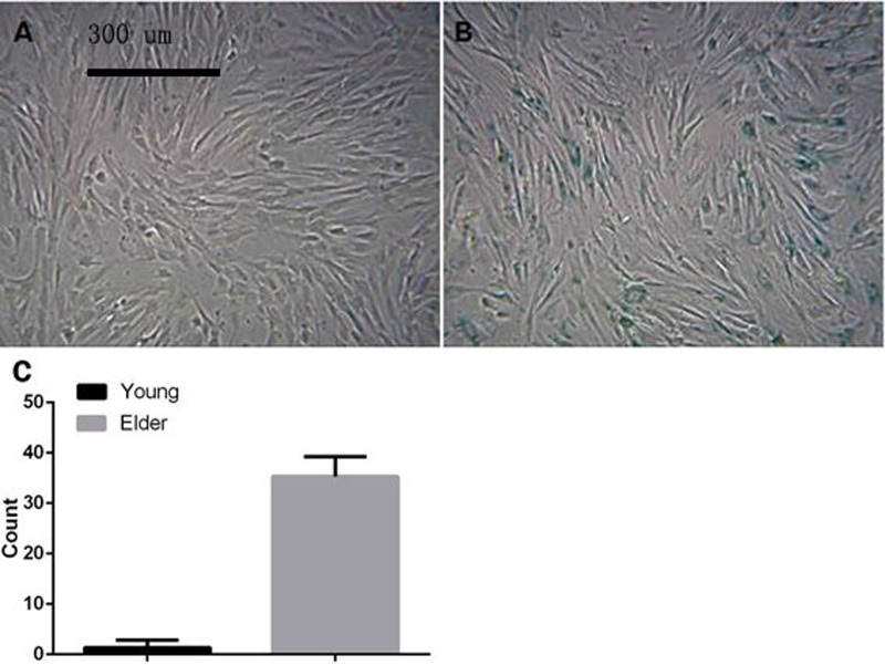
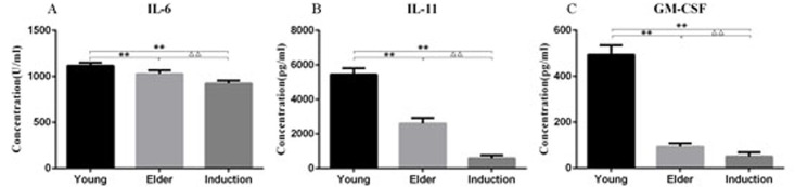
The relationship between bone marrow mesenchymal stem cells (BMSCs) and aging, as well as the antiaging effects of BMSCs, was observed. An aging macaque BMSC model was established. We isolated BMSCs from young and aged macaques and used RT-PCR and Western blot to confirm the aging-related mRNAs and their expression, revealing that TERT, SIRT1 and SIRT6 expression was decreased in the aged BMSCs. The morphology, immunophenotype, differentiation potential, proliferation potential, and antiaging effects of aged and young BMSCs on 293T cells were compared. The expression of aging-related genes and the difference between the secreted cytokines in natural aging and induced aging BMSCs were observed. The transcriptome of peripheral blood mononuclear cells from macaques was analyzed by high-throughput sequencing. Finally, the transcriptional characteristics and regulatory mechanisms of gene transcription in aging macaques were investigated.

Keywords: bone marrow mesenchymal stem cells; cytokines; macaque; senescence; transcriptome sequencing.


Conflict of interest statement

CONFLICTS OF INTEREST: The authors declare that they have no conflicts of interest.

Figures

Figure 1
Figure 1
Cell morphology of young and aged macaque BMSCs (×100). (A, B) show the primary BMSCs of young and aged macaques, respectively; (C, D) show the P3 BMSCs of young and aged macaques, respectively. With prolonged in vitro culture, some cells in the aging group became broad, and the number of polygonal and irregularly shaped cells began to increase.
Figure 2
Figure 2
Macaque BMSCs differentiated into multiple cells. (A, B) show the osteogenic induction of young and aged macaque BMSCs, respectively (×40); (C, D) show the adipogenic induction of young and aged macaque BMSCs, respectively (×200); (E, F) show the cartilage induction of young and aged macaque BMSCs, respectively. (×100); (G) We used ImageJ 1.37c software to quantitate the osteogenic, adipogenic and chondrogenic results. In the aged group, less osteogenic, adipogenic and chondrogenic markers were produced than in the young group (*p<0.05, n=3).
Figure 3
Figure 3
Proliferation curve of BMSCs from aged and young macaques as determined by the CCK-8 method. It was observed that compared with that in the young group, the expansion rate of P3 BMSCs in the aged group was significantly lower, and the proliferative ability was significantly reduced.
Figure 4
Figure 4
Macaque P3 BMSCs stained with senescence-associated SA-β-gal (A) and (B) show young macaque and aged macaque P3 BMSCs, respectively (×100). The results showed that the percentage of positive BMSC staining in the young group was 1.33 ± 1.51%, which was significantly lower than that in the aged group (35.33 ± 3.88%, p<0.01), suggesting that the BMSCs in aged macaques exhibit cell senescence earlier than those in young macaques.
Figure 5
Figure 5
293T cells treated with different concentrations of t-BHP and stained with SA-β-gal (×100). BMSCs were treated with t-BHP at concentrations of 0 μmol/L (A), 100 μmol/L (B), 200 μmol/L (C), 300 μmol/L (D), 400 μmol/L (E), and 500 μmol/L (F). We have quantified the apoptosis rate in (G). Induction with 200 μmol/L t-BHP for 6 h was performed to establish the aging macaque BMSC model.
Figure 6
Figure 6
Relative expression levels of telomeres and TCAB1 in 293T cells by RT-PCR. (A) shows the senescence-induced 293T cells cocultured with BMSCs. (B) shows the control 293T cells cocultured with BMSCs. (C) shows the senescence-induced 293T cells. (D) shows the control 293T cells. After the 293T cells were cocultured with BMSCs for 5 d, the mRNA levels of telomeres in the control group and the induced senescence group were significantly higher than those measured before coculture (P < 0.05, Figure 6A), and the mRNA levels of TCAB1 were also significantly increased (P < 0.05, Figure 6B).
Figure 7
Figure 7
Relative expression of TCAB1 in 293T cells. (A) shows TCAB1 expression in senescence-induced 293T cells cocultured with BMSCs. (B) shows TCAB1 expression in control 293T cells cocultured with BMSCs. (C) shows TCAB1 expression in senescence-induced 293T cells, and (D) shows TCAB1 expression in control 293T cells. (E) shows the Western blots with the quantitated results. TCAB1 expression was weakly positive before induction and positive after induction.
Figure 8
Figure 8
BMSCs treated with different concentrations of t-BHP and stained with SA-β-gal (×100). The concentration of t-BHP used to treat BMSCs is 100 μmol/L (A), 200 μmol/L (B), 300 μmol/L (C), 400 μmol/L (D), and 500 μmol/L (E), and 600 μmol/L (F). The aging macaque BMSC model was established by inducing cells with 200 μmol/L t-BHP for 6 h.
Figure 9
Figure 9
Cytokine levels in the culture supernatant of the young, aged and induced aged macaque BMSCs. IL-6, IL-11 and GM-CSF levels were significantly decreased in the elderly group and the induced aging group compared with the juvenile group (P < 0.01). IL-6, IL-11 and GM-CSF levels were also significantly lower in the induced aging group than in the elderly group (P < 0.01).
Figure 10
Figure 10
RT-PCR detection of the relative expression of TERT, TCAB1, P21, SIRT1 and SIRT6 in BMSCs. The results showed that TERT, SIRT1 and SIRT6 expression levels were significantly lower in the aged cell group than in the juvenile cell group (P < 0.01). P21 expression was significantly increased (P < 0.01), and TCAB1 expression was significantly decreased (P < 0.05). Compared with those in the juvenile cell group, TERT and SIRT6 levels were significantly decreased (P < 0.01) and P21 levels were significantly increased (P < 0.01) in the induced senescence group. TERT expression was significantly decreased in the induced aging group compared with the aged cell group (P < 0.01), and SIRT6 expression was significantly decreased (P < 0.05).
Figure 11
Figure 11
Expression of aging-associated proteins in the young cell group, aged cell group and induced aged BMSC group. The levels of TCAB1 and SIRT6 were relatively low in the aged and induced aging groups compared with the young group. p21 and p53 expression levels were relatively high in the aged and induced aging groups. *p<0.05 compared to the other two groups.
Figure 12
Figure 12
Heat map of the mRNA clustering analysis of macaque mononuclear cells from juvenile and old groups. Samples 1, 2, and 3 correspond to the elderly group, and samples_7, 8, and 9 correspond to the young group. According to the analysis, a total of 5,711 differentially expressed GO terms were identified, of which 2,636 were downregulated and 3,075 were upregulated.
Figure 13
Figure 13
GO enrichment analysis of differentially expressed mRNA in macaque mononuclear cells from the juvenile and old age groups. (A) Downregulated GO terms. (B) Upregulated GO terms. Red indicates the biological process-related GO term; green indicates the cell component-related GO term; and blue indicates the molecular function-related GO term. The top 30 bar graphs of the GO enrichment analysis are shown in Figure 13A and 13B. The top 30 downregulated and upregulated GO terms are listed in Table 7 according to their P-value and sorted from small to large.
Figure 14
Figure 14
Enrichment analysis of KEGG pathways in differentially expressed mRNA in macaque mononuclear cells from the juvenile and old age groups. The enrichment analysis showed that the target gene was mainly enriched in 322 pathways, of which 170 were upregulated and 152 were downregulated, resulting in a top-enriched KEGG pathway map (A), downregulated KEGG pathways; and (B), upregulated KEGG pathways.

Similar articles

Cited by

References

    1. Mooney KM, Morgan AE, Mc Auley MT. Aging and computational systems biology. Wiley Interdiscip Rev Syst Biol Med. 2016;8:123–139. doi: 10.1002/wsbm.1328. - DOI - PubMed
    1. Hayflick L. The cell biology of aging. J Invest Dermatol. 1979;73:8–14. doi: 10.1111/1523-1747.ep12532752. - DOI - PubMed
    1. Sahin E, Depinho RA. Linking functional decline of telomeres, mitochondria and stem cells during ageing. Nature. 2010;464:520–528. doi: 10.1038/nature08982. - DOI - PMC - PubMed
    1. Schultz MB, Sinclair DA. When stem cells grow old: phenotypes and mechanisms of stem cell aging. Development. 2016;143:3–14. doi: 10.1242/dev.130633. - DOI - PMC - PubMed
    1. Scudellari M. Ageing research: Blood to blood. Nature. 2015;517:426–429. doi: 10.1038/517426a. - DOI - PubMed

Publication types