

A method for automatic forensic facial reconstruction based on dense statistics of soft tissue thickness
- PMID: 30673719
- PMCID: PMC6343874
- DOI: 10.1371/journal.pone.0210257
A method for automatic forensic facial reconstruction based on dense statistics of soft tissue thickness
Abstract
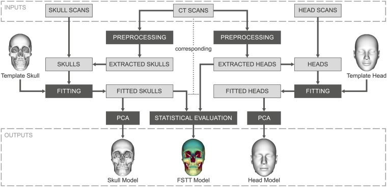
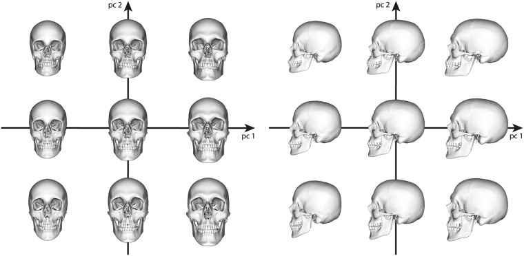
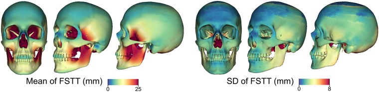
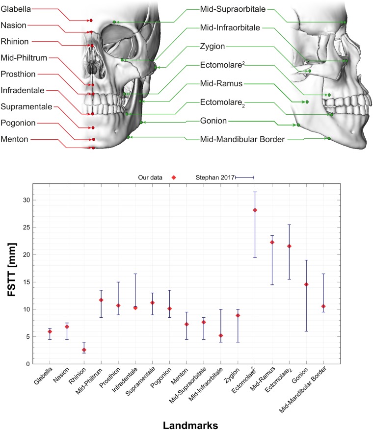
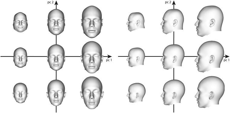
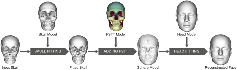
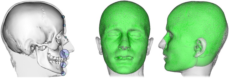
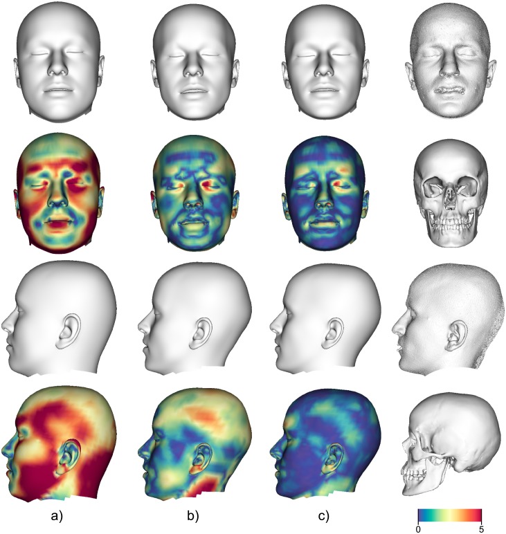
In this paper, we present a method for automated estimation of a human face given a skull remain. Our proposed method is based on three statistical models. A volumetric (tetrahedral) skull model encoding the variations of different skulls, a surface head model encoding the head variations, and a dense statistic of facial soft tissue thickness (FSTT). All data are automatically derived from computed tomography (CT) head scans and optical face scans. In order to obtain a proper dense FSTT statistic, we register a skull model to each skull extracted from a CT scan and determine the FSTT value for each vertex of the skull model towards the associated extracted skin surface. The FSTT values at predefined landmarks from our statistic are well in agreement with data from the literature. To recover a face from a skull remain, we first fit our skull model to the given skull. Next, we generate spheres with radius of the respective FSTT value obtained from our statistic at each vertex of the registered skull. Finally, we fit a head model to the union of all spheres. The proposed automated method enables a probabilistic face-estimation that facilitates forensic recovery even from incomplete skull remains. The FSTT statistic allows the generation of plausible head variants, which can be adjusted intuitively using principal component analysis. We validate our face recovery process using an anonymized head CT scan. The estimation generated from the given skull visually compares well with the skin surface extracted from the CT scan itself.
Conflict of interest statement
The authors have declared that no competing interests exist.
Figures
Similar articles
-
Densely calculated facial soft tissue thickness for craniofacial reconstruction in Chinese adults.Forensic Sci Int. 2016 Sep;266:573.e1-573.e12. doi: 10.1016/j.forsciint.2016.07.017. Epub 2016 Jul 27. Forensic Sci Int. 2016. PMID: 27544400
-
Open-Source Tools for Dense Facial Tissue Depth Mapping of Computed Tomography Models.Hum Biol. 2018 Jan;90(1):63-76. Hum Biol. 2018. PMID: 30387384
-
Radiographic assessment of facial soft tissue thickness in South Indian population--An anthropologic study.J Forensic Leg Med. 2016 Apr;39:161-8. doi: 10.1016/j.jflm.2016.01.032. Epub 2016 Feb 6. J Forensic Leg Med. 2016. PMID: 26924726
-
Systematic review on forensic craniofacial reconstruction. I. facial soft-tissue thickness.Forensic Sci Rev. 2023 Jul;35(2):107-136. Forensic Sci Rev. 2023. PMID: 37531497
-
Facial soft tissue thicknesses in craniofacial identification: Data collection protocols and associated measurement errors.Forensic Sci Int. 2019 Nov;304:109965. doi: 10.1016/j.forsciint.2019.109965. Epub 2019 Oct 2. Forensic Sci Int. 2019. PMID: 31610333 Review.
Cited by
-
Using Computed Tomography (CT) Data to Build 3D Resources for Forensic Craniofacial Identification.Adv Exp Med Biol. 2021;1317:53-74. doi: 10.1007/978-3-030-61125-5_4. Adv Exp Med Biol. 2021. PMID: 33945132
-
Joint multi-ancestry and admixed GWAS reveals the complex genetics behind human cranial vault shape.Nat Commun. 2023 Nov 16;14(1):7436. doi: 10.1038/s41467-023-43237-8. Nat Commun. 2023. PMID: 37973980 Free PMC article. Review.
-
Ethnic-Guided Soft Tissue Cephalometric Analysis on Lambani Indian Inhabitants for Forensic Facial Reconstruction.Cureus. 2022 Mar 23;14(3):e23430. doi: 10.7759/cureus.23430. eCollection 2022 Mar. Cureus. 2022. PMID: 35475086 Free PMC article.
-
Using 21st-Century Technologies to Determine the Cognitive Capabilities of a 11,000-Year-Old Perak Man Who Had Brachymesophalangia Type A2.Malays J Med Sci. 2021 Feb;28(1):1-8. doi: 10.21315/mjms2021.28.1.1. Epub 2021 Feb 24. Malays J Med Sci. 2021. PMID: 33679214 Free PMC article.
-
Advancements in craniofacial reconstruction: approaches and applications in forensics.Forensic Sci Med Pathol. 2025 Jun 6. doi: 10.1007/s12024-025-01031-6. Online ahead of print. Forensic Sci Med Pathol. 2025. PMID: 40478413 Review.
References
-
- Claes P, Vandermeulen D, De Greef S, Willems G, Suetens P. Statistically Deformable Face Models for Cranio-Facial Reconstruction. Journal of Computing and Information Technology. 2006;14:21–30. 10.2498/cit.2006.01.03 - DOI
-
- Tu P, Book R, Liu X, Krahnstoever N, Adrian C, Williams P. Automatic Face Recognition from Skeletal Remains. In: IEEE Conference on Computer Vision and Pattern Recognition (CVPR); 2007. p. 1–7.
-
- Romeiro R, Marroquim R, Esperança C, Breda A, Figueredo CM. Forensic Facial Reconstruction Using Mesh Template Deformation with Detail Transfer over HRBF. In: 27th SIBGRAPI Conference on Graphics, Patterns and Images; 2014. p. 266–273.
Publication types
MeSH terms
LinkOut - more resources
Full Text Sources
Medical
