. 2019 Apr 1;76(4):412-419.
doi: 10.1001/jamaneurol.2018.4882.

Spaceflight-Associated Brain White Matter Microstructural Changes and Intracranial Fluid Redistribution

Affiliations

Spaceflight-Associated Brain White Matter Microstructural Changes and Intracranial Fluid Redistribution

Jessica K Lee et al. JAMA Neurol. .

Abstract

Importance: Spaceflight results in transient balance declines and brain morphologic changes; to our knowledge, the effect on brain white matter as measured by diffusion magnetic resonance imaging (dMRI), after correcting for extracellular fluid shifts, has not been examined.

Objective: To map spaceflight-induced intracranial extracellular free water (FW) shifts and to evaluate changes in brain white matter diffusion measures in astronauts.

Design, setting and participants: We performed retrospective, longitudinal analyses on dMRI data collected between 2010 and 2015. Of the 26 astronauts' dMRI scans released by the National Aeronautics and Space Administration Lifetime Surveillance of Astronaut Health, 15 had both preflight and postflight dMRI scans and were included in the final analyses. Data were analyzed between 2015 and 2018.

Interventions or exposures: Seven astronauts completed a space shuttle mission (≤30 days) and 8 completed a long-duration International Space Station mission (≤200 days).

Main outcomes and measures: The dMRI scans were acquired for clinical monitoring; in this retrospective analysis, we analyzed brain FW and white matter diffusion metrics corrected for FW. We also obtained scores from computerized dynamic posturography tests of balance to assess brain-behavior associations.

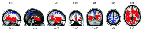
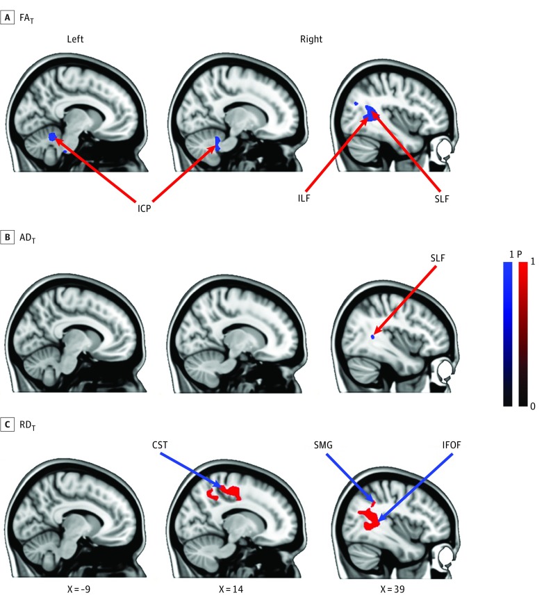
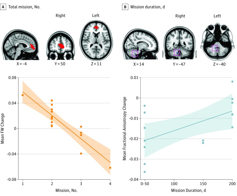
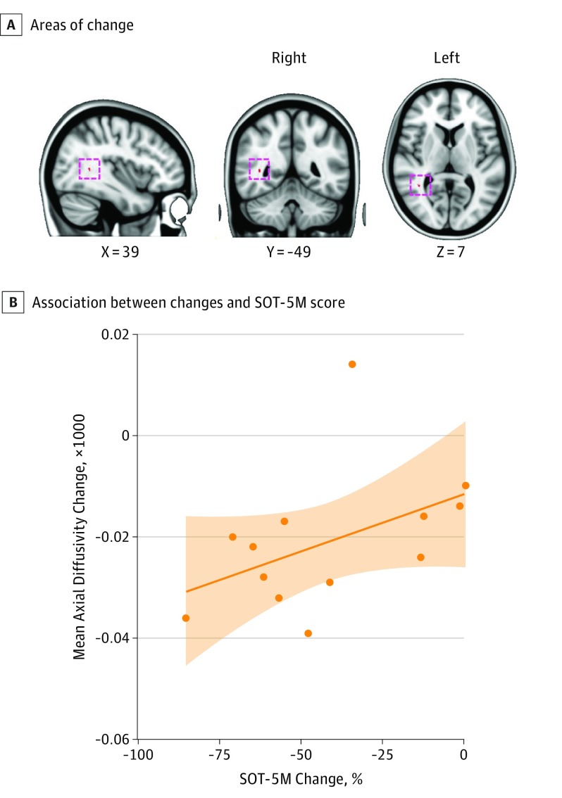
Results: Of the 15 astronauts included, the median (SD) age was 47.2 (1.5) years; 12 were men, and 3 were women. We found a significant, widespread increase in FW volume in the frontal, temporal, and occipital lobes from before spaceflight to after spaceflight. There was also a significant decrease in FW in the posterior aspect of the vertex. All FW changes were significant and ranged from approximately 2.5% to 4.0% across brain regions. We observed white matter changes in the right superior and inferior longitudinal fasciculi, the corticospinal tract, and cerebellar peduncles. All white matter changes were significant and ranged from approximately 0.75% to 1.25%. Spaceflight mission duration was associated with cerebellar white matter change, and white matter changes in the superior longitudinal fasciculus were associated with the balance changes seen in the astronauts from before spaceflight to after spaceflight.

Conclusions and relevance: Free water redistribution with spaceflight likely reflects headward fluid shifts occurring in microgravity as well as an upward shift of the brain within the skull. White matter changes were of a greater magnitude than those typically seen during the same period with healthy aging. Future, prospective assessments are required to better understand the recovery time and behavioral consequences of these brain changes.


Conflict of interest statement

Conflict of Interest Disclosures: None reported.

Figures

Figure 1.
Figure 1.. Preflight to Postflight Change in Free Water (FW) in Astronauts (P < .05; Familywise Error Corrected)
Clusters in red indicate areas where FW significantly increased, and clusters in blue indicate where FW significantly decreased as a function of spaceflight. The results are overlaid on the Montreal Neurological Institute standard space brain template.
Figure 2.
Figure 2.. Association of Spaceflight With White Matter Microstructure as Measured by Changes From Before Spaceflight to After Spaceflight for Free Water (FW)–Corrected Fractional Anisotropy (FAT), Axial Diffusivity (ADT), and Radial Diffusivity (RDT)
A and B, Blue gradients show regions in which the FAT and ADT values significantly decreased as a function of spaceflight (P < .05; familywise error corrected). The red gradient (C) shows regions in which the RDT significantly increased as a function of spaceflight (P < .05, familywise error corrected). The results are overlaid on the Montreal Neurological Institute standard space brain template. CST indicates corticospinal tract; ICP, inferior cerebellar peduncle; ILF, inferior longitudinal fasciculus; SLF, superior longitudinal fasciculus; SMG, supramarginal gyrus; IFOF, inferior-occipital fasciculus.
Figure 3.
Figure 3.. Association of Spaceflight With Free Water (FW) Distribution and Fractional Anisotropy (FAT) Measures
The clusters show brain areas where there was a significant association between the preflight to postflight change in FW and the total count of missions (A) and spaceflight-associated FA change and the mission duration (B) (P < .05; familywise error corrected). The results are overlaid on the Montreal Neurological Institute standard space brain template. The orange scatterplot depicts the mean change in FW of the cluster shown as a function of the number of missions. The blue scatterplot shows the mean change in FA of the cluster shown as a function of the current mission duration. The mission duration is expressed in bins of 50-day increments to prevent identification of individuals based on flight duration.
Figure 4.
Figure 4.. Association Between Spaceflight-Induced Changes of Axial Diffusivity (ADT) and Sensory Organization Test (SOT) 5M Balance Score
A, The red cluster (cluster size 13 mm3) shows the area in which the change in AD values was significantly associated with the change in SOT-5M performance (P < .05; familywise error corrected). B, The scatterplot depicts the association between the mean change of ADT value in the cluster shown in the upper panel and the difference in equilibrium score (expressed in percentage) of SOT-5M from preflight to postflight. The shaded area indicates the 95% confidence limits. The results are overlaid on the Montreal Neurological Institute standard space brain template.

Comment in

References

    1. Koppelmans V, Bloomberg JJ, Mulavara AP, Seidler RD. Brain structural plasticity with spaceflight. NPJ Microgravity. 2016;2(1):2. - PMC - PubMed
    1. Roberts DR, Albrecht MH, Collins HR, et al. . Effects of spaceflight on astronaut brain structure as indicated on MRI. N Engl J Med. 2017;377(18):1746-1753. doi:10.1056/NEJMoa1705129 - DOI - PubMed
    1. Alperin N, Bagci AM, Lee SH. Spaceflight-induced changes in white matter hyperintensity burden in astronauts. Neurology. 2017;89(21):2187-2191. doi:10.1212/WNL.0000000000004475 - DOI - PubMed
    1. Vernooij MW, Ikram MA, Vrooman HA, et al. . White matter microstructural integrity and cognitive function in a general elderly population. Arch Gen Psychiatry. 2009;66(5):545-553. doi:10.1001/archgenpsychiatry.2009.5 - DOI - PubMed
    1. Seidler RD, Bernard JA, Burutolu TB, et al. . Motor control and aging: links to age-related brain structural, functional, and biochemical effects. Neurosci Biobehav Rev. 2010;34(5):721-733. doi:10.1016/j.neubiorev.2009.10.005 - DOI - PMC - PubMed

Publication types

MeSH terms