. 2019 Mar 15;79(6):1165-1177.
doi: 10.1158/0008-5472.CAN-18-0921. Epub 2019 Jan 23.

An ARC-Regulated IL1β/Cox-2/PGE2/β-Catenin/ARC Circuit Controls Leukemia-Microenvironment Interactions and Confers Drug Resistance in AML

Affiliations

An ARC-Regulated IL1β/Cox-2/PGE2/β-Catenin/ARC Circuit Controls Leukemia-Microenvironment Interactions and Confers Drug Resistance in AML

Bing Z Carter et al. Cancer Res. .

Abstract

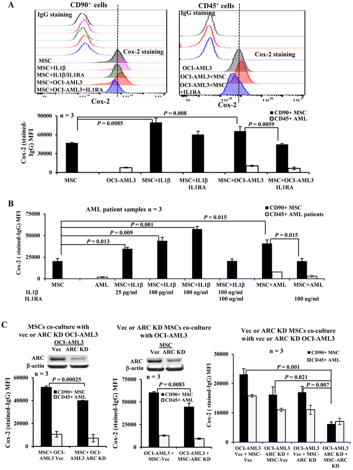
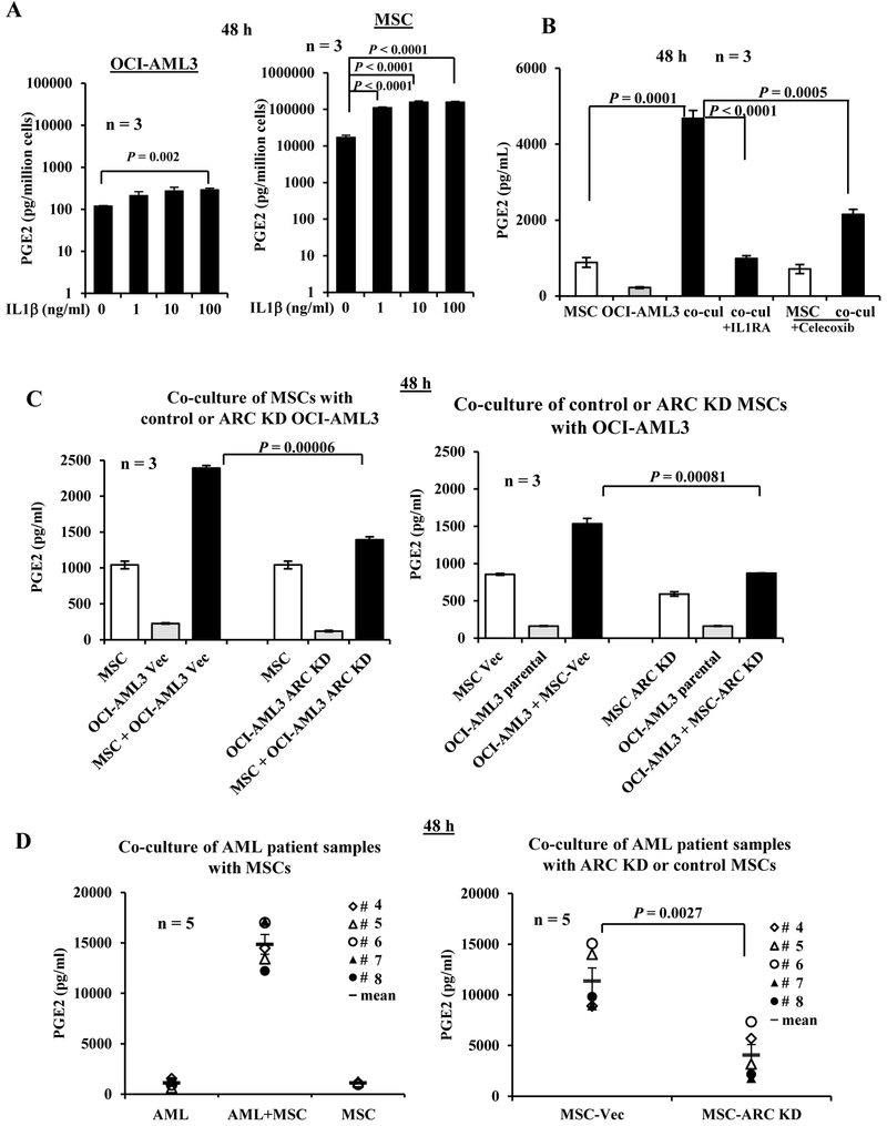
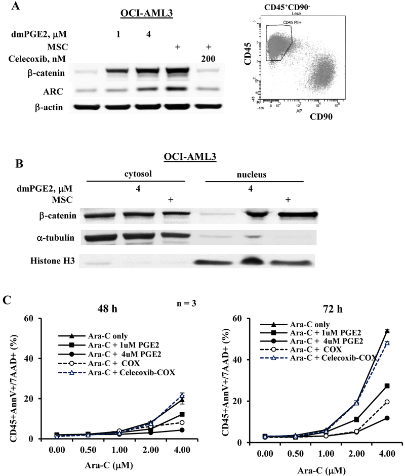
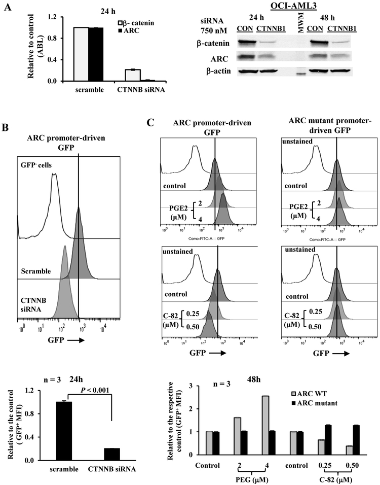
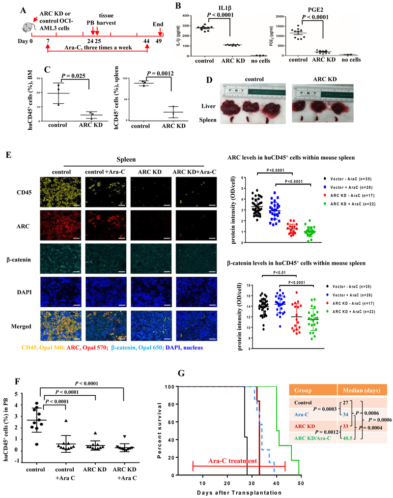
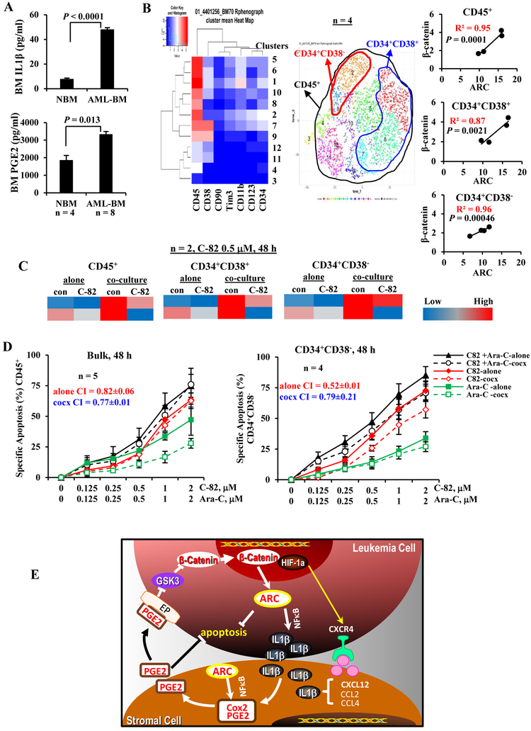
The apoptosis repressor with caspase recruitment domain (ARC) protein is a strong independent adverse prognostic marker in acute myeloid leukemia (AML). We previously reported that ARC regulates leukemia-microenvironment interactions through the NFκB/IL1β signaling network. Malignant cells have been reported to release IL1β, which induces PGE2 synthesis in mesenchymal stromal cells (MSC), in turn activating β-catenin signaling and inducing the cancer stem cell phenotype. Although Cox-2 and its enzymatic product PGE2 play major roles in inflammation and cancer, the regulation and role of PGE2 in AML are largely unknown. Here, we report that AML-MSC cocultures greatly increase Cox-2 expression in MSC and PGE2 production in an ARC/IL1β-dependent manner. PGE2 induced the expression of β-catenin, which regulated ARC and augmented chemoresistance in AML cells; inhibition of β-catenin decreased ARC and sensitized AML cells to chemotherapy. NOD/SCIDIL2RγNull-3/GM/SF mice transplanted with ARC-knockdown AML cells had significantly lower leukemia burden, lower serum levels of IL1β/PGE2, and lower tissue human ARC and β-catenin levels, prolonged survival, and increased sensitivity to chemotherapy than controls. Collectively, we present a new mechanism of action of antiapoptotic ARC by which ARC regulates PGE2 production in the tumor microenvironment and microenvironment-mediated chemoresistance in AML.Significance: The antiapoptotic protein ARC promotes AML aggressiveness by enabling detrimental cross-talk with bone marrow mesenchymal stromal cells.


Conflict of interest statement

Conflicts of interests: None

Figures

Figure 1.
Figure 1.
AML-MSC co-cultures increase Cox-2 in MSCs in an IL1β- and ARC-dependent manner. A. Cox-2 expression in MSCs (CD90+) and OCI-AML3 (CD45+) cells cultured alone or co-cultures, without or with IL1β (100 ng/ml) and/or ILIRA (100 ng/ml) treatments for 48 h. The upper panel is the histogram of one representative experiment and the lower panel is the results of 3 independent experiments with 3 different MSCs. B. Cox-2 expression in MSCs (CD90+) and primary patient samples (CD45+) (n = 3, Table 1, samples 1-3) cultured alone or co-cultures, without or with IL1β (25 pg/ml, 100 pg/ml, or 100 ng/ml) and/or ILIRA (100 ng/ml) treatments for 48 h. The same MSC was used for all three co-cultures. C. Cox-2 expression in MSC and OCI-AML3 cells by flow cytometry under MSCs co-cultured with control or ARC KD OCI-AML3 (left), control or ARC KD MSCs co-cultured with OCI-AML3 (center), or control or ARC KD MSCs co-cultured with control or ARC KD OCI-AML3 (right) for 48 h. Vec, vector control. Three independent experiments were performed.
Figure 2.
Figure 2.
AML-MSC co-cultures increase secreted PGE2 in an IL1β- and ARC-dependent manner. A. OCI-AML3 cells or MSCs were cultured in the absence or presence of IL1β (1 to 100 ng/ml) for 48 h. B. OCI-AML3 and MSCs were cultured alone or co-cultured (co-cul) without or with IL1RA (100 ng/ml) or Celecoxib (200 nM) for 48 h. C. MSCs were co-cultured with vector control or ARC KD OCI-AML3 (left) and vector control or ARC KD MSC were co-cultured with OCI-AML3 cells (right) for 48 h. D. Cells from AML patient samples (n = 5, Table 1, samples 4-8) were co-cultured with MSCs (left) and with ARC KD or control MSCs (right) for 48 h. PGE2 levels in the supernatant were determined by ELISA. Vec, vector. Cell line experiments were done in triplicates. Three MSCs were used for A and B.
Figure 3.
Figure 3.
AML cells treated dmPGE2 or co-cultured with MSCs express increased β-catenin and ARC and are more resistance to chemotherapy. A. OCI-AML3 cells were treated with dmPGE2 (1 or 4 μM) or co-cultured with MSCs without or with Cox-2 inhibitor Celecoxib (200 nM) for 48 h. β-catenin and ARC protein levels were determined by western blot. For co-culture experiments, OCI-AML3 cells were FACS-sorted after co-culture and west blot was done using the lysate from the sorted CD45+CD90 cells as shown in the histogram. B. OCI-AML3 cells were treated with dmPGE2 (4 μM) or co-cultured with MSCs for 48 h. Cytosolic and nuclear β-catenin levels were determined by western blot. C. OCI-AML cells were treated with Ara-C with or without dmPGE2 (1 or 4 μM) or with MSC co-culture in the absence or presence of Cox-2 inhibitor Celecoxib (200 nM) for 48 and 72 h. Apoptosis was assessed by flow cytometry. COX, co-culture.
Figure 4.
Figure 4.
β-catenin regulates ARC expression in AML cells. A. OCI-AML3 cells were transfected with a control siRNA (scramble) or smart pool siRNAs against β-catenin gene (CTNNB1 siRNA) (750 nM) by Amaxa electroporation. The RNA (24 h) and protein (24 and 48 h) levels of β-catenin and ARC were determined by RT-PCR and western blot, respectively, after transfection. CON, scramble control. MWM, molecular weight marker. B. OCI-AML3 cells transduced with ARC promoter-driven GFP reporter gene were transfected with scramble control or β-catenin siRNAs and GFP levels were determined 24 h after transfection. C. OCI-AML3 cells transduced with ARC promoter (WT)- or ARC mutant promoter-driven GFP reporter gene were treated with PGE2 or C-82 for 48 h. For B and C, histograms (top panels) are representative results from one experiment and bar grafts (lower panels) are results of triplicates. GFP levels were determined by flow cytometry. Untransduced OCI-AML3 cells were used as a negative control for GFP.
Figure 5.
Figure 5.
Inhibition of ARC in AML cells reduces serum IL1β and PGE2 levels, decreases leukemia burden in various tissues, prolongs survivals, and sensitizes to Ara-C in NSGS mice. A. In vivo experiment scheme. B. IL1β and PGE2 levels in serum of mice harboring control or ARC KD OCI-AML3 cells determined by ELISA. C. huCD45 positivity in BM and spleen of mice harboring control or ARC KD OCI-AML3 cells determined by flow cytometry. D. Liver and spleen from mice harboring control or ARC KD OCI-AML3 cells. E. Expression of ARC and β-catenin in spleen huCD45+ cells of mice harboring control or ARC KD OCI-AML3 cells with or without Ara-C treatment, determined by Opal/TSA multiplex IHC staining and Vectra multispectral imaging analysis. Left, huCD45 (yellow), ARC (red), β-catenin (cyan), DAPI (blue), and merged imaging (objective lens ×20, scale bar 50 µm). Fluorophores for CD45, ARC, and β-catenin are Opal 540, Opal 570 and Opal 650, respectively. Right, the quantitation of ARC and β-catenin expression in huCD45+ cells of mouse spleen: 35 fields in control cell-injected mice (7,164 to 10,873 cells/field, total 319,431 cells), 26 fields in control cell-injected mice with Ara-C treatment (2,635 to 4,777 cells/field, total 99,727 cells), 17 fields in ARC KD cell-injected mice (610 to 5,021 cells/fields, total 37,206 cells), and 22 fields in ARC KD cell-injected mice with Ara-C treatment (248 to 3,927 cells/field, total 45,431 cells); respectively. F. huCD45 positivity in PB of mice harboring control or ARC KD OCI-AML3 cells with or without Ara-C treatment by flow cytometry. G. Survival of mice injected with control or ARC KD OCI-AML3 cells without or with Ara-C treatment.
Figures 6.
Figures 6.
PGE2/β-catenin/ARC cascade and targeting in primary AML samples and the proposed mechanism of ARC action. A. IL1β and PGE2 levels in BM samples from AML patients (n = 8) (Table 1, samples 9-16) and normal controls (NBM, n = 4) by ELISA. B. Correlation of ARC and β-catenin protein levels, determined by CyTOF in various BM cell populations of AML patients (n = 4) (Table 1, samples 17-20). C. Levels of ARC protein, determined by CyTOF in AML patient samples (n = 2) (Table 1, samples 21 and 22) treated with β-catenin inhibitor C-82 (0.5 μM) without or with MSC co-culture for 48 h. Protein levels determined by CyTOF are expressed as Arcsinh-transformed counts. D. AML patient samples were treated with C-82, Ara-C, or both for 48 h. Apoptosis was determined in blasts (n = 5) (Table 1, samples 23-27) and CD34+CD38 (n = 4) (Table 1, samples 23-26) cells. cocx, co-culture. E. Proposed mechanism of action: ARC, regulated by β-catenin, mediates leukemia stromal interaction through ARC-IL1β/Cox-2/PGE2/β-catenin circuit.

References

    1. Carter BZ, Qiu YH, Zhang N, Coombes KR, Mak DH, Thomas DA, et al. Expression of ARC (apoptosis repressor with caspase recruitment domain), an antiapoptotic protein, is strongly prognostic in AML. Blood. 2011;117:780–7. - PMC - PubMed
    1. Mak PY, Mak DH, Mu H, Shi Y, Ruvolo P, Ruvolo V, et al. Apoptosis repressor with caspase recruitment domain is regulated by MAPK/PI3K and confers drug resistance and survival advantage to AML. Apoptosis. 2014;19:698–707. - PMC - PubMed
    1. Carter BZ, Mak PY, Chen Y, Mak DH, Mu H, Jacamo R, et al. Anti-apoptotic ARC protein confers chemoresistance by controlling leukemia-microenvironment interactions through a NFkappaB/IL1beta signaling network. Oncotarget. 2016;7(15):20054–67. - PMC - PubMed
    1. Morrison SJ, Spradling AC. Stem cells and niches: mechanisms that promote stem cell maintenance throughout life. Cell. 2008;132:598–611. - PMC - PubMed
    1. Konopleva M, Tabe Y, Zeng Z, Andreeff M. Therapeutic targeting of microenvironmental interactions in leukemia: mechanisms and approaches. Drug Resist Updat. 2009;12:103–13. - PMC - PubMed

Publication types

MeSH terms