. 2019 Mar 22;294(12):4359-4367.
doi: 10.1074/jbc.RA118.006477. Epub 2019 Jan 23.

The phenylketonuria-associated substitution R68S converts phenylalanine hydroxylase to a constitutively active enzyme but reduces its stability

Affiliations

The phenylketonuria-associated substitution R68S converts phenylalanine hydroxylase to a constitutively active enzyme but reduces its stability

Crystal A Khan et al. J Biol Chem. .

Abstract

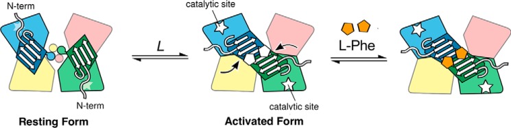
The naturally occurring R68S substitution of phenylalanine hydroxylase (PheH) causes phenylketonuria (PKU). However, the molecular basis for how the R68S variant leads to PKU remains unclear. Kinetic characterization of R68S PheH establishes that the enzyme is fully active in the absence of allosteric binding of phenylalanine, in contrast to the WT enzyme. Analytical ultracentrifugation establishes that the isolated regulatory domain of R68S PheH is predominantly monomeric in the absence of phenylalanine and dimerizes in its presence, similar to the regulatory domain of the WT enzyme. Fluorescence and small-angle X-ray scattering analyses establish that the overall conformation of the resting form of R68S PheH is different from that of the WT enzyme. The data are consistent with the substitution disrupting the interface between the catalytic and regulatory domains of the enzyme, shifting the equilibrium between the resting and activated forms ∼200-fold, so that the resting form of R68S PheH is ∼70% in the activated conformation. However, R68S PheH loses activity 2 orders of magnitude more rapidly than the WT enzyme at 37 °C and is significantly more sensitive to proteolysis. We propose that, even though this substitution converts the enzyme to a constitutively active enzyme, it results in PKU because of the decrease in protein stability.

Keywords: allosteric regulation; allostery; analytical ultracentrifugation; enzyme kinetics; enzyme stability; hydroxylase; phenylalanine hydroxylase; phenylketonuria; protein conformation; protein structure; small-angle X-ray scattering (SAXS).


Conflict of interest statement

The authors declare that they have no conflicts of interest with the contents of this article

Figures

Figure 1.
Figure 1.
Model for allosteric regulation of phenylalanine hydroxylase (8, 14). Catalytic domains are shown in blue, magenta, yellow, and green; the C-terminal helices are depicted as circles; and the regulatory domains corresponding to the blue and green catalytic domains are shown as trapezoids. The allosteric sites for phenylalanine at the interface of the regulatory domain dimer in the activated form are shown as pentagons.
Figure 2.
Figure 2.
Fluorescence emission spectra of WT PheH (A) and R68S PheH (B) in the absence (dashed line) and presence (solid line) of 1 mm phenylalanine. All samples were excited at 295 nm at 23 °C, pH 7.5. The data for the WT enzyme are from Khan and Fitzpatrick (14).
Figure 3.
Figure 3.
Van Holde-Weischet analysis of R68S RDPheH(25–117). Shown are WT RDPheH(25–117) (black) and R68S RDPheH(25–117) (red) in the absence (solid circles) and presence (open circles) of 1 mm phenylalanine. The RDPheH(25–117) data are from Zhang et al. (13).
Figure 4.
Figure 4.
Kratky representations of the SEC-SAXS intensities of tetrameric PheH R68S and WT PheH in the absence (A) and in the presence (B) of phenylalanine. The WT data are from Meisburger et al. (8).
Figure 5.
Figure 5.
Phenylalanine dependence of the SAXS profile of R68S PheH. Inset, change in the scattering curves of R68S PheH upon binding phenylalanine fit to Equation 1. a.u., arbitrary units.
Figure 6.
Figure 6.
Stability of R68S (●) and WT PheH (□) at 37 °C. The enzymes were incubated at 37 °C in in 0.2 m Hepes, pH 7.0, and the remaining activity was determined at the indicated times. The data are from the averages of three separate experiments. The line is from a fit of the data to an exponential decay with an end point of zero. Error bars, S.D.
Figure 7.
Figure 7.
SDS-PAGE of limited proteolysis by trypsin of WT and R68S PheH in the absence and presence of phenylalanine by trypsin. The enzymes (0.8 mg/ml) were incubated with 16 μg/ml trypsin at 23 °C and quenched at the indicated times with 1 mg/ml phenylmethylsulfonyl fluoride. When denoted, WT and R68S PheH samples were incubated with a final concentration of 1 mm phenylalanine for 3 min at 23 °C prior to the reaction.
Figure 8.
Figure 8.
Location of Arg-68 in the PheH structure: Arg-68 (cyan) in the isolated regulatory domain with phenylalanine bound (green) (PDB code 5FII) (A) and Arg-68 in the resting form of intact PheH with adjacent subunits colored in blue and tan (PDB code 5EGQ) (B).
Figure 9.
Figure 9.
Evaluation of atomic models for PheH R68S in the resting state. A, three candidate models for tetrameric R68S were evaluated: the crystallographic structure of the inactive WT PheH (PDB code 5FGJ) (I); a minimum ensemble selected from a pool of randomly generated structures with undocked regulatory domains (II); and a mixture of inactive and active WT conformations (III) (8). The catalytic and tetramerization domains are gray, and the regulatory domains are red. B, scattering curves predicted from models I, II, and III (solid lines, top panel) are compared with the SAXS profile of PheH R68S in the absence of phenylalanine (blue, gray, and magenta circles; top). The SAXS profile of PheH R68S in the presence of 1 mm phenylalanine is shown for reference (orange circles; top). Curves are offset for clarity. The residuals (bottom; corresponding colors) are largest for model I (χ2 = 7.4), intermediate for model II (χ2 = 5.6), and smallest for model III (χ2 = 2.8).

Similar articles

Cited by

References

    1. Fitzpatrick P. F. (2003) Mechanism of aromatic amino acid hydroxylation. Biochemistry 42, 14083–14091 10.1021/bi035656u - DOI - PMC - PubMed
    1. Shiman R., Jones S. H., and Gray D. W. (1990) Mechanism of phenylalanine regulation of phenylalanine hydroxylase. J. Biol. Chem. 265, 11633–11642 - PubMed
    1. Fitzpatrick P. F. (2012) Allosteric regulation of phenylalanine hydroxylase. Arch. Biochem. Biophys. 519, 194–201 10.1016/j.abb.2011.09.012 - DOI - PMC - PubMed
    1. Flydal M. I., and Martinez A. (2013) Phenylalanine hydroxylase: function, structure, and regulation. IUBMB Life 65, 341–349 10.1002/iub.1150 - DOI - PubMed
    1. Fitzpatrick P. F. (2015) Structural insights into the regulation of aromatic amino acid hydroxylation. Curr. Opin. Struct. Biol. 35, 1–6 10.1016/j.sbi.2015.07.004 - DOI - PMC - PubMed

Publication types

Substances