. 2019 Jan 23;11(476):eaat1199.
doi: 10.1126/scitranslmed.aat1199.

Hypertrophic cardiomyopathy mutations in MYBPC3 dysregulate myosin

Affiliations

Hypertrophic cardiomyopathy mutations in MYBPC3 dysregulate myosin

Christopher N Toepfer et al. Sci Transl Med. .

Abstract

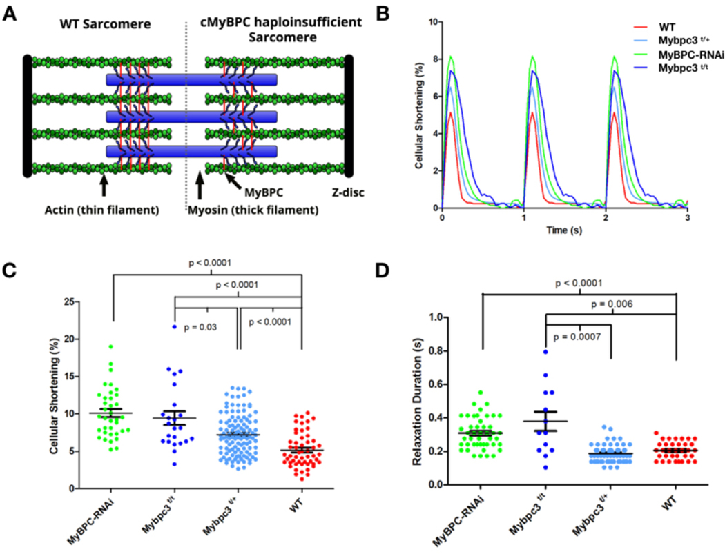
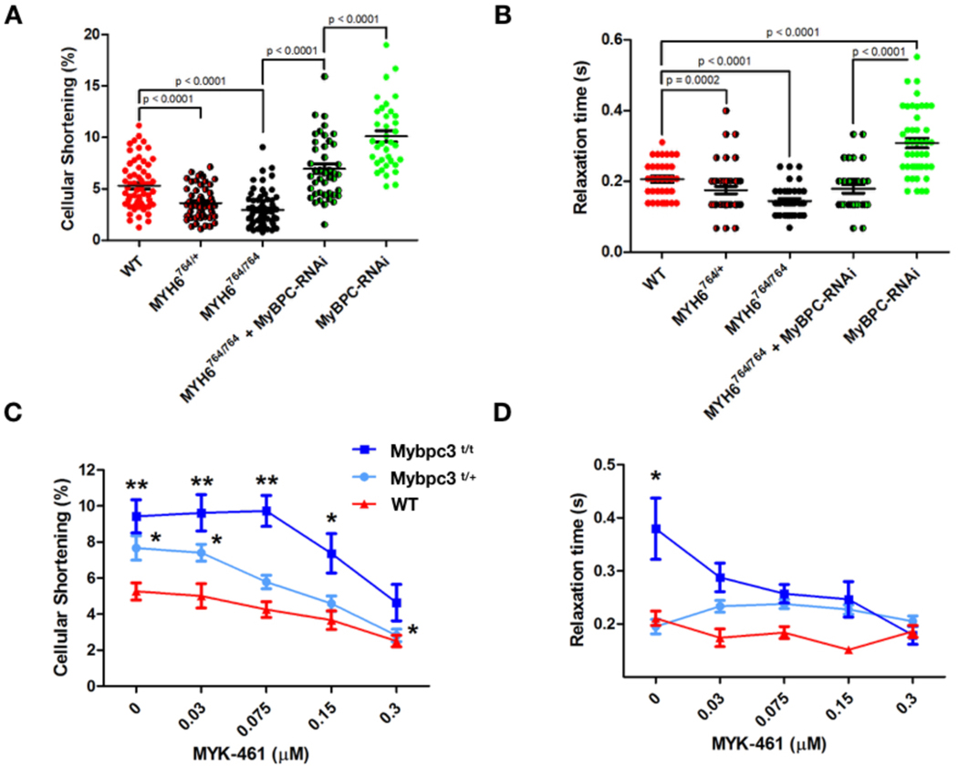
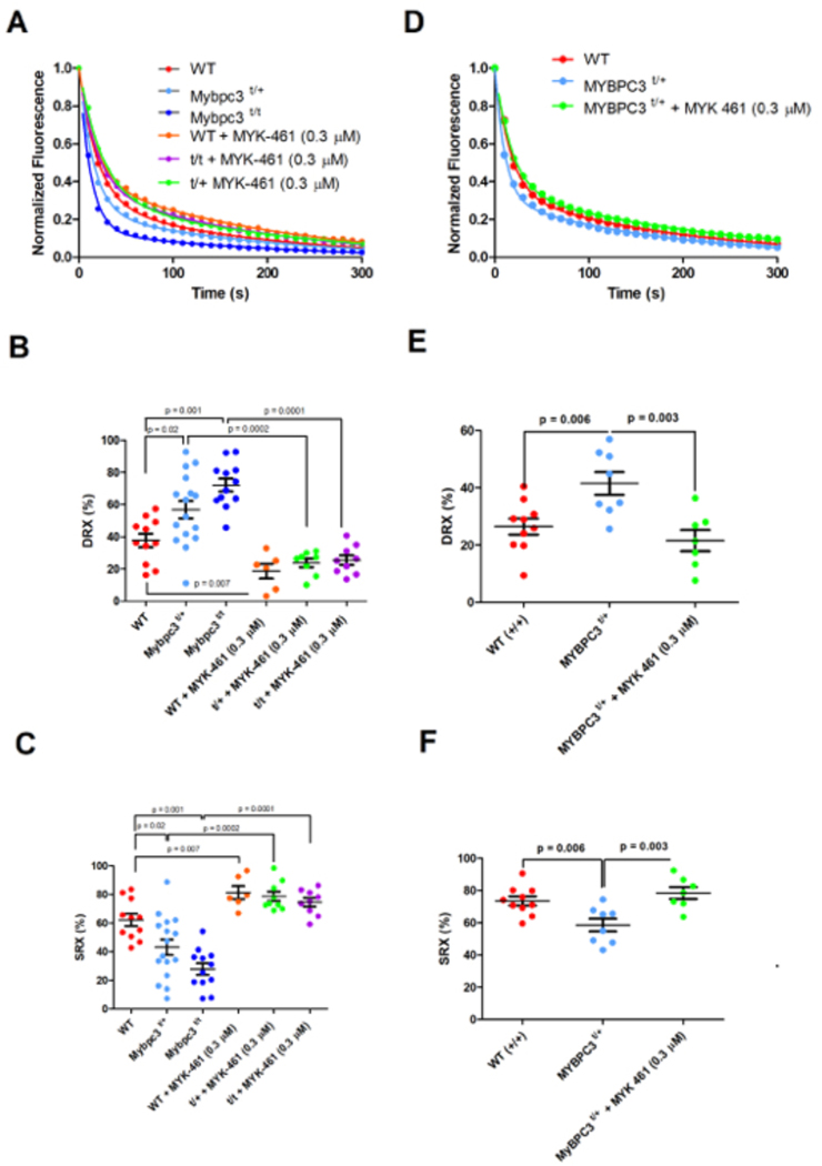
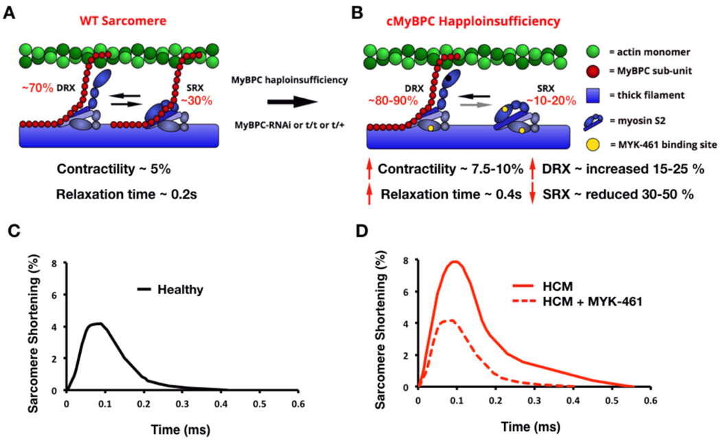
The mechanisms by which truncating mutations in MYBPC3 (encoding cardiac myosin-binding protein C; cMyBPC) or myosin missense mutations cause hypercontractility and poor relaxation in hypertrophic cardiomyopathy (HCM) are incompletely understood. Using genetic and biochemical approaches, we explored how depletion of cMyBPC altered sarcomere function. We demonstrated that stepwise loss of cMyBPC resulted in reciprocal augmentation of myosin contractility. Direct attenuation of myosin function, via a damaging missense variant (F764L) that causes dilated cardiomyopathy (DCM), normalized the increased contractility from cMyBPC depletion. Depletion of cMyBPC also altered dynamic myosin conformations during relaxation, enhancing the myosin state that enables ATP hydrolysis and thin filament interactions while reducing the super relaxed conformation associated with energy conservation. MYK-461, a pharmacologic inhibitor of myosin ATPase, rescued relaxation deficits and restored normal contractility in mouse and human cardiomyocytes with MYBPC3 mutations. These data define dosage-dependent effects of cMyBPC on myosin that occur across the cardiac cycle as the pathophysiologic mechanisms by which MYBPC3 truncations cause HCM. Therapeutic strategies to attenuate cMyBPC activity may rescue depressed cardiac contractility in patients with DCM, whereas inhibiting myosin by MYK-461 should benefit the substantial proportion of patients with HCM with MYBPC3 mutations.


Conflict of interest statement

Competing Financial Interest

C.E.S. and J.G.S. are founders and own shares in Myokardia Inc., a startup company that is developing therapeutics that target the sarcomere.

Figures

Figure 1:
Figure 1:. Contractile characterization of cMyBPC mouse models.
A) A schematic depiction of the WT sarcomere with normal cMyBPC integration (left half) and the consequences of mutations that deplete cMyBPC quantities in the sarcomere (right half). B) Representative contractile waveforms from isolated cardiomyocytes paced at 1Hz. Sarcomere lengths of isolated cardiomyocytes were tracked to define the percentage shortening per cell and duration of relaxation. Each trace is the averaged waveform across all cells analyzed for each treatment group. C) Comparisons of cellular shortening of isolated cardiomyocytes from four mice with different genotypes. (Cells analyzed: MyBPC-RNAi = 36; Mybpc3t/t = 23; Mybpc3t/+ = 118,WT= 53.) Data is plot as mean ± SEM. D) Measures of duration from peak contraction to relaxation in seconds plot as mean ± SEM (Cells analyzed: MyBPC-RNAi = 34; Mybpc3t/t = 13; Mybpc3t/+= 61,WT = 30).
Figure 2:
Figure 2:. Genetic and pharmacological manipulation of cardiomyocytes depleted for cMyBPC.
A) Sarcomere contractility in isolated cardiomyocytes from WT, Myh6764/+, Myh6764/764, Myh6764/764 + MyBPC-RNAi, and MyBPC-RNAi mice. (Cells analyzed; WT = 63, Myh6764/+ = 55, Myh6764/764 = 71, Myh6764/764 + MyBPC-RNAi = 43, MyBPC-RNAi = 36) B) Sarcomere relaxation of isolated cardiomyocytes from WT, Myh6764/+, Myh6764/764, Myh6764/764 treated with MyBPC-RNAi, and MyBPC-RNAi mice. Individual data points are plot with mean ± SEM indicated. All significant p values are indicated on the graph. (Cells analyzed; WT = 30, Myh6764/+ = 45, Myh6764/764 = 29, Myh6764/764 + MyBPC-RNAi = 31, MyBPC-RNAi = 45) C) Sarcomere contractility of cardiomyocytes treated with 0.03 – 0.3 μM MYK-461. More than 20 cardiomyocytes were analyzed for each drug concentration and treatment group) D) Sarcomere relaxation of cardiomyocytes treated with 0.03 – 0.3 μM MYK-461. All data is displayed as mean ± SEM. *p < 0.01 and **p < 0.0001 denote comparisons with WT without MYK-461.
Figure 3:
Figure 3:. Mant-ATP assessment and correction of SRX and DRX ratios in mouse and human myocardium.
A) Raw average Mant-ATP fluorescence decay curves plot from fluorescence decay due to dark ATP wash, acquisition duration 5 minutes. Data points are the mean of ~12 separate experiments from 3 separate individuals in each genotype/treatment group. Data is fit by a double exponential decay to assess ratios of DRX and SRX heads (Methods). B) Plot of the initial rapid decay amplitude corresponding to DRX heads. C) Plot of the second exponents slow decay amplitude corresponding to SRX heads. D) Average Mant-ATP fluorescence decay curves of unrelated human hearts: three without HCM (WT) and three HCM heart with MYBPC3 t/+ mutations. Each curve is the average of 12 experiments from three separate samples in each treatment group. Data is fit by double exponential decay to assess ratios of DRX and SRX heads in the myocardium. E) Plot of the initial rapid decay amplitude corresponding to DRX heads. F) Plot of the second exponents slow decay amplitude corresponding to SRX heads. All data is presented in each panel, plotted as mean ± SEM with significances indicated with p values.
Figure 4:
Figure 4:. Schematic of the mechanism by which haploinsufficiency of cMyBPC causes HCM.
A) Schematic of a WT sarcomere with normal cMyBPC quantities, and physiologic contractility and relaxation due to appropriate proportions myosins in state of super relaxation (SRX) with low energy consumption or disordered relaxation (DRX) with high energy consumption. B) Schematic of an HCM sarcomere with reduced cMyBPC quantities that dysregulates the proportions of myosins in DRX (increased) and SRX (reduced). The increased proportion of DRX myosins causes inappropriate sarcomere hypercontractility. Yellow denotes the approximate interaction site of MYK-461 on myosin, which abates the hypercontractile phenotype and shifts the myosin DRX:SRX equilibrium back toward normal. C) Contractile waveform of an individual cardiomyocyte isolated from a healthy individual, showing normal sarcomere shortening and normal relaxation duration. D) Contractile waveform from a cardiomyocyte isolated from a HCM patient with cMyBPC haploinsufficiency, showing hypercontractility with increased sarcomere shortening and slowed relaxation. MYK-461 normalizes the HCM phenotypes of hypercontractility by restoring physiologic balance of myosin DRX:SRX.

Comment in

Similar articles

Cited by

References

    1. Maron BJ, Gardin JM, Flack JM, Gidding SS, Kurosaki TT and Bild DE. Prevalence of hypertrophic cardiomyopathy in a general population of young adults. Echocardiographic analysis of 4111 subjects in the CARDIA Study. Coronary Artery Risk Development in (Young) Adults. Circulation. 1995;92:785–9. - PubMed
    1. Maron BJ, Casey SA, Poliac LC, Gohman TE, Almquist AK and Aeppli DM. Clinical course of hypertrophic cardiomyopathy in a regional United States cohort. JAMA. 1999;281:650–5. - PubMed
    1. Green EM, Wakimoto H, Anderson RL, Evanchik MJ, Gorham JM, Harrison BC, Henze M, Kawas R, Oslob JD, Rodriguez HM, Song Y, Wan W, Leinwand LA, Spudich JA, McDowell RS, Seidman JG and Seidman CE. A small-molecule inhibitor of sarcomere contractility suppresses hypertrophic cardiomyopathy in mice. Science. 2016;351:617–21. - PMC - PubMed
    1. Walsh R, Thomson KL, Ware JS, Funke BH, Woodley J, McGuire KJ, Mazzarotto F, Blair E, Seller A, Taylor JC, Minikel EV, Exome Aggregation C, MacArthur DG, Farrall M, Cook SA and Watkins H. Reassessment of Mendelian gene pathogenicity using 7,855 cardiomyopathy cases and 60,706 reference samples. Genet Med. 2017;19:192–203. - PMC - PubMed
    1. Moolman-Smook JC, De Lange WJ, Bruwer EC, Brink PA and Corfield VA. The origins of hypertrophic cardiomyopathy-causing mutations in two South African subpopulations: a unique profile of both independent and founder events. Am J Hum Genet. 1999;65:1308–20. - PMC - PubMed

Publication types

LinkOut - more resources