. 2019 Jan 23;11(476):eaav1620.
doi: 10.1126/scitranslmed.aav1620.

A recombinant human protein targeting HER2 overcomes drug resistance in HER2-positive breast cancer

Affiliations

A recombinant human protein targeting HER2 overcomes drug resistance in HER2-positive breast cancer

Lu Yang et al. Sci Transl Med. .

Abstract

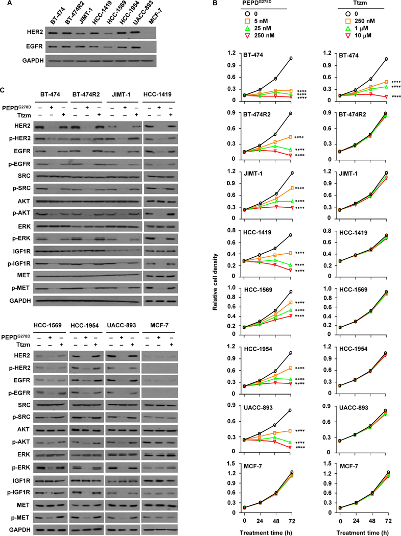
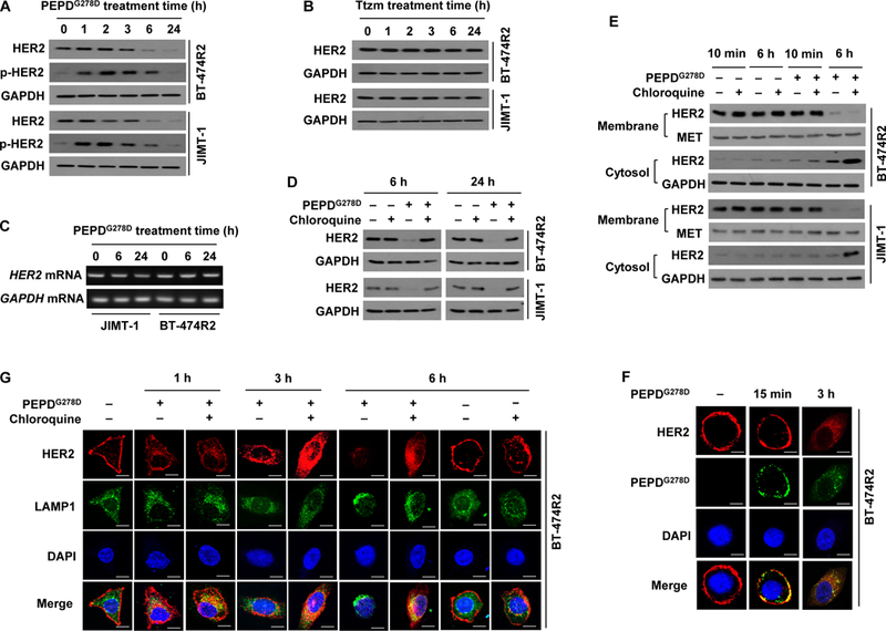
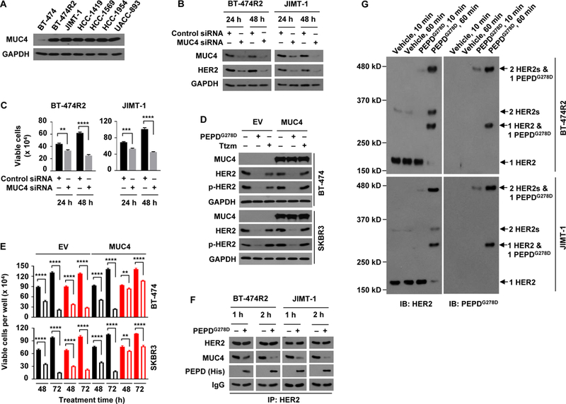
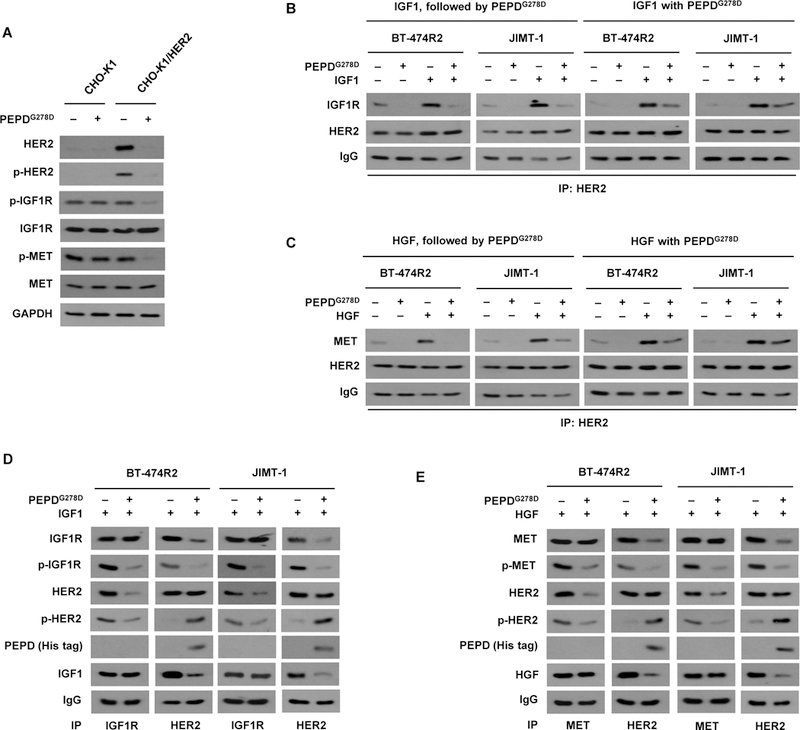
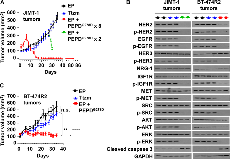
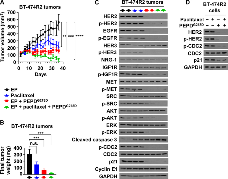
Resistance to current human epidermal growth factor receptor 2 (HER2) inhibitors, such as trastuzumab (Ttzm), is a major unresolved clinical problem in HER2-positive breast cancer (HER2-BC). Because HER2 remains overexpressed in drug-resistant HER2-BC cells, we investigated whether PEPDG278D can overcome the resistance. PEPDG278D is a recombinant enzymatically inactive mutant of human peptidase D, which strongly inhibits HER2 in cancer cells by binding to its extracellular domain. Here, we show that PEPDG278D is highly active in preclinical models of HER2-BC resistant to Ttzm and other HER2 inhibitors and also enhances the therapeutic efficacy of paclitaxel. The therapeutic activity is underscored by its ability to bind to HER2 and free it from protection by mucin 4, disrupt its interplay with other receptor tyrosine kinases, and subsequently direct HER2 for degradation. PEPDG278D also down-regulates epidermal growth factor receptor, which contributes to drug resistance in HER2-BC. In contrast, Ttzm, whose therapeutic activity also depends on its binding to the extracellular domain of HER2, cannot perform any of these functions of PEPDG278D PEPDG278D inhibits HER2-BC cells and tumors that carry clinically relevant molecular changes that confer resistance to Ttzm. Our results show that HER2 remains a critical target in drug-resistant HER2-BC and that PEPDG278D is a promising agent for overcoming drug resistance in this disease.


Figures

Fig. 1.
Fig. 1.. PEPDG278D targets HER2-BC cells resistant to Ttzm.
(A) IB analysis of whole cell lysates from untreated cells. (B) Effects of PEPDG278D and Ttzm on cell proliferation, measured by MTT assay. Each value is mean ± SD (n=3). ****P<0.0001 by one-way ANOVA, followed by Tukey test for comparison to the control. (C) IB analysis of whole cell lysates from cells treated with vehicle, PEPDG278D (25 nM) or Ttzm (1 μM) for 48 hours. The following phosphorylation sites were measured: pY1221/1222-HER2, pY1173-EGFR, pY416-SRC, pS473-AKT, pT202/Y204-ERK, pY1131-IGF1R, and pY1234/1235-MET. Glyceraldehyde 3-phosphate dehydrogenase (GAPDH) was measured as a loading control here and elsewhere.
Fig. 2.
Fig. 2.. PEPDG278D, but not Ttzm, induces internalization and lysosomal degradation of HER2 in Ttzm-resistant HER2-BC cells.
(A and B) IB analysis of whole cell lysates from cells treated with PEPDG278D (25 nM, A) or Ttzm (1 μM, B) for 0–24 hours. p-HER2: pY1221/1222-HER2. (C) RT-PCR analysis of gene expression in cells treated with PEPDG278D (25 nM) for 0–24 hours. (D) IB analysis of whole cell lysates from cells treated with vehicle, PEPDG278D (25 nM), and/or chloroquine (25 μM) for 6 or 24 hours. (E) IB analysis of membrane and cytosol fractions from cells treated with vehicle, PEPDG278D (25 nM) and/or chloroquine (25 μM) for 10 min or 6 hours, using MET and GAPDH as loading controls. (F) Confocal fluorescence staining images of HER2, PEPDG278D (staining its His tag) and nuclei (DAPI) in BT-474R2 cells treated with PEPDG278D (25 nM) for 0 or 15 min or 3 hours. Scale bar: 10 μm. (G) Confocal fluorescence staining images of HER2, LAMP1 (lysosome) and nuclei (DAPI) in BT-474R2 cells with or without treatment with PEPDG278D (25 nM) and/or chloroquine (25 μM) for 0–6 hours. Scale bar, 10 μm.
Fig. 3.
Fig. 3.. PEPDG278D breaks MUC4 protection of HER2 in Ttzm-resistant HER2-BC cells.
(A) IB analysis of whole cell lysates from untreated cells. (B and C) IB analysis of whole cell lysates (B) and trypan blue viability analysis (C) of cells after treatment with siRNA for 24 or 48 hours. (D and E) Cells were transfected with empty vector (EV) or MUC4, and 24 hours later treated with vehicle, PEPDG278D (25 nM) or Ttzm (1 μM), followed by IB analysis of whole cell lysates at 48 hours (D) and trypan blue viability analysis at 48 and 72 hours (E). Filled and unfilled black bars represent vehicle and PEPDG278D, respectively; filled and unfilled red bars represent vehicle and Ttzm, respectively. p-HER2: pY1221/1222-HER2. (F) IB analysis of anti-HER2 IP of whole cell lysates from cells treated with vehicle or PEPDG278D (25 nM) for 1 or 2 hours. (G) IB analysis of whole cell lysates from cells treated with vehicle or PEPDG278D (25 nM) for 10 or 60 min and then treated with BS3 (2 mM) for 30 min. Each value in (C and E) is mean ± SD (n=3), **P<0.01, ***P<0.001, P****<0.0001 by paired two-tailed t-test.
Fig. 4.
Fig. 4.. PEPDG278D disrupts HER2-IGF1R and HER2-MET heterodimers.
(A) IB analysis of whole cell lysates from cells which were transfected with an empty vector (CHO-K1) or human HER2 (CHO-K1/HER2) for 24 hours and then treated with vehicle or PEPDG278D (25 nM) for 48 hours. (B) IB analysis of anti-HER2 IP of whole cell lysates from cells treated first with IGF1 (12.5 nM) for 15 min and then PEPDG278D (25 nM) for 1 hour or treated simultaneously with IGF1 (12.5 nM) and PEPDG278D (25 nM) for 1 hour. (C) IB analysis of anti-HER2 IP of whole cell lysates from cells treated first with HGF (0.3 nM) for 15 min and then with PEPDG278D (25 nM) for 1 hour or treated simultaneously with HGF (0.3 nM) and PEPDG278D (25 nM) for 1 hour. (D) IB analysis of anti-IGF1R IP or anti-HER2 IP of whole cell lysates from cells treated with IGF1 (12.5 nM) with or without PEPDG278D (25 nM) for 1 hour. (E) IB analysis of anti-MET IP or anti-HER2 IP of whole cell lysates from cells treated with HGF (0.3 nM) with or without PEPDG278D (25 nM) for 1 hour. Protein phosphorylation sites are described in Fig. 1C legend.
Fig. 5.
Fig. 5.. PEPDG278D inhibits Ttzm-resistant HER2-BC in vivo.
(A) JIMT-1 tumors in SCID mice, treated with EP (daily, days 1–34, n=10), Ttzm (weekly, days 6–34, n=8), or EP + PEPDG278D x 8 (EP daily, days 1–22; PEPDG278D thrice weekly, days 6–22, 8 doses; n=8). In the fourth group, PEPDG278D was added to the EP control on days 27 and 29 (PEPDG278D x 2; n=4). All tumors were harvested 24 hours after the last treatment, but one group of mice (EP + PEPDG278D x 8) became tumor-free and were observed without treatment for 60 days (days 23–82). (B) IB analysis of tumor homogenates (2 tumors/group). See Fig. 1C legend for protein phosphorylation sites. (C) BT-474R2 tumors in nude mice, treated with EP daily (days 1–34, n = 13), Ttzm weekly (days 6–34, n =15), or EP daily (days 1–34) plus PEPDG278D thrice weekly (days 6–34) (n = 12). All tumors were harvested 24 hours after the last treatment. EP, Ttzm and PEPDG278D were dosed ip at 0.5 mg/kg, 10 mg/kg and 4 mg/kg per dose, respectively. Each value in (A and C) is mean ± SEM **P<0.01, ****P<0.0001, by one-way ANOVA followed by Tukey test; n.s., not significant.
Fig. 6.
Fig. 6.. ombination of PEPDG278D and paclitaxel enhances therapeutic outcome.
(A) BT-474R2 tumors in nude mice, treated with EP (0.5 mg/kg daily, days 2–35, n = 8), paclitaxel (20 mg/kg weekly, days 8–35, n = 7), EP plus PEPDG278D (EP at 0.5 mg/kg daily, days 2–35; PEPDG278D at 8 mg/kg thrice weekly, days 8–35; n = 10), or EP plus paclitaxel plus PEPDG278D (EP at 0.5 mg/kg daily, days 2–35; paclitaxel at 20 mg/kg weekly, days 8–35; PEPDG278D at 4 mg/kg thrice weekly, days 8–35; n = 8). (B) Weights of tumors harvested 24 hours after the last treatment. All agents were dosed ip. (C) IB analysis of tumors homogenates (2 tumors/group). See Fig. 1C legend for protein phosphorylation sites, except for pY15-CDC2. (D) IB analysis of whole cell lysates of cells after treatment with vehicle, paclitaxel (25 nM), and/or PEPDG278D (25 nM) for 24 hours. Each value in (A and B) is mean ± SEM. **P<0.01, ***P<0.001, ****P<0.0001, by one-way ANOVA followed by Tukey test; n.s., not significant.

Similar articles

Cited by

References

    1. Choritz H, Busche G, Kreipe H, On behalf of the Study Group HER2 Monitor, Quality assessment of HER2 testing by monitoring of positivity rates. Virchows Arch. 459, 283–289 (2011). - PMC - PubMed
    1. Witton CJ, Reeves JR, Going JJ, Cooke TG, Bartlett JM, Expression of the HER1–4 family of receptor tyrosine kinases in breast cancer. J. Pathol. 200, 290–297 (2003). - PubMed
    1. Paik S, Hazan R, Fisher ER, Sass RE, Fisher B, Redmond C, Schlessinger J, Lippman ME, King CR, Pathologic findings from the National Surgical Adjuvant Breast and Bowel Project: prognostic significance of erbB-2 protein overexpression in primary breast cancer. J. Clin. Oncol. 8, 103–112 (1990). - PubMed
    1. Slamon DJ, Clark GM, Wong SG, Levin WJ, Ullrich A, McGuire WL, Human breast cancer: correlation of relapse and survival with amplification of the HER-2/neu oncogene. Science 235, 177–182 (1987). - PubMed
    1. Romond EH, Perez EA, Bryant J, Suman VJ, Geyer CE Jr., Davidson NE, Tan-Chiu E, Martino S, Paik S, Kaufman PA, Swain SM, Pisansky TM, Fehrenbacher L, Kutteh LA, Vogel VG, Visscher DW, Yothers G, Jenkins RB, Brown AM, Dakhil SR, Mamounas EP, Lingle WL, Klein PM, Ingle JN, Wolmark N, Trastuzumab plus adjuvant chemotherapy for operable HER2-positive breast cancer. N. Engl. J. Med. 353, 1673–1684 (2005). - PubMed

Publication types

MeSH terms