

Molecular mechanism for NLRP6 inflammasome assembly and activation
- PMID: 30674671
- PMCID: PMC6369754
- DOI: 10.1073/pnas.1817221116
Molecular mechanism for NLRP6 inflammasome assembly and activation
Abstract
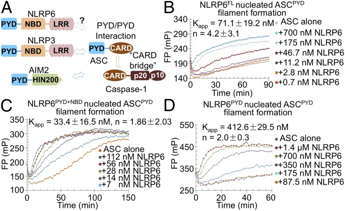
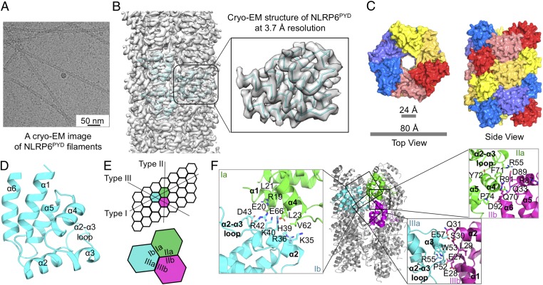
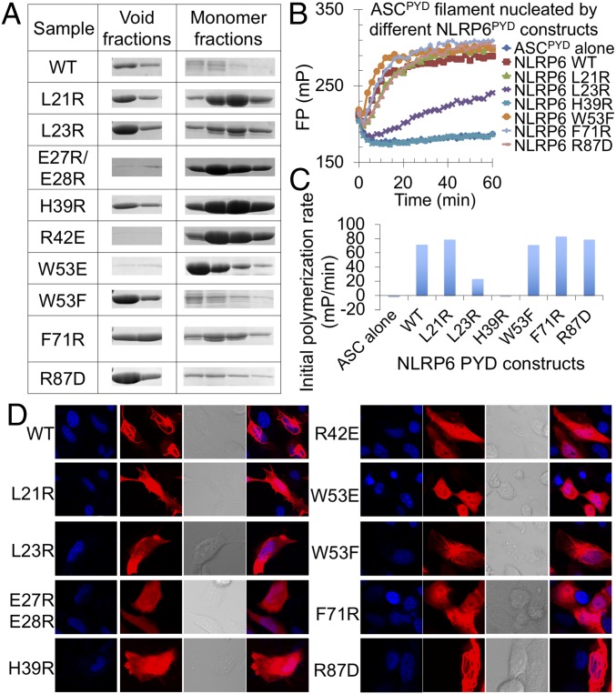
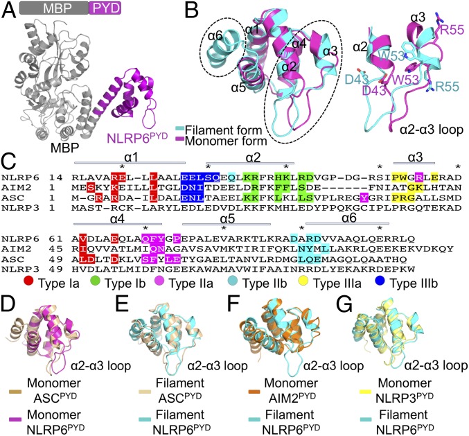
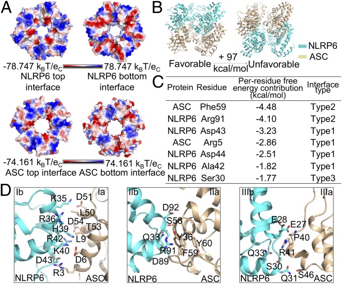
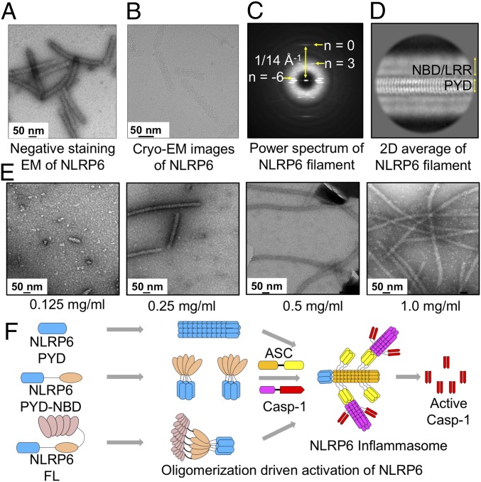
Inflammasomes are large protein complexes that trigger host defense in cells by activating inflammatory caspases for cytokine maturation and pyroptosis. NLRP6 is a sensor protein in the nucleotide-binding domain (NBD) and leucine-rich repeat (LRR)-containing (NLR) inflammasome family that has been shown to play multiple roles in regulating inflammation and host defenses. Despite the significance of the NLRP6 inflammasome, little is known about the molecular mechanism behind its assembly and activation. Here we present cryo-EM and crystal structures of NLRP6 pyrin domain (PYD). We show that NLRP6 PYD alone is able to self-assemble into filamentous structures accompanied by large conformational changes and can recruit the ASC adaptor using PYD-PYD interactions. Using molecular dynamics simulations, we identify the surface that the NLRP6 PYD filament uses to recruit ASC PYD. We further find that full-length NLRP6 assembles in a concentration-dependent manner into wider filaments with a PYD core surrounded by the NBD and the LRR domain. These findings provide a structural understanding of inflammasome assembly by NLRP6 and other members of the NLR family.
Keywords: NLRP6; X-ray crystallography; cryo-EM; inflammasome; innate immunity.
Conflict of interest statement
The authors declare no conflict of interest.
Figures
Similar articles
-
The NLRP6 inflammasome in health and disease.Mucosal Immunol. 2020 May;13(3):388-398. doi: 10.1038/s41385-020-0256-z. Epub 2020 Jan 27. Mucosal Immunol. 2020. PMID: 31988468 Free PMC article. Review.
-
Unified polymerization mechanism for the assembly of ASC-dependent inflammasomes.Cell. 2014 Mar 13;156(6):1193-1206. doi: 10.1016/j.cell.2014.02.008. Cell. 2014. PMID: 24630722 Free PMC article.
-
The NLRP6 inflammasome.Immunology. 2021 Mar;162(3):281-289. doi: 10.1111/imm.13293. Epub 2020 Dec 27. Immunology. 2021. PMID: 33314083 Free PMC article. Review.
-
NLRP6 self-assembles into a linear molecular platform following LPS binding and ATP stimulation.Sci Rep. 2020 Jan 13;10(1):198. doi: 10.1038/s41598-019-57043-0. Sci Rep. 2020. PMID: 31932628 Free PMC article.
-
Crystal structure of the human NLRP9 pyrin domain suggests a distinct mode of inflammasome assembly.FEBS Lett. 2020 Aug;594(15):2383-2395. doi: 10.1002/1873-3468.13865. Epub 2020 Jul 6. FEBS Lett. 2020. PMID: 32542665
Cited by
-
An EDS1-SAG101 Complex Is Essential for TNL-Mediated Immunity in Nicotiana benthamiana.Plant Cell. 2019 Oct;31(10):2456-2474. doi: 10.1105/tpc.19.00099. Epub 2019 Jul 2. Plant Cell. 2019. PMID: 31266900 Free PMC article.
-
Methamphetamine-mediated astrocytic pyroptosis and neuroinflammation involves miR-152-NLRP6 inflammasome signaling axis.Redox Biol. 2025 Mar;80:103517. doi: 10.1016/j.redox.2025.103517. Epub 2025 Jan 25. Redox Biol. 2025. PMID: 39879739 Free PMC article.
-
Liquid-liquid phase separation in innate immunity.Trends Immunol. 2024 Jun;45(6):454-469. doi: 10.1016/j.it.2024.04.009. Epub 2024 May 17. Trends Immunol. 2024. PMID: 38762334 Free PMC article. Review.
-
A structural atlas of death domain fold proteins reveals their versatile roles in biology and function.Proc Natl Acad Sci U S A. 2025 Feb 25;122(8):e2426986122. doi: 10.1073/pnas.2426986122. Epub 2025 Feb 20. Proc Natl Acad Sci U S A. 2025. PMID: 39977327 Free PMC article.
-
Structural and mechanistic elucidation of inflammasome signaling by cryo-EM.Curr Opin Struct Biol. 2019 Oct;58:18-25. doi: 10.1016/j.sbi.2019.03.033. Epub 2019 May 22. Curr Opin Struct Biol. 2019. PMID: 31128494 Free PMC article. Review.
References
-
- Broz P, Dixit VM. Inflammasomes: Mechanism of assembly, regulation and signalling. Nat Rev Immunol. 2016;16:407–420. - PubMed
-
- Lamkanfi M, Dixit VM. Mechanisms and functions of inflammasomes. Cell. 2014;157:1013–1022. - PubMed
-
- Shi J, et al. Inflammatory caspases are innate immune receptors for intracellular LPS. Nature. 2014;514:187–192. - PubMed
Publication types
MeSH terms
Substances
Associated data
- Actions
- Actions
Grants and funding
LinkOut - more resources
Full Text Sources
Other Literature Sources
Molecular Biology Databases
Miscellaneous
