. 2019 Feb 5;116(6):2097-2102.
doi: 10.1073/pnas.1815767116. Epub 2019 Jan 23.

Small molecule ISRIB suppresses the integrated stress response within a defined window of activation

Affiliations

Small molecule ISRIB suppresses the integrated stress response within a defined window of activation

Huib H Rabouw et al. Proc Natl Acad Sci U S A. .

Erratum in

Abstract

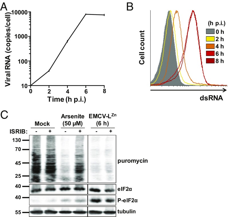
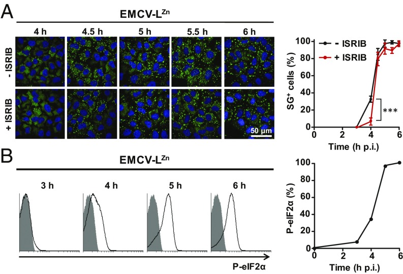
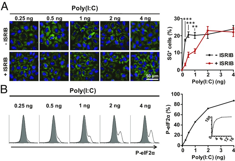
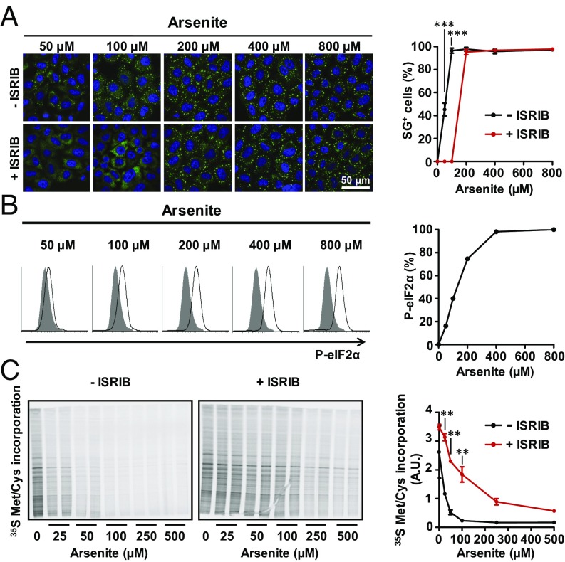
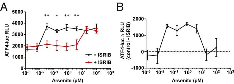
Activation of the integrated stress response (ISR) by a variety of stresses triggers phosphorylation of the α-subunit of translation initiation factor eIF2. P-eIF2α inhibits eIF2B, the guanine nucleotide exchange factor that recycles inactive eIF2•GDP to active eIF2•GTP. eIF2 phosphorylation thereby represses translation. Persistent activation of the ISR has been linked to the development of several neurological disorders, and modulation of the ISR promises new therapeutic strategies. Recently, a small-molecule ISR inhibitor (ISRIB) was identified that rescues translation in the presence of P-eIF2α by facilitating the assembly of more active eIF2B. ISRIB enhances cognitive memory processes and has therapeutic effects in brain-injured mice without displaying overt side effects. While using ISRIB to investigate the ISR in picornavirus-infected cells, we observed that ISRIB rescued translation early in infection when P-eIF2α levels were low, but not late in infection when P-eIF2α levels were high. By treating cells with varying concentrations of poly(I:C) or arsenite to induce the ISR, we provide additional proof that ISRIB is unable to inhibit the ISR when intracellular P-eIF2α concentrations exceed a critical threshold level. Together, our data demonstrate that the effects of pharmacological activation of eIF2B are tuned by P-eIF2α concentration. Thus, ISRIB can mitigate undesirable outcomes of low-level ISR activation that may manifest neurological disease but leaves the cytoprotective effects of acute ISR activation intact. The insensitivity of cells to ISRIB during acute ISR may explain why ISRIB does not cause overt toxic side effects in vivo.

Keywords: ISRIB; P-eIF2; eIF2B; integrated stress response.


Conflict of interest statement

Conflict of interest statement: P.W. [University of California, San Francisco (UCSF) employee] currently holds ISRIB-related patents. These patents are licensed by UCSF to Genentech and Calico.

Figures

Fig. 1.
Fig. 1.
ISRIB does not inhibit virus-induced ISR activity late in infection. (A and B) HeLa-R19 cells were infected at MOI 20 with EMCV-LZn. (A) At the indicated time points, EMCV-LZn genome copies per cell were quantified by qPCR. A representative of two independent experiments is shown. Error bars indicate SEM of triplicate measurements. (B) At the same time points, dsRNA content in EMCV-LZn–infected cells was analyzed by flow cytometry (n = 3). (C) Cells were infected with EMCV-LZn for 6 h or treated with 50 µM arsenite for 1 h. One hour before harvesting, cells were treated with 200 nM ISRIB or left untreated. Fifteen minutes before harvesting, all samples were treated with 20 µg/mL puromycin. Arsenite and EMCV-LZn were kept the cells during these treatments. Subsequently, cells were harvested and analyzed by Western blot, using the indicated antibodies. A representative of two independent experiments is shown.
Fig. 2.
Fig. 2.
ISRIB inhibits only the virus-induced ISR early in infection, when P-eIF2α levels are relatively low. HeLa-R19 cells were infected at MOI 20 with EMCV-LZn. (A) At the indicated time points, cells were fixed in PFA and analyzed by an immunofluorescence assay using antibodies specific to SG marker G3BP1. Percentages of SG positive cells were quantified from at least four images. Representative images are shown on the Left, quantification is shown on the Right. Error bars indicate SEM. Statistical significance was analyzed by a two-way ANOVA, with Bonferroni post hoc test (***P < 0.001). (B) At the indicated time points, cells were harvested and the level of P-eIF2α was analyzed by flow cytometry. Results are shown as histograms (Left) and the percentage increase in mean fluorescence intensity is shown on the Right. Mock-infected cells are set at 0% induction; maximum P-eIF2α level was set at 100% induction. Shown is a representative of two independent experiments.
Fig. 3.
Fig. 3.
ISRIB fails to counteract high poly(I:C)-induced P-eIF2α levels. HeLa-R19 cells were transfected with a total of 100 ng RNA, out of which the indicated amounts of poly(I:C), supplemented with total cellular RNA. (A) Six hours posttransfection, cells were fixed in PFA and SG formation was analyzed by an immunofluorescence assay. Shown are representative images (Left) and quantifications of at least four images per sample (Right). Error bars indicate SEM. Statistical significance was analyzed by a two-way ANOVA, with Bonferroni post hoc test (**P < 0.01; ***P < 0.001). (B) Six hours posttransfection, P-eIF2α levels in the transfected cell populations were analyzed by flow cytometry. Results are shown as histograms (Left) and the percentage increase in P-eIF2α mean fluorescence intensity is shown on the Right. Mock-infected cells are set at 0% induction; maximum P-eIF2α level was set at 100% induction. Shown is a representative of two independent experiments.
Fig. 4.
Fig. 4.
ISRIB does not rescue translation in the presence of high P-eIF2α levels irrespective of the eIF2α kinase involved. HeLa-R19 cells were treated with the indicated arsenite concentrations for 1 h. (A) Cells were fixed in PFA and SG formation was analyzed by an immunofluorescence assay, using antibodies specific to G3BP1. Shown are representative images (Left) and quantifications of at least four images per sample (Right). Error bars indicate SEM. Statistical significance was analyzed by a two-way ANOVA, with Bonferroni post hoc test (***P < 0.001). (B) P-eIF2α levels were analyzed by flow cytometry. Results are shown as histograms (Left) and the percentage increase in P-eIF2α mean fluorescence intensity is shown on the Right. Mock-infected cells are set at 0% induction; maximum P-eIF2α level was set at 100% induction. Shown is a representative of three independent experiments. (C) Cells were treated for 30 min with the indicated arsenite concentrations, and subsequently translation was pulse labeled using 35S Met/Cys for another 90 min in medium containing the same arsenite concentrations. 35S incorporation into newly synthesized proteins was analyzed using a phosphor imager (Left) and quantified using ImageJ software (Right). Error bars indicate SEM of duplicate measurements. A representative of two independent experiments is shown. Statistical significance was analyzed by a two-way ANOVA, with Bonferroni post hoc test (**P < 0.01).
Fig. 5.
Fig. 5.
ISRIB interferes with ATF4 expression only within a defined window of P-eIF2α concentrations. HEK293T cells stably expressing the ATF4-luc reporter were treated with the indicated amounts of arsenite for 2 h in the presence or absence of ISRIB. Shown are the relative luminescence units (RLU) values (A), plotted once more as the difference in RLU values between cells treated with ISRIB and without ISRIB (B). Error bars indicate SEM of triplicate measurements. A representative of two independent experiments is shown. Statistical significance was analyzed by a two-way ANOVA, with Bonferroni post hoc test (*P < 0.05; **P < 0.01).

Similar articles

Cited by

References

    1. Arimoto K, Fukuda H, Imajoh-Ohmi S, Saito H, Takekawa M. Formation of stress granules inhibits apoptosis by suppressing stress-responsive MAPK pathways. Nat Cell Biol. 2008;10:1324–1332. - PubMed
    1. Takahara T, Maeda T. Transient sequestration of TORC1 into stress granules during heat stress. Mol Cell. 2012;47:242–252. - PubMed
    1. Pain VM. Initiation of protein synthesis in eukaryotic cells. Eur J Biochem. 1996;236:747–771. - PubMed
    1. Hinnebusch AG, Lorsch JR. The mechanism of eukaryotic translation initiation: New insights and challenges. Cold Spring Harb Perspect Biol. 2012;4:a011544. - PMC - PubMed
    1. Levin D, London IM. Regulation of protein synthesis: Activation by double-stranded RNA of a protein kinase that phosphorylates eukaryotic initiation factor 2. Proc Natl Acad Sci USA. 1978;75:1121–1125. - PMC - PubMed

Publication types

MeSH terms