. 2019 Jan 24;4(2):e123837.
doi: 10.1172/jci.insight.123837.

Carbon irradiation overcomes glioma radioresistance by eradicating stem cells and forming an antiangiogenic and immunopermissive niche

Affiliations

Carbon irradiation overcomes glioma radioresistance by eradicating stem cells and forming an antiangiogenic and immunopermissive niche

Sara Chiblak et al. JCI Insight. .

Abstract

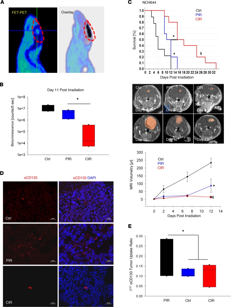
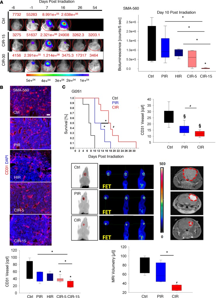
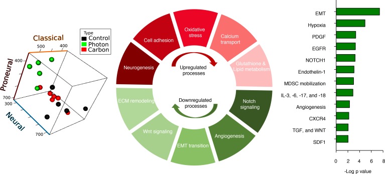
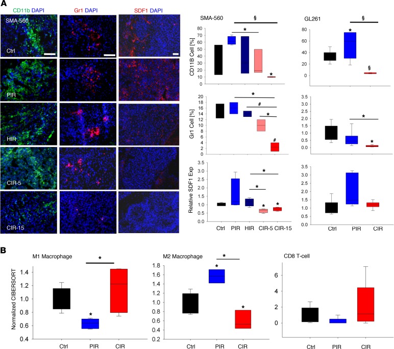
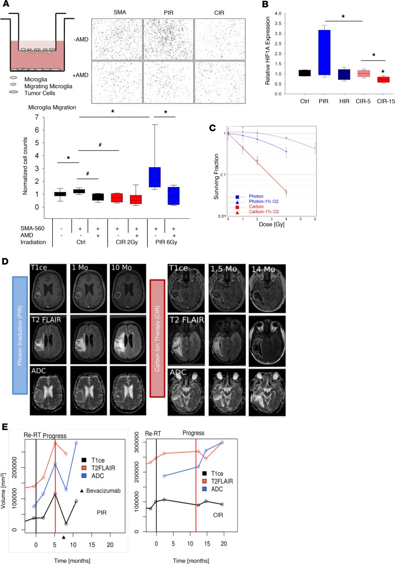
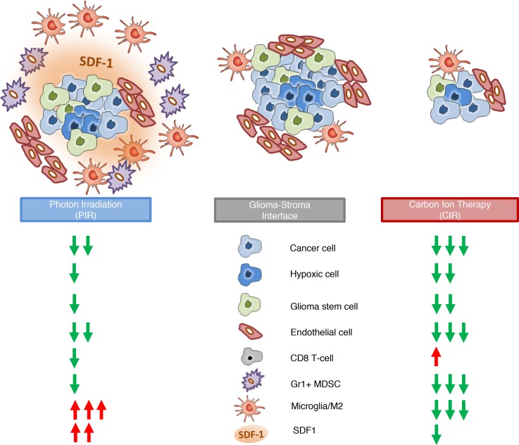
Tumor radioresistance leading to local therapy failure remains a major obstacle for successful treatment of high-grade glioma. We hypothesized that distinct radiobiological features of particle therapy with carbon ions may circumvent glioma radioresistance. We demonstrate that carbon irradiation (CIR) efficiently eradicates radioresistant patient-derived glioma stem cells (GSCs), leading to growth inhibition and prolonged survival. The impact of CIR at the tumor-stroma interface was further investigated in 2 syngeneic mouse and 2 orthotopic GSC xenograft models. Intriguingly, tumor regressions and long-term local controls were observed at doses greater than or equal to 15-Gy CIR. Fractionated CIR further prolonged survival. The enhanced relative biological effectiveness of CIR in vivo was attributed to its potent antiangiogenic effects and eradication of radioresistant hypoxic tumor cells. Blockade of the HIF1-α/stromal cell-derived factor 1/CXCR4 axis by CIR reduced the recruitment of microglia and myeloid-derived suppressor cells (CD11b+Gr1+). Consequently, CIR abrogated M2-like immune polarization and enhanced the influx of CD8+ cells, generating an immunopermissive niche. We report that radiotherapy with carbon ions could surmount several central glioma resistance mechanisms by eradicating hypoxic and stem cell-like tumor cells, as well as modulating the glioma niche toward an antiangiogenic and less immunosuppressive state. Conclusively, potentially novel rationales for CIR in conquering glioma radioresistance are provided.

Keywords: Brain cancer; Mouse models; Oncology; Therapeutics.


Conflict of interest statement

Conflict of interest: The authors declare that they have no conflict of interest.

Figures

Figure 1
Figure 1. Effect of CIR on patient-derived, GSC-enriched xenograft models.
Establishment of small animal CIR in orthotopic glioma models. (A) Verification of administered dose by overlay of CIR-induced secondary positrons (C11 micro-PET, black) and tumor uptake of 18F-fluoro-ethyl-tyrosine (FET) PET, circled in red at the intersection of the horizontal and vertical lines, indicating precision treatment. (B) Significant inhibition of tumor growth after CIR (5 × 1.5 Gy) versus PIR (5 × 5 Gy) and sham-treated control in an orthotopic xenograft model derived from patient NCH441 GSCs (n = 3). Tumor growth was monitored longitudinally by bioluminescence (see also supplemental material). Ctrl: control. (C) CIR (6 Gy, n = 10) significantly prolonged animal survival compared with PIR (15 Gy, n = 10), both administered in 3 consecutive daily fractions in the NCH644 GSC model. Kaplan-Meier survival estimates and tumor growth kinetic via MRI-based tumor volumetry are shown. (D and E) PIR (n = 5) led to a relative enrichment of the GSC population compared with control (n = 3) and CIR (n = 5) as determined by NCH644 tumor I131α-CD133 uptake ratio and biodistribution studies (supplemental material). The lack of selection for α-CD133+ GSCs (red) is also elucidated by representative immunofluorescence staining. Nuclei counterstained using DAPI (blue). Box and whisker plots represent median, interquartile, minimum, and maximum of all data points. §P < 0.0001, *P < 0.05 versus control, when shown over a box, or versus the indicated irradiation treatment. Scale bar: 50 μm.
Figure 2
Figure 2. Beneficial effect of CIR in syngeneic, orthotopic murine models.
(A) Comparison of PIR, HIR, and CIR in an intracranial syngeneic SMA-560 murine glioma model. Tumor control at single doses ≥ 15-Gy CIR was observed. Tumor growth (n = 2/study arm) was longitudinally monitored by bioluminescence at –6, –1, 7, 16, 26, and 54 days after irradiation. Next, SMA-560 tumors were irradiated with 15-Gy PIR (n = 12), 15-Gy HIR (n = 12), 15-Gy CIR (CIR-15, n = 12), or 5-Gy CIR (CIR-5, n = 12). Tumor growth inhibition was significant only for the CIR-15 group versus all other arms at day 10 after irradiation as determined by bioluminescence. Representative photomicrographs of tumor vascular staining by anti-endothelial CD31 marker (red) are shown. (B) Nuclei counterstained using DAPI (blue). cpf, counts per field. (C) Likewise, in the Gl261 glioma model, irradiation with 6-Gy CIR significantly prolonged survival (control n = 8, PIR n = 8, CIR n = 8) compared with 15-Gy PIR, both administered in 3 consecutive daily fractions. This effect also correlated with reduced microvascular density (MVD) (n = 6/study arm), metabolism by micro–FET PET imaging (tumor margins delineated in yellow), and tumor size measured by volumetry using T1-weighted contrast-enhanced (T1CE) MRI (n = 4) (hyperintense gross tumor area is contoured by red dashed lines).Bioluminescence, MRI tumor volumes, and CD31+ blood vessels per field data are represented as box-and-whisker plots showing median, interquartile, the minimum and maximum of all data points. §P < 0.0001, #P < 0.001, *P < 0.05 versus control, when shown over a box, or versus the indicated irradiation treatment. Scale bar: 50 μm.
Figure 3
Figure 3. Transcriptomic switches associated with carbon ion irradiation in glioma models.
Transcriptome analysis of in vivo Gl261 tumor samples (n = 5 control, photon, n = 6 carbon) revealed that sham-treated control tumors represent predominantly a “classical” gene expression subtype. Conventional radiotherapy (photon) led to a uniform switch of this gene expression subtype toward a “neural” phenotype. In contrast, Gl261 tumors treated with carbon ions (carbon) mainly retained their gene expression subtype and were classified in the prognostically more favorable “classical” subtype. Graphical depiction of subtype changes in the proneural-neural-classical space for PIR versus CIR is shown (see also supplemental material). In the SMA-560 model (n = 4/study arm), 595 differentially expressed transcripts as a function of radiation quality were identified (P < 0.01 after Bonferroni’s multiple testing correction). Significantly enriched pathways identified among the 265 upregulated (red), and 323 downregulated (green) transcripts after CIR are presented. Bars represent negative log P values, representing the probability for enrichment based on gene ontology mapping to arise by chance. Of note, numerous gene ontologies related to tumor microenvironment (hypoxia, angiogenesis), growth factors, and cytokines, as well as modulators of an immunosuppressive niche (SDF1/CXCR4, TGF, MdSC mobilization), were found to contain genes with inverse regulation pattern, i.e., decreased expression after carbon ion exposure whereas upregulation by conventional PIR. SDF1: stromal cell–derived factor 1.
Figure 4
Figure 4. CIR generates an immunopermissive glioma niche.
(A) In line with these observations, reduced influx of CD11b+ microglia and myeloid cells, including Gr1+ MdSCs, after CIR was confirmed and correlated with decreased SDF1 levels (SMA-560 n = 3–4, Gl261 n = 6). Exp: expression. (B) In silico cell sorting identified a switch in macrophages/microglia polarization toward an M2 phenotype after PIR, which was accompanied by reduced abundance of CD8+ T cells (control n = 5, PIR n = 3–5, CIR n = 3–5). However, a reverse trend was found after CIR, indicating lack of immunosuppressive signal and accelerated glioma immunity. Box and whisker plots represent median, interquartile, minimum, and maximum of all data points. §P < 0.0001, #P < 0.001, *P < 0.05 versus control, when shown over a box, or versus the indicated irradiation treatment. Scale bar: 50 μm (CD11b, Gr1).
Figure 5
Figure 5. Functional validation of CIR effects on tumor hypoxia, HIF1-α/SDF1/CXCR4 axis, and tumor recruitment of microglia.
(A) Irradiation of tumor cells (bottom) enhanced tumor-induced migration of microglia (embryonic stem cell–derived microglia, in transwell insert). This effect was functionally reversed by inhibition of the SDF1/CXCR4 pathway (AMD3100). In contrast, reduced influx of microglia was found after irradiation of tumor cells with carbon ions. Representative photomicrographs of infiltrated microglia nuclei on the bottom side of the transwell in coculture with SMA-560 cells in the lower chamber are shown. (B) Enhanced HIF1-α expression in vivo in SMA-560 tumors treated with PIR versus reduced expression after CIR suggest efficient ablation of hypoxic tumor cells by CIR. Box and whisker plots represent median, interquartile, minimum, and maximum of all data points. Indeed, clonogenic survival assay clearly demonstrated overlapping tumor sensitivity to carbon ions independent of the oxygen concentration. (C) In contrast, tumors under mild hypoxia (1% O2, dotted lines) demonstrated relative radioresistance to conventional PIR. Bars indicate OER for photon and carbon ions. (D) Effects of CIR versus PIR in reirradiating patients with HGG. Alteration of tumor pathophysiology determined over time (months, Mo) correlated with improved time to progression in CIR-treated patients. Green: T1CE-defined tumor volume; yellow: T2 fluid-attenuated inversion recovery–defined (FLAIR-defined) tumor volume, apparent diffusion coefficient (ADC). (E) Longitudinal follow-up of different MRI derived tumor volumes. Time point of reirradiation (Re-RT, black vertical line) and detection of tumor progress (Progress, red vertical line), as well as treatment with antiangiogenic agent Bevacizuman (an antibody against VEGF). #P < 0.001, *P < 0.05 versus control, when shown over a box, or versus the indicated irradiation treatment.
Figure 6
Figure 6. Radiotherapy effects at the tumor–stroma interface.
A schematic presentation of the proposed model for cellular and molecular effects of CIR versus PIR based on data reported in this study is shown. Due to their distinct radiobiological effects, CIR is capable of eradicating therapy-resistant tumor subpopulations, e.g., GSCs and hypoxic tumor cells. This observation contrasted with conventional PIR, where these subpopulations were enriched by the therapy-induced selection pressure. Consequently, the concentration of tumor stroma– modulating factors, such as SDF1 released by hypoxic cells, was decreased after CIR, leading to reduced influx of bone marrow–derived immunosuppressive cells (MdSCs) and a less M2-polarized microglia/TAM microenvironment.

References

    1. Debus J, Abdollahi A. For the next trick: new discoveries in radiobiology applied to glioblastoma. Am Soc Clin Oncol Educ Book. 2014:e95–e99. - PubMed
    1. Stupp R, et al. Radiotherapy plus concomitant and adjuvant temozolomide for glioblastoma. N Engl J Med. 2005;352(10):987–996. doi: 10.1056/NEJMoa043330. - DOI - PubMed
    1. Stupp R, et al. Effects of radiotherapy with concomitant and adjuvant temozolomide versus radiotherapy alone on survival in glioblastoma in a randomised phase III study: 5-year analysis of the EORTC-NCIC trial. Lancet Oncol. 2009;10(5):459–466. doi: 10.1016/S1470-2045(09)70025-7. - DOI - PubMed
    1. Wick W, et al. MGMT testing — the challenges for biomarker-based glioma treatment. Nat Rev Neurol. 2014;10(7):372–385. doi: 10.1038/nrneurol.2014.100. - DOI - PubMed
    1. Abdollahi A, Folkman J. Evading tumor evasion: current concepts and perspectives of anti-angiogenic cancer therapy. Drug Resist Updat. 2010;13(1–2):16–28. - PubMed