. 2019 Jan 24;4(2):e124387.
doi: 10.1172/jci.insight.124387.

One-year pilot study on the effects of nitisinone on melanin in patients with OCA-1B

Affiliations

One-year pilot study on the effects of nitisinone on melanin in patients with OCA-1B

David R Adams et al. JCI Insight. .

Abstract

Background: Oculocutaneous albinism (OCA) results in reduced melanin synthesis, skin hypopigmentation, increased risk of UV-induced malignancy, and developmental eye abnormalities affecting vision. No treatments exist. We have shown that oral nitisinone increases ocular and fur pigmentation in a mouse model of one form of albinism, OCA-1B, due to hypomorphic mutations in the Tyrosinase gene.

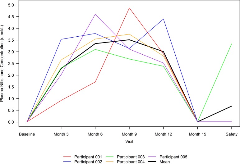
Methods: In this open-label pilot study, 5 adult patients with OCA-1B established baseline measurements of iris, skin, and hair pigmentation and were treated over 12 months with 2 mg/d oral nitisinone. Changes in pigmentation and visual function were evaluated at 3-month intervals.

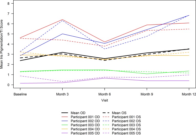
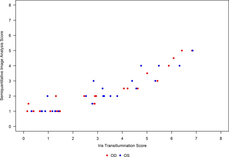
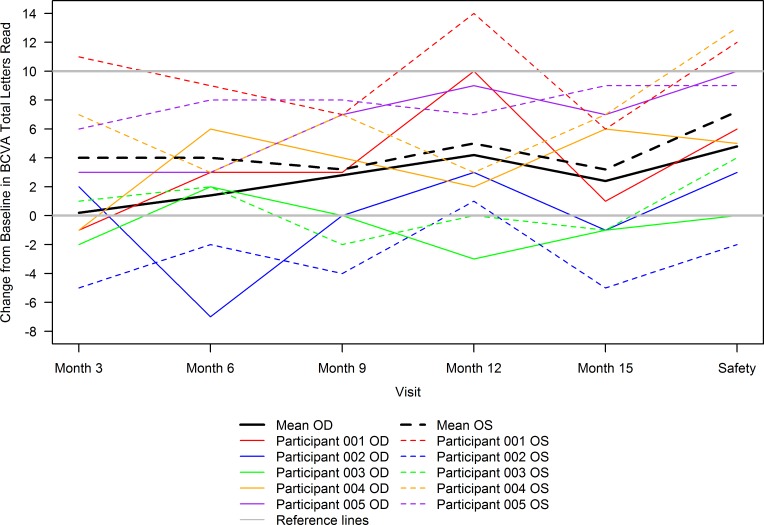
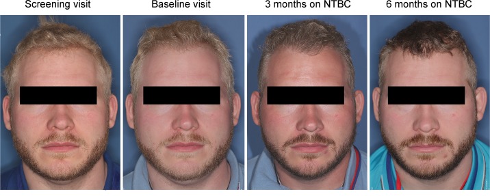
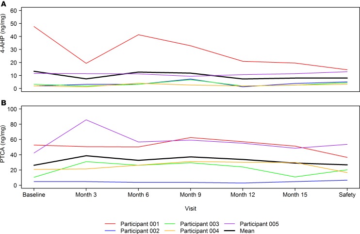
Results: The mean change in iris transillumination, a marker of melanin, from baseline was 1.0 ± 1.54 points, representing no change. The method of iris transillumination grading showed a high intergrader reliability (intraclass correlation coefficient ≥ 0.88 at each visit). The number of letters read (visual acuity) improved significantly at month 12 for both eyes (right eye, OD, mean 4.2 [95% CI, 0.3, 8.1], P = 0.04) and left eye (OS, 5 [1.0, 9.1], P = 0.003). Skin pigmentation on the inner bicep increased (M index increase = 1.72 [0.03, 3.41], P = 0.047). Finally, hair pigmentation increased by both reflectometry (M index [17.3 {4.4, 30.2}, P = 0.01]) and biochemically.

Conclusion: Nitisinone did not result in an increase in iris melanin content but may increase hair and skin pigmentation in patients with OCA-1B. The iris transillumination grading scale used in this study proved robust, with potential for use in future clinical trials.

Clinicaltrials: gov NCT01838655.

Funding: Intramural program of the National Eye Institute.

Keywords: Genetic diseases; Genetics; Ophthalmology.


Conflict of interest statement

Conflict of interest: BPB and WAG are named inventors on the patent “Nitisinone For Treatment of Oculocutaneous Albinism And For Increasing Pigmentation” in the US, Canada, and the European Union” (US 8,822,540 B2).

Figures

Figure 1
Figure 1. Mean 8-point iris pigmentation/transillumination score over time by participant eye and overall.
Mean 8-point iris pigmentation/transillumination (TI) scores were calculated as the mean across all 18 graders for the 2 images of each eye for a participant at each visit. The solid lines correspond to oculus dexter (OD), and dashed lines correspond to oculus sinister (OS) measurements. The colored lines correspond to individual participant measurements, and the black lines correspond to mean of the scores (n = 5) across all participants for each eye. An overall increase in score, implying a decrease in melanin content, was noted when each eye was assessed separately (mean [n = 5] change from baseline at 12 months of OD 1.1 [1.70] and OS 0.9 [1.56] points) and together (data not shown) (mean [n = 10] change from baseline at 12 months of oculus uterque/OU 1.0 [1.54] points; P = 0.01 based on a longitudinal model fit using data through month 12).
Figure 2
Figure 2. Correlation between mean 8-point iris pigmentation/transillumination and semiquantitative image analysis scores.
Mean 8-point iris pigmentation/transillumination (TI)scores were calculated as the mean across all 18 graders for the 2 images of each eye for a participant at each visit. Semiquantitative image analysis scores were calculated as the mean of scores across the 2 images of each eye for a participant at each visit. Oculus dexter (OD) and oculus sinister (OS) values of mean 8-point iris pigmentation/TI and semiquantitative image analysis scores are plotted. These scores were found to be highly correlated with a Spearman’s rank correlation coefficient of 0.9, although the range of semiquantitative image analysis scores assigned was lower than that of mean 8-point iris pigmentation/TI scores.
Figure 3
Figure 3. Change from baseline in best-corrected visual acuity total letters read over time by participant eye and overall.
Gray solid lines are reference lines drawn at no change and 10-letter increase in best-corrected visual acuity (BCVA) total letters read from baseline. All other solid lines correspond to oculus dexter (OD), and dashed lines correspond to oculus sinister (OS) measurements. The colored lines correspond to individual participant measurements, and the black lines correspond to mean (n = 5) across all participants for each eye. In general, an increase in total letters read was seen at all follow-up visits from baseline. Longitudinal models fit using data through safety visit revealed that total letters read significantly improved from baseline at month 12 for OD (mean [95% CI] change from baseline of 4.2 [0.3, 8.1], P = 0.04) and OS (mean [95% CI] change from baseline of 5.0 [1.0, 9.1], P = 0.02) and, subsequently, at safety follow-up visit for OD (mean [95% CI] change from baseline of 4.8 [0.64, 8.96], P = 0.03) and OS (mean [95% CI] change from baseline of 7.2 [2.79, 11.61], P = 0.003).
Figure 4
Figure 4. Subject 003 exhibited the greatest visible change in hair and skin pigmentation.
He reports that he was previously unable to tan but did so by at 3 months, a summer month. Hair and beard darkening remained at 6 months and until the end of the study. The subject provided written consent for having identifiable images published.
Figure 5
Figure 5. Hair melanin content over time by participant and overall.
(A) 4-amino-3-hydroxyphenylalanine (4-AHP), a marker of pheomelanin, over time. The colored lines correspond to individual participant measurements, and the black line corresponds to mean (n = 5) across all participants. A decrease in 4-AHP was noted at both month 12 (mean [SD] of change from baseline of –5.92 [11.68] ng/mg) and the safety follow-up visit (mean [SD] of change from baseline of –5.24 [15.71] ng/mg). (B) Pyrrole-2,3,5-tricarboxylic acid (PTCA), a marker of eumelanin, over time. The colored lines correspond to individual participant measurements, and the black line corresponds to mean (n = 5) across all participants. An increase in PTCA was noted at month 12 (mean [SD] of change from baseline of 7.62 [6.43] ng/mg), with almost no change from baseline at the safety follow-up visit (mean [SD] of change from baseline of 0.52 [11.25] ng/mg).
Figure 6
Figure 6. Plasma nitisinone concentration over time by participant and overall.
The colored lines correspond to individual participant measurements, and the black line corresponds to mean (n = 5) across all participants. Plasma concentrations of nitisinone peaked between months 6 and 12 for participants and dropped to almost nil at month 15, which is 3 months after investigational product (IP) discontinuation. One participant chose the option to restart IP at month 18, before discontinuing it approximately 1.5 months later and completing their safety follow-up visit, resulting in the spike in mean plasma nitisinone concentration at the safety follow-up visit.

References

    1. Montoliu L, et al. Increasing the complexity: new genes and new types of albinism. Pigment Cell Melanoma Res. 2014;27(1):11–18. doi: 10.1111/pcmr.12167. - DOI - PubMed
    1. Grønskov K, Ek J, Brondum-Nielsen K. Oculocutaneous albinism. Orphanet J Rare Dis. 2007;2:43. - PMC - PubMed
    1. Simeonov DR, et al. DNA variations in oculocutaneous albinism: an updated mutation list and current outstanding issues in molecular diagnostics. Hum Mutat. 2013;34(6):827–835. doi: 10.1002/humu.22315. - DOI - PMC - PubMed
    1. Kamaraj B, Purohit R. Mutational analysis of oculocutaneous albinism: a compact review. Biomed Res Int. 2014;2014:905472. - PMC - PubMed
    1. Hutton SM, Spritz RA. Comprehensive analysis of oculocutaneous albinism among non-Hispanic caucasians shows that OCA1 is the most prevalent OCA type. J Invest Dermatol. 2008;128(10):2442–2450. doi: 10.1038/jid.2008.109. - DOI - PMC - PubMed

Associated data