Review
. 2018 May 22;4(2):46.
doi: 10.3390/gels4020046.

Recent Developments in Tough Hydrogels for Biomedical Applications

Affiliations
Review

Recent Developments in Tough Hydrogels for Biomedical Applications

Yuan Liu et al. Gels. .

Abstract

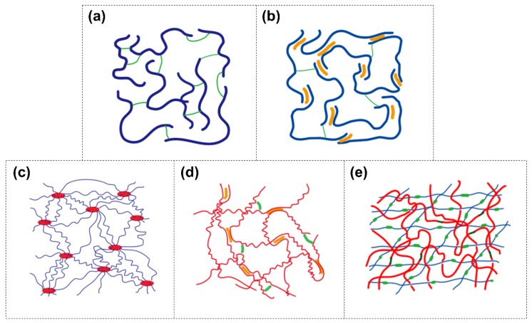
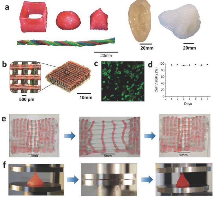
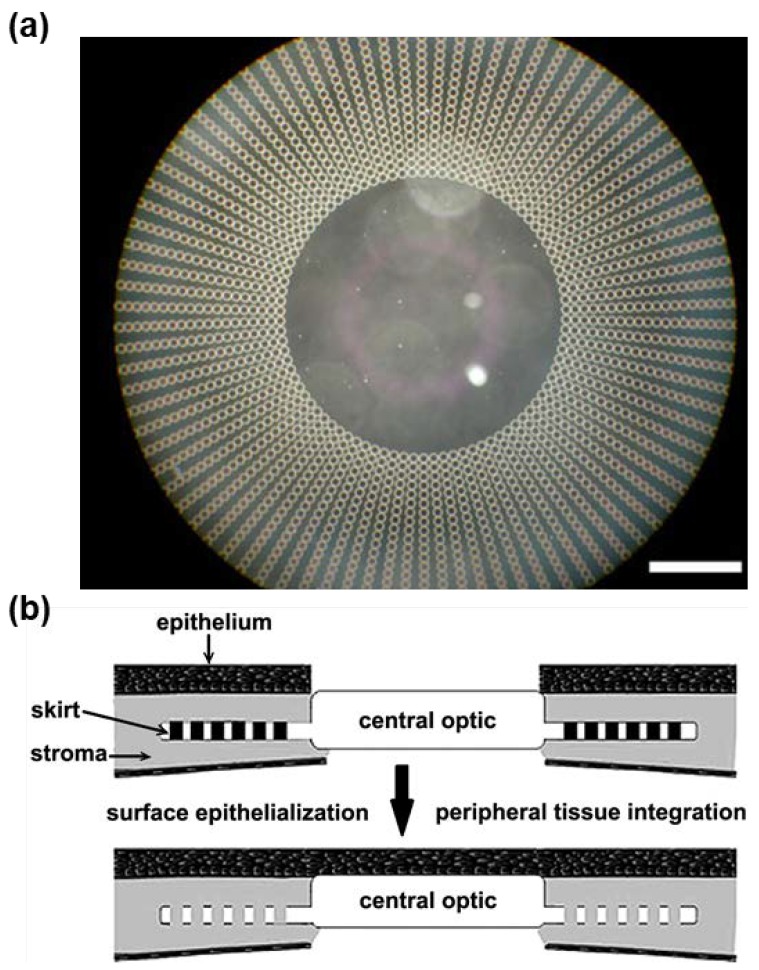
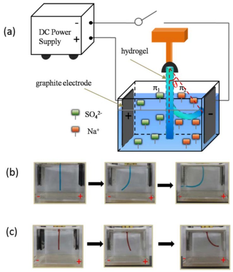
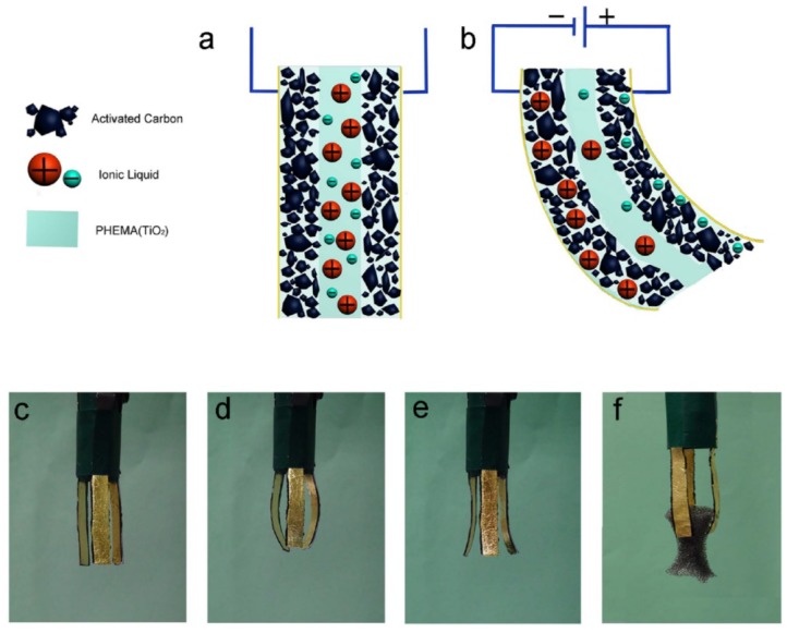
A hydrogel is a three-dimensional polymer network with high water content and has been attractive for many biomedical applications due to its excellent biocompatibility. However, classic hydrogels are mechanically weak and unsuitable for most physiological load-bearing situations. Thus, the development of tough hydrogels used in the biomedical field becomes critical. This work reviews various strategies to fabricate tough hydrogels with the introduction of non-covalent bonds and the construction of stretchable polymer networks and interpenetrated networks, such as the so-called double-network hydrogel. Additionally, the design of tough hydrogels for tissue adhesive, tissue engineering, and soft actuators is reviewed.

Keywords: biomedical applications; soft actuators; tissue adhesives; tissue engineering; tough hydrogels.


Conflict of interest statement

The authors declare no conflict of interest.

Figures

Figure 1
Figure 1
Schematic of (a) the classic covalent single-network hydrogel, (b) dual-crosslinked hydrogel, (c) polymer-intercalated nanocomposite hydrogel, (d) hydrogel with elastomer-like segments and non-covalent bonds, and (e) double-network (DN) hydrogel. Blue and red long lines: polymer backbones; short green lines: covalent crosslinking points; yellow short lines: non-covalent crosslinking points.
Figure 2
Figure 2
Non-covalent and covalent catechol chemistries (adapted from [76], copyright 2016 Springer).
Figure 3
Figure 3
(a) Schematic of hydrogel formed by polymer intercalating into nanoclay sheets with polydopamine. (b) Photographs of hydrogel that sticks to human skin and can be easily removed (reproduced from [37], copyright 2017 ACS).
Figure 4
Figure 4
Chemical structure of tannic acid.
Figure 5
Figure 5
(a) Schematic of poly(ethylene glycol) (PEG) hydrogel incorporated with nanoclay. PEG chains form non-covalent bonds to nanoclay. (b) Photographs of hydrogel that is stretchy and sticks to human skin and metal surfaces in an elongated state (reproduced from [34], copyright 2011 Elsevier).
Figure 6
Figure 6
(a) Schematic of interpenetrating network (IPN) hydrogel coated with tissue adhesive polymer. (b) Photographs of hydrogel gluing liver tissue in an elongated state. (c) Photographs of hydrogel functioning as a tissue sealant and (d) hemostatic wound dressing (reproduced from [101], copyright 2017 Science).
Figure 7
Figure 7
(a) Schematic of the formation of a double network by incorporating proteoglycan aggregate stent (St) molecules into the neutral networks. (b) Schematic of the St molecule, proteoglycan aggregate. (c) Schematic of the monomer and crosslinker used to construct the neutral networks (reproduced from [119], copyright 2014 Wiley).
Figure 8
Figure 8
(a) Schematic of the formation of a cell-laden interpenetrating network (IPN) hydrogel immobilized with RGD peptide sequences. (b) Illustration of chondrocyte outgrowth in a hydrogel with and without RGD via live/dead staining (reproduced from [128], copyright 2014 Elsevier).
Figure 9
Figure 9
(a) Photographs of 3D-printed hydrogels with shapes of a hollow cube, hemisphere, pyramid, twisted bundle, artificial ear, and nose. (b) A photograph of scaffold frame printed by the hydrogel in a meshed fashion. (c,d) Live/dead assay and cell viability of human embryonic kidney (HEK) cells in the scaffold. (e) Photographs of stretching and recovery of the printed hydrogel mesh. (f) Photographs of compression and recovery of the printed hydrogel pyramid (reproduced from [142], copyright 2015 Wiley).
Figure 10
Figure 10
(a) A photograph of the artificial cornea. Scale bar: 1300 μm. (b) Ideal schematic representation of the implanted artificial corneal (reproduced from [161], copyright 2007 Springer).
Figure 11
Figure 11
(a) Schematic of polyionic hydrogel submerged in between two parallel electrode plates in an electrolyte solution. (b) Photographs of the polyanionic hydrogel bending towards the cathode. (c) Photographs of the polycationic hydrogel bending towards the anode (reproduced from [171], copyright 2016 ACS).
Figure 12
Figure 12
(a) Schematic of hydrogel containing ionic liquid sandwiched between two porous activated carbon layers. (b) Schematic of the hydrogel “sandwich” actuated by an applied voltage. (cf) Photographs of a functioning manipulator constructed with multiple pieces of the hydrogel “sandwich” that can grip an object (reproduced from [174], copyright 2014 Springer Nature).
Figure 13
Figure 13
(a) Photograph of a bi-layered hydrogel consisting of polyanionic (blue) and polycationic (white) hydrogels. (b,c) Photographs of a bi-layered hydrogel bending towards the polycationic side in 0.2 M (b) and 0.05 M (c) NaCl solution. The bending angle (θ) is defined as shown. (d) The reversible actuation of hydrogel shuttling between 0.2 and 0.05 M NaCl solutions (reproduced from [178], copyright 2016 RSC).
Figure 14
Figure 14
(a) Schematic of assembling a bi-layered hydraulic actuator consisting of a softer top layer shaped to several serial units and a stiffer bottom layer. (b) Photographs of the bi-layered hydrogel actuated by hydraulic pressure (reproduced from [180]).
Figure 15
Figure 15
Schematic of (a) bending and (b) polypeptide-like twisting of bi-layered hydrogel. (c) Schematic of DNA-like twisting of tri-layered hydrogel. The blue and red parts represent hydrogels with different swelling ratios (reproduced from [181], copyright 2011 RSC).

References

    1. Ghobril C., Grinstaff M. The chemistry and engineering of polymeric hydrogel adhesives for wound closure: A tutorial. Chem. Soc. Rev. 2015;44:1820–1835. doi: 10.1039/C4CS00332B. - DOI - PubMed
    1. Nguyen Q.V., Park J.H., Lee D.S. Injectable polymeric hydrogels for the delivery of therapeutic agents: A review. Eur. Polym. J. 2015;72:602–619. doi: 10.1016/j.eurpolymj.2015.03.016. - DOI
    1. Matricardi P., Di Meo C., Coviello T., Hennink W.E., Alhaique F. Interpenetrating polymer networks polysaccharide hydrogels for drug delivery and tissue engineering. Adv. Drug Deliv. Rev. 2013;65:1172–1187. doi: 10.1016/j.addr.2013.04.002. - DOI - PubMed
    1. Hoffman A.S. Hydrogels for biomedical applications. Adv. Drug Deliv. Rev. 2012;64:18–23. doi: 10.1016/j.addr.2012.09.010. - DOI - PubMed
    1. Vashist A., Vashist A., Gupta Y., Ahmad S. Recent advances in hydrogel based drug delivery systems for the human body. J. Mater. Chem. B. 2014;2:147–166. doi: 10.1039/C3TB21016B. - DOI - PubMed

LinkOut - more resources