

Concepts for Developing Physical Gels of Chitosan and of Chitosan Derivatives
- PMID: 30674843
- PMCID: PMC6209275
- DOI: 10.3390/gels4030067
Concepts for Developing Physical Gels of Chitosan and of Chitosan Derivatives
Abstract
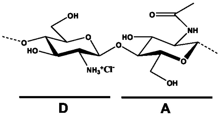
Chitosan macro- and micro/nano-gels have gained increasing attention in recent years, especially in the biomedical field, given the well-documented low toxicity, degradability, and non-immunogenicity of this unique biopolymer. In this review we aim at recapitulating the recent gelling concepts for developing chitosan-based physical gels. Specifically, we describe how nowadays it is relatively simple to prepare networks endowed with different sizes and shapes simply by exploiting physical interactions, namely (i) hydrophobic effects and hydrogen bonds-mostly governed by chitosan chemical composition-and (ii) electrostatic interactions, mainly ensured by physical/chemical chitosan features, such as the degree of acetylation and molecular weight, and external parameters, such as pH and ionic strength. Particular emphasis is dedicated to potential applications of this set of materials, especially in tissue engineering and drug delivery sectors. Lastly, we report on chitosan derivatives and their ability to form gels. Additionally, we discuss the recent findings on a lactose-modified chitosan named Chitlac, which has proved to form attractive gels both at the macro- and at the nano-scale.
Keywords: Chitlac; chitosan; chitosan-derivatives; drug delivery; gel; gelling mechanisms; physical interactions; tissue engineering.
Conflict of interest statement
The authors declare no conflict of interest.
Figures
References
-
- Lapasin R. Rheological characterization of hydrogels. In: Matricardi P., Alhaique F., Coviello T., editors. Polysaccharide Hydrogels: Characterization and Biomedical Applications. Pan Stanford; Singapore: 2005. pp. 83–137.
-
- Kim T.G., Shin H., Lim D.W. Biomimetic scaffolds for tissue engineering. Adv. Funct. Mater. 2012;22:2446–2468. doi: 10.1002/adfm.201103083. - DOI
-
- Croisier F., Jérôme C. Chitosan-based biomaterials for tissue engineering. Eur. Polym. J. 2013;49:780–792. doi: 10.1016/j.eurpolymj.2012.12.009. - DOI
Publication types
Grants and funding
LinkOut - more resources
Full Text Sources
Other Literature Sources
