. 2018 Nov 21;4(4):85.
doi: 10.3390/gels4040085.

Viscoelastic Oxidized Alginates with Reversible Imine Type Crosslinks: Self-Healing, Injectable, and Bioprintable Hydrogels

Affiliations

Viscoelastic Oxidized Alginates with Reversible Imine Type Crosslinks: Self-Healing, Injectable, and Bioprintable Hydrogels

Shahzad Hafeez et al. Gels. .

Abstract

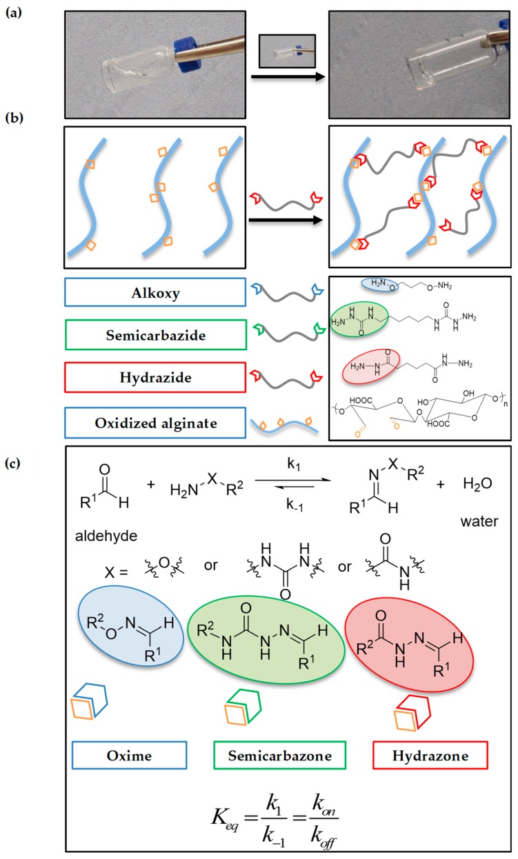
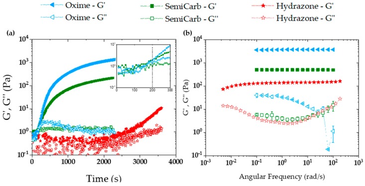
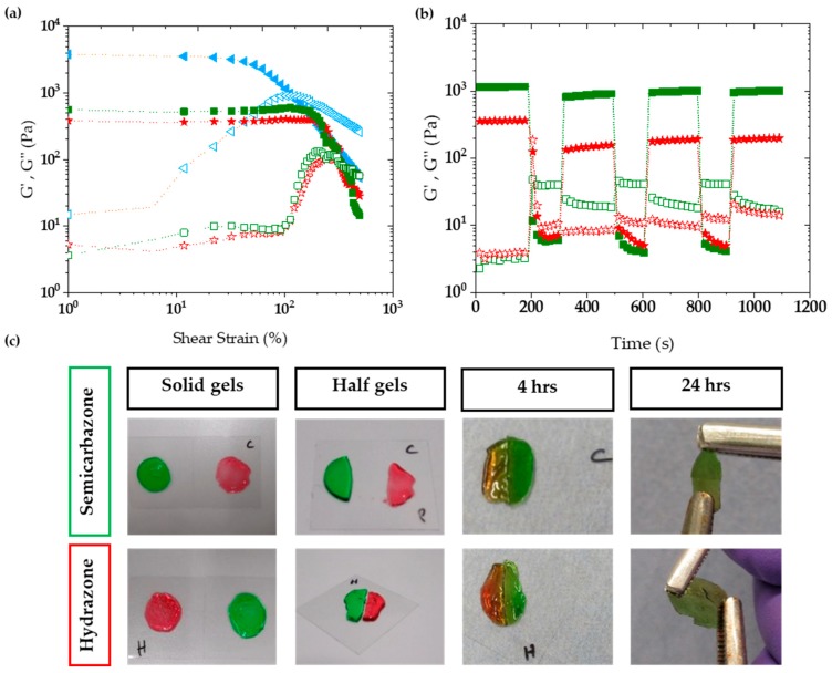
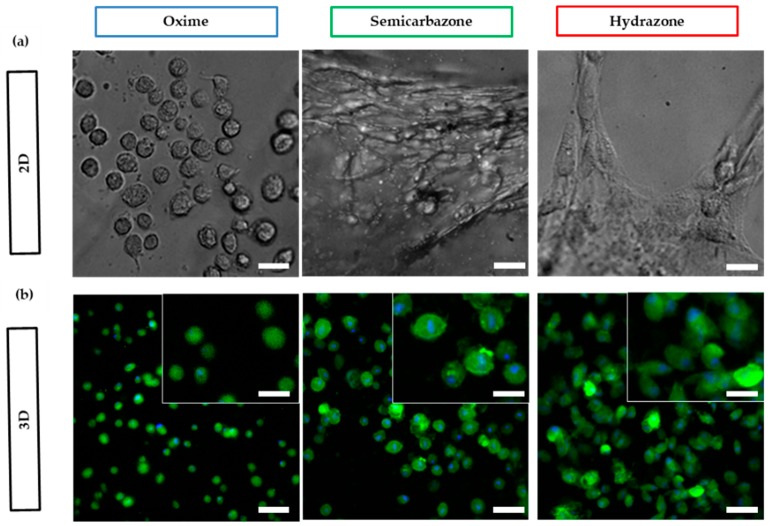
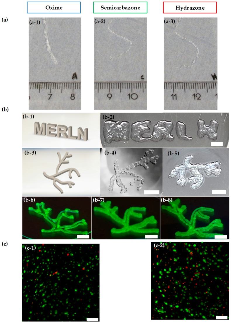
Bioprinting techniques allow for the recreation of 3D tissue-like structures. By deposition of hydrogels combined with cells (bioinks) in a spatially controlled way, one can create complex and multiscale structures. Despite this promise, the ability to deposit customizable cell-laden structures for soft tissues is still limited. Traditionally, bioprinting relies on hydrogels comprised of covalent or mostly static crosslinks. Yet, soft tissues and the extracellular matrix (ECM) possess viscoelastic properties, which can be more appropriately mimicked with hydrogels containing reversible crosslinks. In this study, we have investigated aldehyde containing oxidized alginate (ox-alg), combined with different cross-linkers, to develop a small library of viscoelastic, self-healing, and bioprintable hydrogels. By using distinctly different imine-type dynamic covalent chemistries (DCvC), (oxime, semicarbazone, and hydrazone), rational tuning of rheological and mechanical properties was possible. While all materials showed biocompatibility, we observed that the nature of imine type crosslink had a marked influence on hydrogel stiffness, viscoelasticity, self-healing, cell morphology, and printability. The semicarbazone and hydrazone crosslinks were found to be viscoelastic, self-healing, and printable-without the need for additional Ca2+ crosslinking-while also promoting the adhesion and spreading of fibroblasts. In contrast, the oxime cross-linked gels were found to be mostly elastic and showed neither self-healing, suitable printability, nor fibroblast spreading. The semicarbazone and hydrazone gels hold great potential as dynamic 3D cell culture systems, for therapeutics and cell delivery, and a newer generation of smart bioinks.

Keywords: bioprinting (BP); dynamic covalent chemistry (DCvC); dynamic hydrogel; hydrazone; oxidized alginate (ox-alg); oxime; reversible bonds; semicarbazone; tissue engineering; viscoelastic.


Conflict of interest statement

The authors declare no conflict of interest.

Figures

Figure 1
Figure 1
Scheme of hydrogel formation. (a) Representative gel formation for a hydrazone gel. (b) Schematic illustrating the crosslinking of alginate chains using dihydrazide cross-linker, the structure of oxidized alginate and the structures of cross-linkers utilized in this study. (c) A general reaction scheme for reversible imine type bonds formation and chemical structures of oxime, semicarbazone and hydrazones.
Figure 2
Figure 2
Hydrogel formation kinetics and viscoelasticity exhibited by a series of 2% (w/v) 10% ox-alg samples prepared with different cross-linkers (equimolar). (a) Time sweep using the three different cross-linkers; aminooxy, semicarbazide, and hydrazide for evaluating hydrogels formation kinetics measured at 1% strain and 10 rad/s. (b) Frequency sweep for the same three hydrogels measured at 1% strain.
Figure 3
Figure 3
Demonstration of strain to rupture the network and the self-healing capacity of 10% ox-alg samples prepared with semicarbazide and hydrazide cross-linkers. (a) Strain sweeps from 0% to 800% strain using oxime, semicarbazone, hydrazone and hydrogels, measured at 10 rad/s. (b) self-healing capacity with semicarbazide and hydrazide cross-linkers. The hydrogels were submitted to three strain cycles. Initially a low strain (1%) was applied, followed by three cycles of high strain (600%), to rupture the network, and low strain (1%), to allow recovery. The semicarbazone and hydrazone gels rapidly self-heal. (c) Macroscopic self-healing of the hydrogels. From left to right, the colored gels were formed (12 mm diameter, 2 mm thickness), cut in half, left to self-heal for four hours, and then tested at 24 h.
Figure 4
Figure 4
Cell viability: (a) Image showing live cells (in green) after seven days of encapsulation in gels indicating that great majority of cells are live and gels did not cause cytotoxicity, Scale bar: 200 µm and 25 µm for inset images. (b) Cell metabolic activity recorded after 12, 24, 96, and 168 h. The values reported are an average of n = 3, ± standard deviation. * and ** indicates p < 0.05 and p < 0.01 (Student’s t-test, independent sample populations). For cell aggr condition, cells were cultured in pellets, a standard for chondrocyte cell culture.
Figure 5
Figure 5
Fibroblasts spreading morphologies in oxime RGD ligated gels, (a) on top of (2D) and (b) within (3D) oxime, semicarbazone and hydrazone cross-linked gels after 24 h. Green color represents actin staining and blue color represent nucleus staining. scale bar: 25 µm for 2D, 50 µm for 3D images, and 25 µm for 3D insets.
Figure 6
Figure 6
Injectability and bioprintability: (a) 10% ox-alg gels with oxime (a-1), semicarbazone (a-2), and hydrazone (a-3) cross-linkers were extruded through a 25G needle. Semicarbazone and hydrazone showed injectability, and to our surprise oxime gels were also injectable, although they were not self-healing. (b) Printed MERLN and vascular tree structures using hydrazone crosslinks; 2% (w/v) of the 10% ox-alg used for (b-2b-6) and 3% (w/v) and 4% (w/v) of 5% ox-alg used for (b-7) and (b-8), respectively. From the top left: 3D model of MERLN structure (b-1), printed MERLN structure (b-2, 6 mm thickness), 3D model of vascular tree (b-3), printed vascular tree (b-4, 2 mm thickness), printed vascular tree (b-5, 6 mm thickness), printed vascular tree (b-6b-8) where the network was manually disrupted (fluorescein included for visibility), scale bar: 5 mm. (c) 10% ox-alg hydrazone gels after 24 h, (c-1): Without printing and (c-2): After bioprinting. Green color represents live cells and red color represents dead cells, Scale bar: 200 µm.

References

    1. Moroni L., Burdick J.A., Highley C., Lee S.J., Morimoto Y., Takeuchi S., Yoo J.J. Biofabrication strategies for 3D in vitro models and regenerative medicine. Nat. Rev. Mater. 2018;3:21–37. doi: 10.1038/s41578-018-0006-y. - DOI - PMC - PubMed
    1. Moroni L., Boland T., Burdick J.A., De Maria C., Derby B., Forgacs G., Groll J., Li Q., Malda J., Mironov V.A., et al. Biofabrication: A Guide to Technology and Terminology. Trends Biotechnol. 2018;36:384–402. doi: 10.1016/j.tibtech.2017.10.015. - DOI - PubMed
    1. Bose S., Vahabzadeh S., Bandyopadhyay A., Keck W.M. Bone tissue engineering using 3D printing. Biochem. Pharmacol. 2013;16:496–504. doi: 10.1016/j.mattod.2013.11.017. - DOI
    1. Chia H.N., Wu B.M. Recent advances in 3D printing of biomaterials. J. Biol. Eng. 2015;9:1–14. doi: 10.1186/s13036-015-0001-4. - DOI - PMC - PubMed
    1. Chua C.K., Yeong W.Y., An J. Special issue: 3D printing for biomedical engineering. Materials. 2017;10:243. doi: 10.3390/ma10030243. - DOI - PMC - PubMed

LinkOut - more resources