. 2019 Jan 15;10(2):34.
doi: 10.1038/s41419-018-1169-6.

Sirt3 suppresses calcium oxalate-induced renal tubular epithelial cell injury via modification of FoxO3a-mediated autophagy

Affiliations

Sirt3 suppresses calcium oxalate-induced renal tubular epithelial cell injury via modification of FoxO3a-mediated autophagy

Yonghan Peng et al. Cell Death Dis. .

Retraction in

Abstract

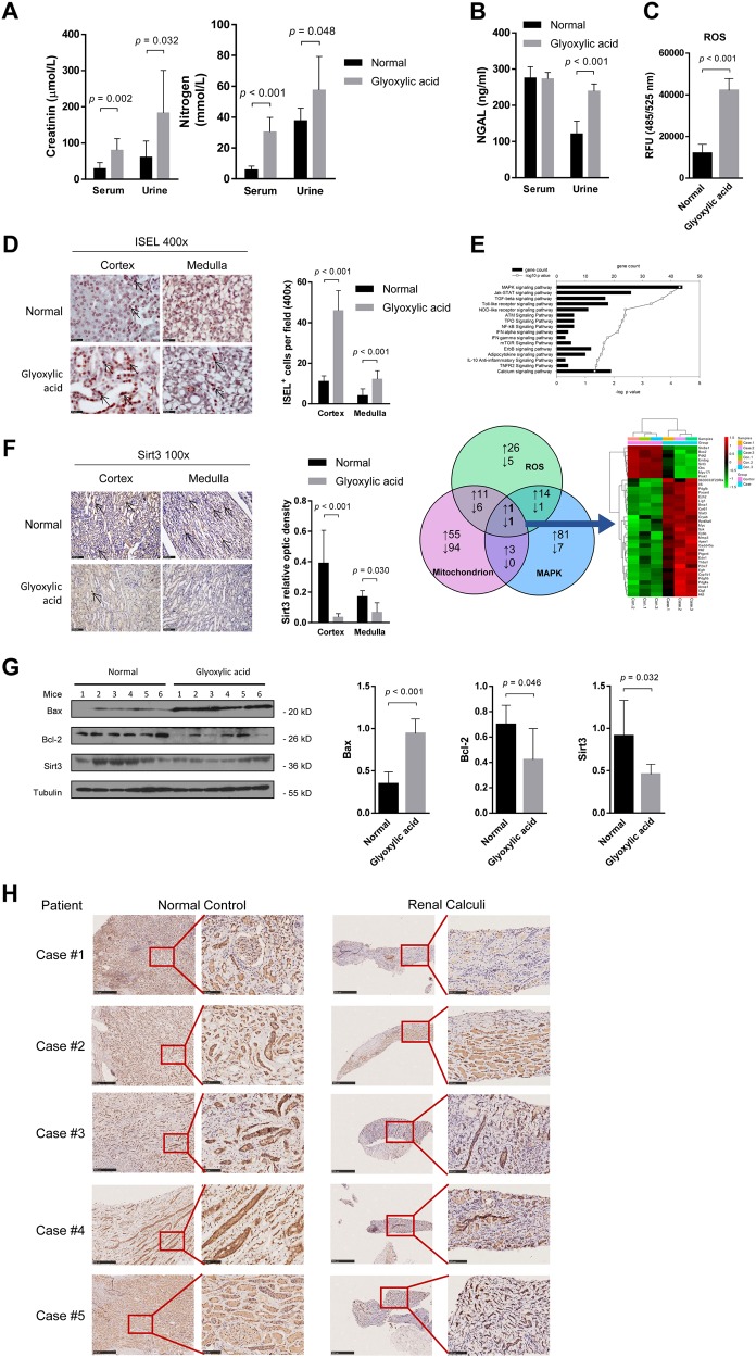
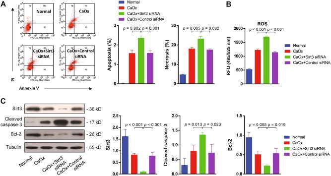
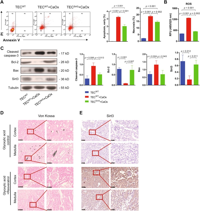
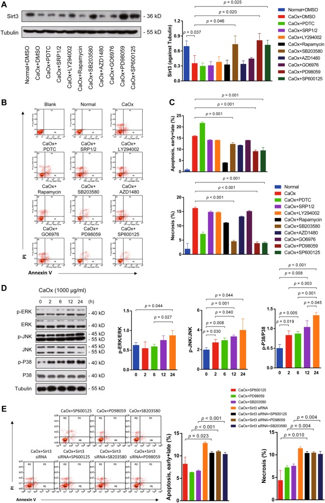
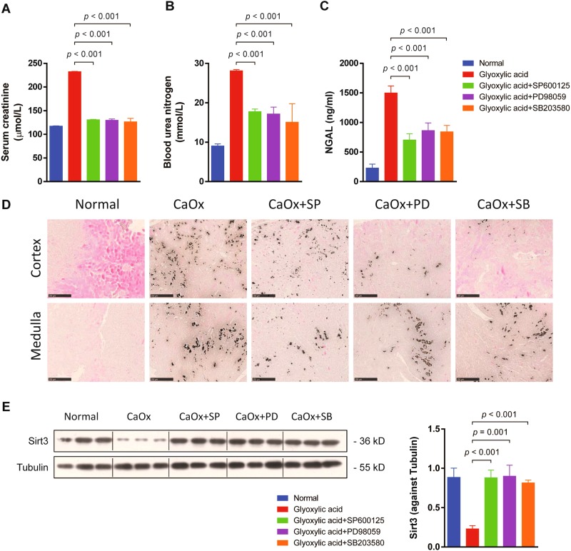
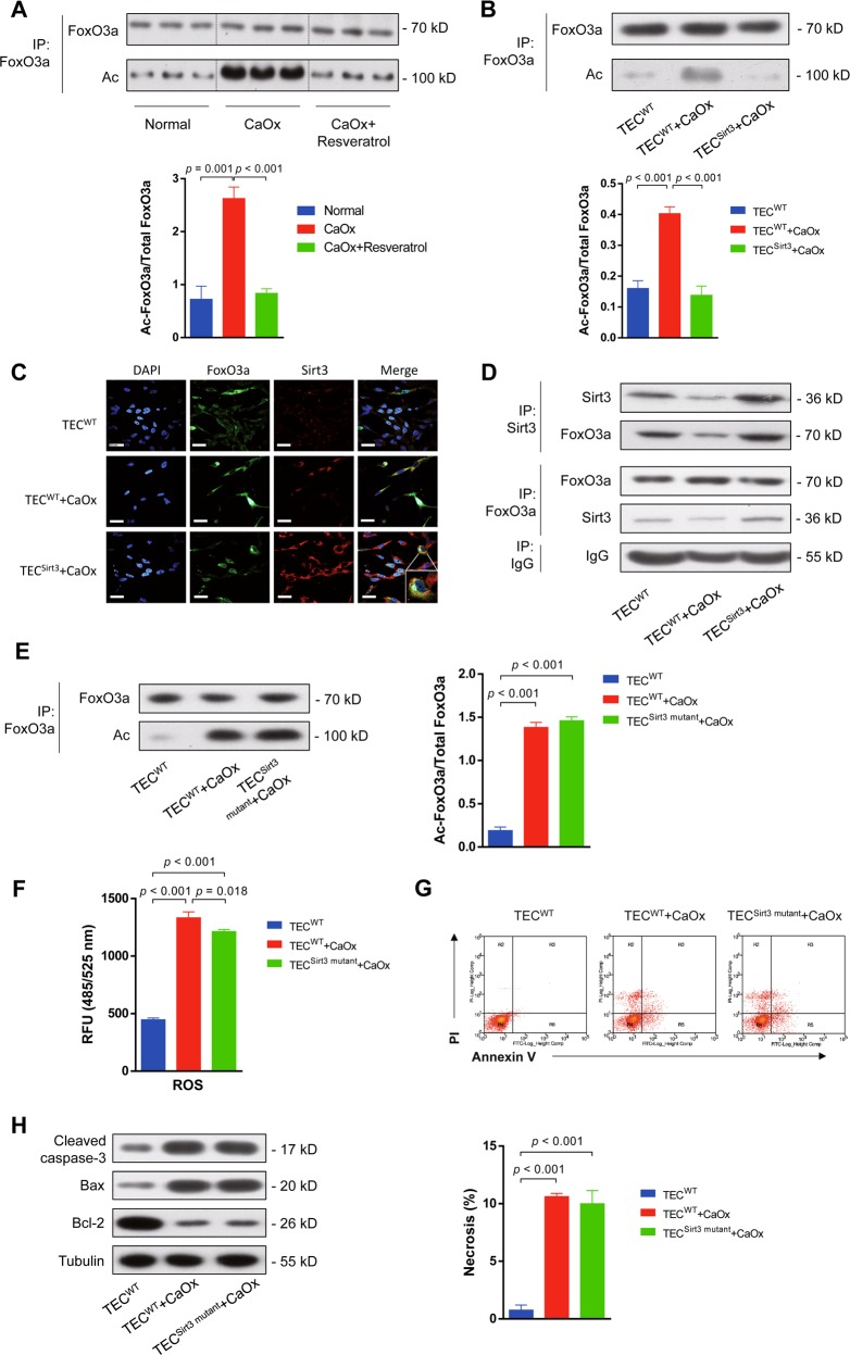
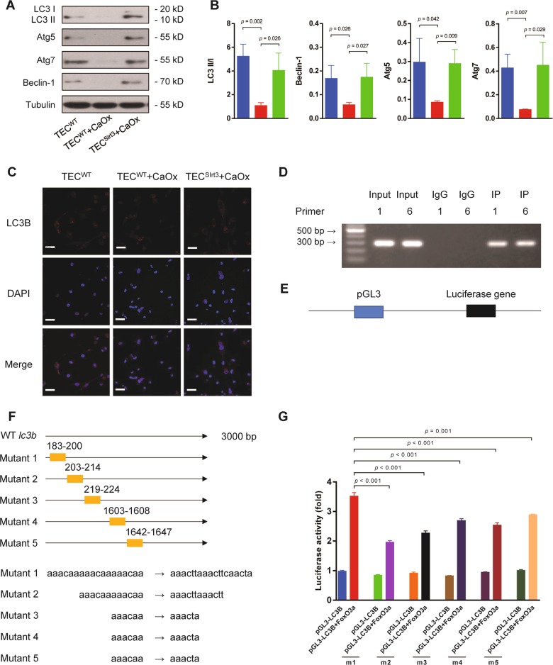
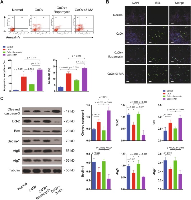
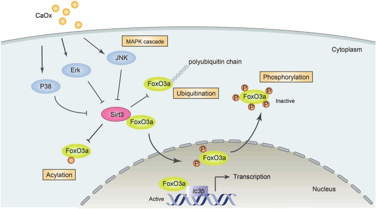
High oxalic acid and calcium oxalate (CaOx)-induced renal tubular epithelial cell (TEC) injury plays a key role in nephrolithiasis. However, the mechanism remains unknown. Gene array analysis of the mice nephrolithiasis model indicated significant downregulation of sirtuin 3 (Sirt3) and activation of mitogen-activated protein kinase (MAPK) pathway. Kidney biopsy tissues of renal calculi patients also showed decreased Sirt3 expression. Silencing Sirt3 exacerbated oxidative stress and TEC death under CaOx stimulation. Restoring Sirt3 expression by overexpression or enhancing its activity protected renal function and reduced TEC death both in vitro and in vivo. Inhibiting the MAPK pathway resulted in upregulation of Sirt3 expression, preservation of renal function and decreased cell death both in vitro and in vivo. Furthermore, Sirt3 could upregulate FoxO3a activity post-translationally via deacetylation, dephosphorylation and deubiquitination. FoxO3a was found to interact with the promoter region of LC3B and to increase its expression, enhancing TEC autophagy and suppressing cell apoptosis and necrosis. Taken together, our results indicate that the MAPK/Sirt3/FoxO3a pathway modulates renal TEC death and autophagy in TEC injury.


Conflict of interest statement

The authors declare that they have no conflict of interest.

Figures

Fig. 1
Fig. 1. Sirt3 expression was inhibited in nephrolithiasis in vivo.
a In the glyoxylic acid-induced nephrolithiasis model, creatinine and urea nitrogen levels in both serum and urine were significantly increased relative to the control levels. b Glyoxylic acid treatment increased the urine NGAL level, but the serum NGAL level showed no difference between the glyoxylic acid and control treatments. c ROS activity in the kidney was upregulated in the glyoxylic acid group compared to the control group. d The in situ end labelling (ISEL) assay showed more apoptotic cells in the kidney following glyoxylic acid treatment. Scale bar: 50 μm. e The top pathways of differentially expressed (DE) genes between normal and glyoxylic acid-treated murine kidney samples are shown in the histogram. The Venn diagram shows the number of upregulated (upward arrow) and downregulated (downward arrow) DE genes associated with the mitochondrion, ROS and the MAPK cascade. Heat map of the fold change in expression (ratio of the normalized intensities). Among the 29 upregulated and eight downregulated genes, Sirt3 was the only gene in all three subgroups that was downregulated (Case: glyoxylic acid-treated mice model; Control: saline-treated control mice). f In the glyoxylic acid group, the Sirt3 expression level was significantly decreased in both the cortex and medulla (n = 5). Scale bar: 250 μm. g Western blot analysis showed decreased Sirt3 and Bcl-2 levels and increased Bax levels in the glyoxylic acid group. h Sirt3 expression was downregulated in renal calculi patients compared with normal control; left: normal control samples from radical resection of the kidney; right: kidney needle biopsy tissues. Data are shown as the means ± S.D.; n = 5 patients per group. Scale bar: 500 μm (left); 100 μm (right)
Fig. 2
Fig. 2. Suppressing Sirt3 expression exacerbated CaOx-induced cell death in vitro.
a Flow cytometry analysis showed that silencing sirt3 increased TEC apoptosis and necrosis in the presence of CaOx. b ROS activity was significantly upregulated by Sirt3 siRNA transfection. c Western blot showed the expression of Sirt3, cleaved caspase-3 and Bcl-2 with Sirt3 or control siRNA transfection. Sirt3 siRNA transfection effectively inhibited Sirt3 protein expression. In the Sirt3 siRNA group, cleaved caspase-3 expression was significantly increased, and Bcl-2 expression was decreased. Data are shown as the means ± S.D.; n = 3 per group
Fig. 3
Fig. 3. Overexpression Sirt3 or enhancing its activity rescued the kidney from CaOx-induced cell death in vitro and in vivo.
a Flow cytometry showed that overexpression of sirt3 blocked CaOx-induced apoptosis and necrosis of TEC. b Overexpression of sirt3 significantly reduced ROS levels under CaOx stimulation. c Sirt3 protein expression was rescued in the sirt3-overexpressing group under CaOx stimulation. The cleaved caspase-3 and Bax levels were significantly decreased, and the Bcl-2 level was increased in the sirt3-overexpressing group compared to the non-transfected control group. d The von Kossa staining results revealed that resveratrol reduced calcium deposits in the medulla of the murine nephrolithiasis model. Scale bar: 100 μm (left); 50 μm (right). e Sirt3 expression in both the renal cortex and medulla was increased by resveratrol treatment. Data are shown as the means ± S.D.; n = 6 mice per group. Scale bar: 100 μm (left); 50 μm (right)
Fig. 4
Fig. 4. Suppression of Sirt3 expression in nephrolithiasis in vitro was dependent on activation of the MAPK pathway.
a SB203580 (P38 inhibitor), PD98059 (JNK inhibitor) and SP6000125 (ERK inhibitor) significantly increased Sirt3 expression, while PDTC (NF-κB inhibitor), SRP1/2 (mTOR inhibitor), Ly294002 (PI3K inhibitor), rapamycin (mTOR inhibitor), AZD1480 (JakStat inhibitor) and GO6976 (PKCα/PKCβ1 inhibitor) decreased Sirt3 expression. b These inhibitors were used to determine the pathways responsible for apoptosis and necrosis under CaOx stimulation. c SB203580 (P38 inhibitor), PD98059 (JNK inhibitor) and SP6000125 (ERK inhibitor) significantly reduced apoptosis and necrosis. d The phosphorylation levels of ERK, JNK and P38 were increased in a time-dependent manner under CaOx stimulation. e TEC apoptosis and necrosis were increased with sirt3 knockdown. Data are shown as the means ± S.D.; n = 3 per group
Fig. 5
Fig. 5. Inhibition of the MAPK pathway ameliorated glyoxylic acid-induced nephrolithiasis and kidney injury in vivo.
Mice were treated with SB203580, PD98059 and SP6000125 in vivo to inhibit the MAPK pathway in a nephrolithiasis model. ac Inhibition of the MAPK pathway decreased serum creatinine, blood urea nitrogen and serum NGAL levels. d The von Kossa staining revealed that SB203580, PD98059 or SP6000125 treatment reduced calcium deposits in the kidney. e The expression levels of Sirt3 were significantly increased by SB203580, PD98059 or SP6000125 treatment (n = 3). Scale bar: 250 μm. Data are shown as the means ± S.D.; n = 6 mice per group
Fig. 6
Fig. 6. Sirt3 directly deacetylated FoxO3a to protect TEC from apoptosis and necrosis.
a Immunoprecipitation assays demonstrated that Sirt3 overexpression via resveratrol treatment decreased acetylated FoxO3a expression in the kidney in vivo. b Similarly, overexpression of Sirt3 in TEC in vitro significantly decreased the FoxO3a acetylation level under CaOx stimulation. c Immunofluorescence assays indicated that Sirt3 interacted with FoxO3a most abundantly around the nucleus (DAPI: blue, FoxO3a: green and Sirt3: red). Scale bar: 60 μm. d Co-immunoprecipitation (co-IP) assays confirmed the interaction between Sirt3 and FoxO3a. e Overexpression of a catalytic mutant of Sirt3 (Sirt3H248Y) did not influence FoxO3a acetylation. f Overexpression of Sirt3H248Y did not reduce ROS activity in TEC. g Overexpression of Sirt3H248Y did not decrease TEC necrosis under CaOx stimulation. h Overexpression of Sirt3H248Y increased cleaved caspase-3 and Bax expression and reduced Bcl-2 expression. Data are shown as the means ± S.D.; n = 6 mice per group for an in vivo experiment. n = 3 per group for an in vitro experiment
Fig. 7
Fig. 7. The Sirt3/FoxO3a pathway promoted autophagy by transcriptionally regulating LC3B in TEC.
a The expression of the autophagy-associated proteins Beclin-1, Atg5 and Atg7 and the LC3 II/I ratio were detected by western blot. b The expression of Beclin-1, Atg5 and Atg7 and the LC3 II/I ratio were significantly upregulated by Sirt3 overexpression compared to the non-transfected control. c Immunofluorescence assays indicated LC3B upregulation with Sirt3 overexpression (DAPI: blue, LC3B: red). Scale bar: 60 μm. d Two pairs of lc3b primers were selected. e The LC3B promoter region in renal TEC DNA was inserted into the pGL3-enhancer luciferase reporter vector. f Five pGL3LC3B plasmids with different mutation regions were constructed. g The luciferase activity assay showed that all five mutated regions regulated the transcription of lc3b and that the first region played the most important role in the regulation of lc3b transcription. Data are shown as the means ± S.D.; n = 3 per group
Fig. 8
Fig. 8. Enhancement of autophagy ameliorated TEC injury under CaOx stimulation.
a Flow cytometry was performed on TEC treated with the autophagy agonist rapamycin or the autophagy antagonist 3-MA. Both apoptosis and necrosis of TEC were significantly decreased when autophagy was induced but were increased when autophagy was inhibited. b TEC apoptosis was examined by the TUNEL assay. Induction of autophagy with rapamycin reduced apoptosis, and inhibition of autophagy increased apoptosis. Scale bar: 150 μm. c Autophagy- and apoptosis-associated proteins were detected by western blot. In the rapamycin-treated group, the cleaved caspase-3 and Bax levels were reduced, and the anti-apoptotic protein Bcl-2 was upregulated. Beclin-1, Atg5 and Atg7 expression levels were upregulated by rapamycin treatment and downregulated by 3-MA. Data are shown as the means ± S.D.; n = 3 per group
Fig. 9
Fig. 9. Schematic diagram demonstrating the mechanism of CaOx-mediated TEC injury.
In nephrolithiasis, CaOx activates the MAPK pathway members P38, ERK and JNK. Then, Sirt3 expression is inhibited. As a result, acetylation, ubiquitination and phosphorylation of FoxO3a are enhanced, leading to FoxO3a inactivation and dysfunction. Because functional FoxO3a binds to the promoter of lc3b and promotes autophagy, TEC autophagy is inhibited in nephrolithiasis

References

    1. Sorokin I, et al. Epidemiology of stone disease across the world. World J. Urol. 2017;35:1301–1320. doi: 10.1007/s00345-017-2008-6. - DOI - PubMed
    1. Pearle, M. S., Calhoun, E. A., Curhan, G. C. & Urologic Diseases of America Project. Urologic diseases in America project: urolithiasis. J. Urol.173, 848–857 (2005). - PubMed
    1. Worcester EM, Coe FL. Clinical practice. Calcium kidney stones. N. Eng. J. Med. 2010;363:954–963. doi: 10.1056/NEJMcp1001011. - DOI - PMC - PubMed
    1. Evan AP, et al. Renal histopathology and crystal deposits in patients with small bowel resection and calcium oxalate stone disease. Kidney Int. 2010;78:310–317. doi: 10.1038/ki.2010.131. - DOI - PMC - PubMed
    1. Evan AP, et al. Randall’s plaque of patients with nephrolithiasis begins in basement membranes of thin loops of Henle. J. Clin. Invest. 2003;111:607–616. doi: 10.1172/JCI17038. - DOI - PMC - PubMed

Publication types