

ISL1 predicts poor outcomes for patients with gastric cancer and drives tumor progression through binding to the ZEB1 promoter together with SETD7
- PMID: 30674889
- PMCID: PMC6393520
- DOI: 10.1038/s41419-018-1278-2
ISL1 predicts poor outcomes for patients with gastric cancer and drives tumor progression through binding to the ZEB1 promoter together with SETD7
Abstract
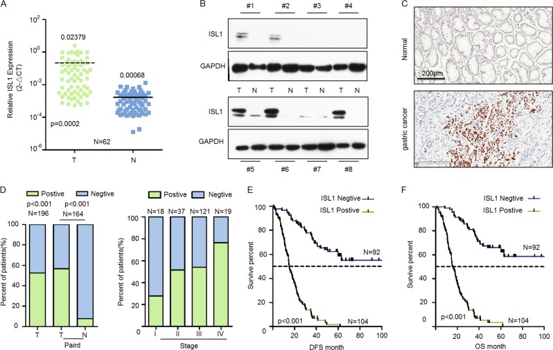
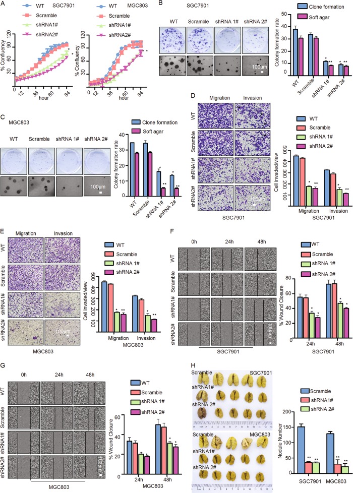
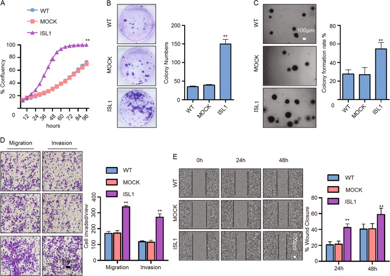
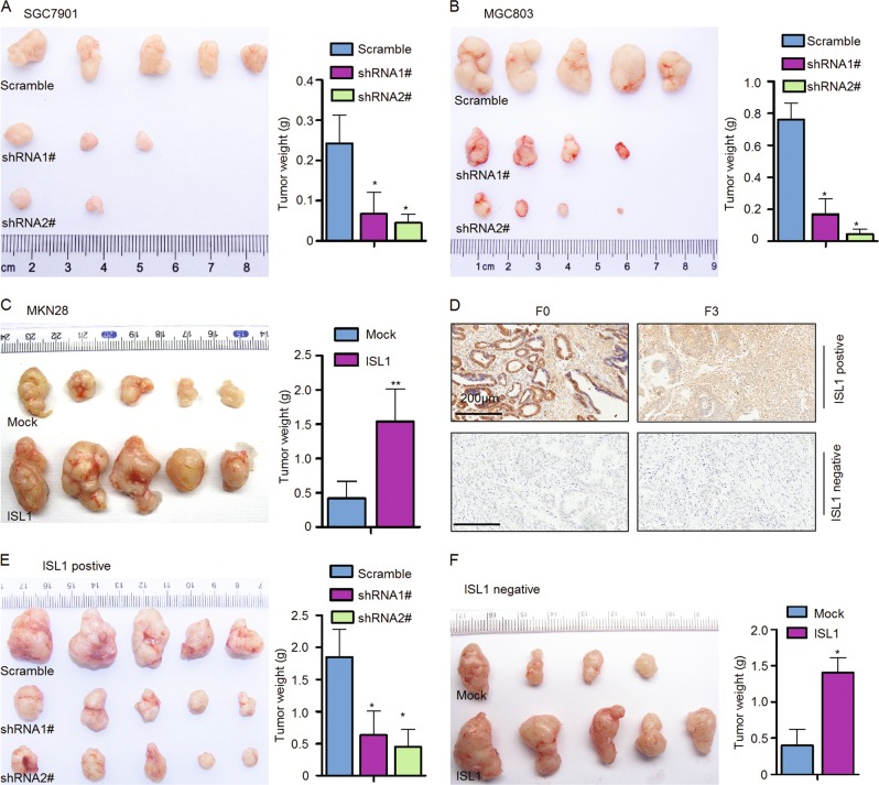
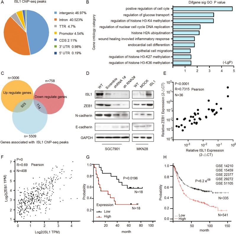
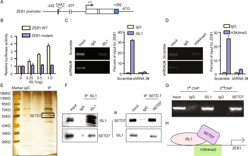
ISL1, a LIM-homeodomain transcription factor, serves as a biomarker of metastasis in multiple tumors. However, the function and underlying mechanisms of ISL1 in gastric cancer (GC) have not been fully elucidated. Here we found that ISL1 was frequently overexpressed in GC FFPE samples (104/196, 53.06%), and associated with worse clinical outcomes. Furthermore, the overexpression of ISL1 and loss-of-function of ISL1 influenced cell proliferation, invasion and migration in vitro and in vivo, including GC patient-derived xenograft models. We used ChIP-seq and RNA-seq to identify that ISL1 influenced the regulation of H3K4 methylation and bound to ZEB1, a key regulator of the epithelial-mesenchymal transition (EMT). Meanwhile, we validated ISL1 as activating ZEB1 promoter through influencing H3K4me3. We confirmed that a complex between ISL1 and SETD7 (a histone H3K4-specific methyltransferase) can directly bind to the ZEB1 promoter to activate its expression in GC cells by immunoprecipitation, mass spectrometry, and ChIP-re-ChIP. Moreover, ZEB1 expression was significantly positively correlated with ISL1 and was positively associated with a worse outcome in primary GC specimens. Our paper uncovers a molecular mechanism of ISL1 promoting metastasis of GC through binding to the ZEB1 promoter together with co-factor SETD7. ISL1 might be a potential prognostic biomarker of GC.
Conflict of interest statement
The authors declare that they have no conflict of interest.
Figures
Similar articles
-
The calcium pump PMCA4 prevents epithelial-mesenchymal transition by inhibiting NFATc1-ZEB1 pathway in gastric cancer.Biochim Biophys Acta Mol Cell Res. 2020 Dec;1867(12):118833. doi: 10.1016/j.bbamcr.2020.118833. Epub 2020 Aug 27. Biochim Biophys Acta Mol Cell Res. 2020. PMID: 32860837
-
ISL1, a novel regulator of CCNB1, CCNB2 and c-MYC genes, promotes gastric cancer cell proliferation and tumor growth.Oncotarget. 2016 Jun 14;7(24):36489-36500. doi: 10.18632/oncotarget.9269. Oncotarget. 2016. PMID: 27183908 Free PMC article.
-
MicroRNA-574-3p regulates epithelial mesenchymal transition and cisplatin resistance via targeting ZEB1 in human gastric carcinoma cells.Gene. 2019 Jun 5;700:110-119. doi: 10.1016/j.gene.2019.03.043. Epub 2019 Mar 24. Gene. 2019. PMID: 30917930
-
Zinc Finger Proteins in the War on Gastric Cancer: Molecular Mechanism and Clinical Potential.Cells. 2023 May 4;12(9):1314. doi: 10.3390/cells12091314. Cells. 2023. PMID: 37174714 Free PMC article. Review.
-
A Systematic Review to Define the Multi-Faceted Role of Lysine Methyltransferase SETD7 in Cancer.Cancers (Basel). 2022 Mar 10;14(6):1414. doi: 10.3390/cancers14061414. Cancers (Basel). 2022. PMID: 35326563 Free PMC article. Review.
Cited by
-
Functions of SETD7 during development, homeostasis and cancer.Stem Cell Investig. 2019 Sep 2;6:26. doi: 10.21037/sci.2019.06.10. eCollection 2019. Stem Cell Investig. 2019. PMID: 31620473 Free PMC article. No abstract available.
-
Sex-Specific Genetic Associations for Barrett's Esophagus and Esophageal Adenocarcinoma.Gastroenterology. 2020 Dec;159(6):2065-2076.e1. doi: 10.1053/j.gastro.2020.08.052. Epub 2020 Sep 9. Gastroenterology. 2020. PMID: 32918910 Free PMC article.
-
SDCBP-AS1 destabilizes β-catenin by regulating ubiquitination and SUMOylation of hnRNP K to suppress gastric tumorigenicity and metastasis.Cancer Commun (Lond). 2022 Nov;42(11):1141-1161. doi: 10.1002/cac2.12367. Epub 2022 Oct 9. Cancer Commun (Lond). 2022. PMID: 36209503 Free PMC article.
-
LIM homeodomain transcription factor Isl1 affects urethral epithelium differentiation and apoptosis via Shh.Cell Death Dis. 2019 Sep 26;10(10):713. doi: 10.1038/s41419-019-1952-z. Cell Death Dis. 2019. PMID: 31558700 Free PMC article.
-
Insulin gene enhancer protein 1 mediates glycolysis and tumorigenesis of gastric cancer through regulating glucose transporter 4.Cancer Commun (Lond). 2021 Mar;41(3):258-272. doi: 10.1002/cac2.12141. Epub 2021 Feb 11. Cancer Commun (Lond). 2021. PMID: 33570246 Free PMC article.
References
Publication types
MeSH terms
Substances
LinkOut - more resources
Full Text Sources
Medical
Molecular Biology Databases
Miscellaneous
