. 2019 Jan 23;9(1):411.
doi: 10.1038/s41598-018-36558-y.

Toxicity and possible mechanisms of action of honokiol from Magnolia denudata seeds against four mosquito species

Affiliations

Toxicity and possible mechanisms of action of honokiol from Magnolia denudata seeds against four mosquito species

Zhangqian Wang et al. Sci Rep. .

Abstract

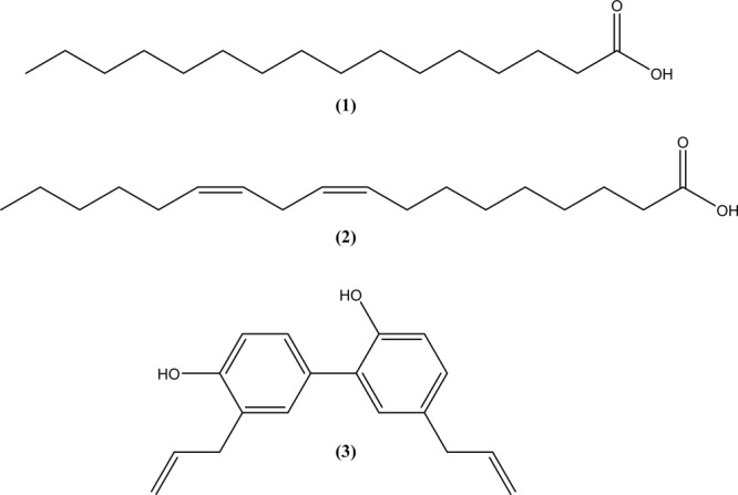
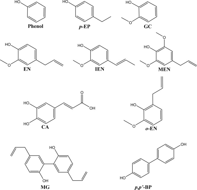
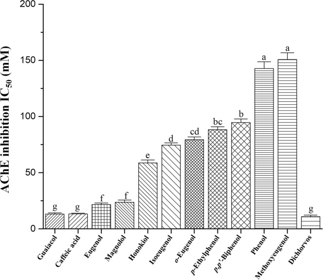
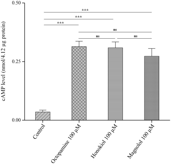
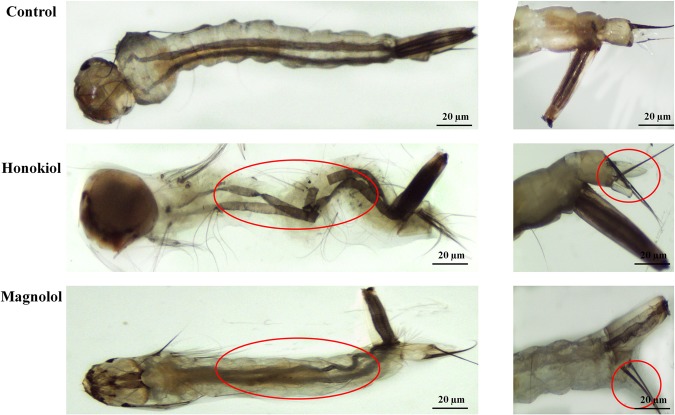
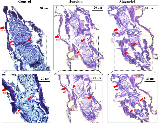
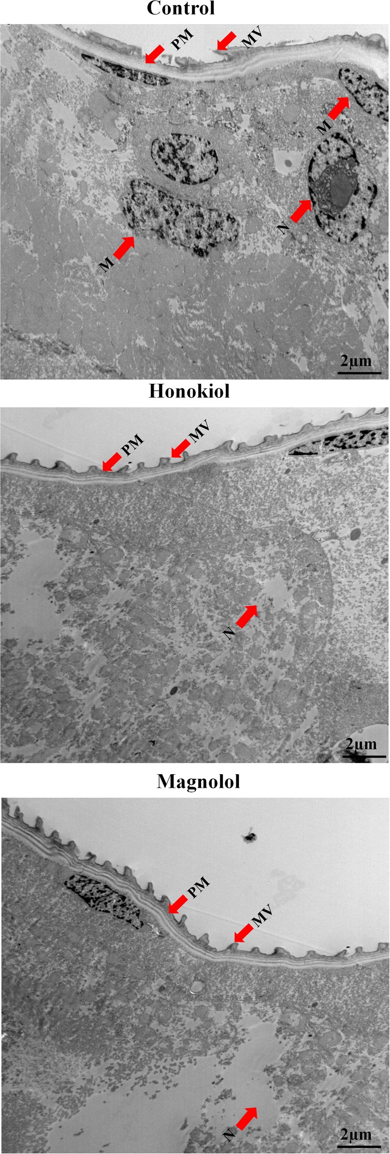
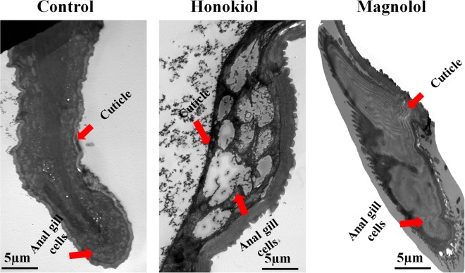
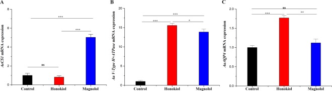
This study was performed to determine the toxicity and possible mechanism of the larvicidal action of honokiol, extracted from Magnolia denudata seeds, and its 10 related compounds against third-instar larvae of insecticide-susceptible Culex pipiens pallens, Aedes aegypti, and Aedes albopictus and Anopheles sinensis resistant to deltamethrin and temephos. Honokiol (LC50, 6.13-7.37 mg/L) was highly effective against larvae of all of the four mosquito species, although the toxicity of the compound was lower than that of the synthetic larvicide temephos. Structure-activity relationship analyses indicated that electron donor and/or bulky groups at the ortho or para positions of the phenol were required for toxicity. Honokiol moderately inhibited acetylcholinesterase and caused a considerable increase in cyclic AMP levels, indicating that it might act on both acetylcholinesterase and octopaminergic receptors. Microscopy analysis clearly indicated that honokiol was mainly targeted to the midgut epithelium and anal gills, resulting in variably dramatic degenerative responses of the midgut through sequential epithelial disorganization. Honokiol did not affect the AeCS1 mRNA expression level in Ae. aegypti larvae, but did enhance expression of the genes encoding vacuolar-type H+-ATPase and aquaporin 4, indicating that it may disturb the Na+, Cl- and K+ co-transport systems. These results demonstrate that honokiol merits further study as a potential larvicide, with a specific target site, and as a lead molecule for the control of mosquito populations.


Conflict of interest statement

The authors declare no competing interests.

Figures

Figure 1
Figure 1
Structures of palmitic acid, linoleic acid, and honokiol. These compounds were identified in the seeds of Magnolia denudata in this study. The chemical formula of the saturated fatty acid palmitic acid (1) is C16H32O2, with a molar mass of 256.43 g/mol. The chemical formula of the unsaturated fatty acid linoleic acid (2) is C18H32O2, with a molar mass of 280.45 g/mol. The chemical formula of the neolignan honokiol (3) is C18H18O2, with a molar mass of 266.33 g/mol.
Figure 2
Figure 2
Structures of 10 structurally related compounds of honokiol. Phenol, p-ethylphenol (p-EP), guaiacol (GC), eugenol (EN), isoeugenol (IEN), caffeic acid (CA), o-eugenol (o-EN), magnolol (MG), methoxyeugenol (MEN), and p,p′-biphenol (p,p′-BP).
Figure 3
Figure 3
Inhibitory effects on acetylcholinesterase. Inhibition of acetylcholinesterase (AChE) extracted from third-instar Aedes aegypti larvae by isolated neolignan honokiol, magnolol, a structural isomer of honokiol, and nine structurally related phenol compounds of honokiol was determined by acetylthiocholine iodide hydrolysis at 30 °C and pH 8.0 as described in the Methods section. Each bar represents the mean ± standard error of triplicate samples from three independent experiments (P = 0.05, using Bonferroni’s multiple comparisons test).
Figure 4
Figure 4
Effects on cyclic AMP levels. A whole-body homogenate from third-instar Aedes aegypti larvae was tested for adenylate cyclase activity, as described in the Methods section, in the presence of honokiol (100 µM) and magnolol (100 µM). The effects of the neolignans on cyclic AMP (cAMP) levels of the homogenate were compared with those of octopamine (100 µM) alone. Data were expressed as nmol/4.12 μg protein. Each bar represents the mean ± standard error of duplicate samples from three independent experiments (***P < 0.001; ns, no significant difference, using Bonferroni’s multiple comparisons test).
Figure 5
Figure 5
Light micrographs of midgut, thorax, and anal gill parts of larval Aedes aegypti. Third-instar Ae. aegypti larvae were placed into paper cups containing a methanol–Triton X-100 solution in distilled water with an LC50 of honokiol (6.5 mg/L) or magnolol (25 mg/L) for 24 h. The morphology of the whole body of the larvae was observed with a stereo microscope (35× magnification). (A) Untreated control larvae showed normal appearances of typical structures, with well-developed, distinguished head, thorax, and abdominal regions. Honokiol-treated larvae were completely damaged with indistinct appearances, particularly in the thorax and abdominal region segments. In particular, the midgut region was completely ruptured, and the midgut contents oozed out from the body. Magnolol-treated larvae showed shrunken bodies with no distinguishable abdominal segments. Intrinsic body fluid contents became dark, and there were unusually elongated digestive tracts with damaged interior tissues. (B) Control larvae showed well-developed anal gills and anal gill cells. In larvae treated with honokiol, the anal gill from one side was swollen and reduced in length and the other one was completely damaged. In larvae treated with magnolol, the anal gill region was completely damaged, with outer cuticle membranes with indistinct anal gills and no internal body fluid. All experiments were performed in duplicate, and 20 mosquito larvae were used in each replicate. More than 10 live larvae from control and treated groups were randomly collected and used for analysis.
Figure 6
Figure 6
Histology of midgut regions of larval Aedes aegypti. Third-instar Ae. aegypti larvae were placed into paper cups containing a methanol–Triton X-100 solution in distilled water with an LC50 of honokiol (6.5 mg/L) or magnolol (25 mg/L) for 24 h. Carson’s trichrome staining was performed as described in the Methods section. Observations were taken of 15 larvae under the microscope. (A) Control larvae. The well-developed midgut regions of control larvae showed distinct midgut epithelium (ME) and peritrophic membranes (PMs), which enclosed the rich lumen contents (LCs) in the midgut region. (B) Honokiol-treated larvae. The midgut regions of the larvae showed a damaged midgut epithelial layer with smashed PMs consisting of lumen debris. (C) Magnolol-treated larvae. The midgut regions of the larvae showed indistinguishable midgut epithelial layers as well as PMs with damaged LC.
Figure 7
Figure 7
Transmission electronic micrographs of midgut regions of larval Aedes aegypti. Third-instar Ae. aegypti larvae were placed into paper cups containing a methanol–Triton X-100 solution in distilled water with an LC50 of honokiol (6.5 mg/L) or magnolol (25 mg/L) for 24 h. (A) Control larvae. A transmission electron micrograph revealed that the peritrophic membrane (PM) in the midgut regions of the larvae enclosed the midgut lumen contents (LCs) and consisted of numerous microvilli (MV) from the outside. The LC consists of the cytoplasm and numerous cell organelles surrounded by a plasma membrane. There was no damage to cell organelles in the cytoplasm, including prominent large central nuclei (approximately 15 µm diameter), mitochondria, and other cell organelles. (B) Honokiol-treated larvae. The neolignan destroyed all cellular material and extruded masses in the cytoplasm. In particular, mitochondria and large prominent nuclei were indistinguishable from damaged cellular contents. There was no distinct nuclear material inside the nucleus. (C) Magnolol-treated larvae. These showed complete disappearance of the nucleus, as well as cytoplasmic contents. Nuclei and mitochondria were completely absent. Other cytoplasmic organelles were damaged with indistinct appearances.
Figure 8
Figure 8
Transmission electronic micrographs of the anal gill regions of larval Aedes aegypti. Third-instar Ae. aegypti larvae were placed into paper cups containing a methanol–Triton X-100 solution in distilled water with an LC50 of honokiol (6.5 mg/L) or magnolol (25 mg/L) for 24 h. (A) Control larvae. Transmission electronic micrography (TEM) analysis revealed that the anal gills of the control larva were surrounded by thick cuticles and that the inner surfaces of the cytoplasm became fuller, with anal gill cells and fluid-filled tracheoles. (B) Honokiol-treated larvae. Histopathological observation of the larvae indicated cytoplasmic disruption in anal gills with damaged anal gill cells. (C) Magnolol-treated larvae. TEM showed damaged outer membranes of thick cuticles, which led to internal cytoplasmic destruction.
Figure 9
Figure 9
Effects of two neolignans on the expression levels of AeCS, AaV-type H+ ATPase, and AaAQP4 mRNA. Third-instar Ae. aegypti larvae were placed into paper cups containing a methanol–Triton X-100 solution in distilled water with an LC50 of honokiol (6.5 mg/L) or magnolol (25 mg/L) for 24 h. Total RNA was extracted from the anal gills (for AaAQP4 and AaV-type H+-ATPase) and midguts (for AeCS1) of 50 larvae. Real-time quantitative reverse transcription polymerase chain reaction was performed to determine the levels of AeCS1, AaV-type H+-ATPase, and AaAQP4 mRNA. Specific AeCS1, AaV-type H+-ATPase, and AaAQP4, and Aarps7 coding sequence primers were used to amplify AeCS1, AaV-type H+-ATPase, AaAQP4, and Aarps7 DNA, as described in the Methods section. (A) Midgut specific chitin synthase AeCS1 gene expression was slightly inhibited in honokiol-treated larvae, whereas the gene expression level was significantly increased in magnolol-treated larvae. (B) AaV-type H+-ATPase gene expression was increased in honokiol- and magnolol-treated larvae compared to control larvae. (C) The AaAQP4 gene expression level was significantly increased in honokiol-treated larval Ae. aegypti compared to control larvae. Magnolol did not affect the AaAQP4 mRNA expression level. The mRNA expression levels were normalized to constitutive expression of mRNA of the housekeeping gene Aarps7 and analyzed by the 2−ΔΔCT method. Each bar represents the mean ± SE of duplicate samples run in three independent experiments (***P < 0.001; **P < 0.01; *P < 0.05; ns, no significant difference, using Bonferroni’s multiple comparisons test).
Figure 10
Figure 10
Procedures to isolate the mosquito larvicidal constituents. The Magnolia denudata seed methanol extract was sequentially partitioned into hexane-, chloroform-, ethyl acetate-, butanol-, and water-soluble portions. The hexane-soluble fraction was the most biologically active fraction, and medium-pressure liquid chromatography and high-performance liquid chromatography were performed. Each fraction (10–100 mg/L) was tested in a contact mortality bioassay against third-instar Culex pipiens pallens and Aedes agypti larvae to isolate the active constituents from the fraction.

Similar articles

Cited by

References

    1. Moritz UGK, et al. The global distribution of the arbovirus vectors Aedes aegypti and Ae. albopictus. eLife. 2015;4:e08347. doi: 10.7554/eLife.08347. - DOI - PMC - PubMed
    1. Sinka M, et al. The dominant Anopheles vectors of human malaria in the Asia-Pacific region: occurrence data, distribution maps and bionomic précis. Parasit. Vectors. 2011;4:89. doi: 10.1186/1756-3305-4-89. - DOI - PMC - PubMed
    1. Pages N, et al. Scientific review on mosquitoes and mosquito-borne disease. EFSA Supporting Publications. 2009;6:1–96. doi: 10.2903/sp.efsa.2009.EN-7. - DOI
    1. Musso D, Gubler DJ. Zika virus. Clin. Microbiol. Rev. 2016;29:487–524. doi: 10.1128/CMR.00072-15. - DOI - PMC - PubMed
    1. Confalonieri, U. et al. Human health in Climate Change 2007: Impacts, Adaptation and Vulnerability (eds Parry, M. L. et al.) 391–431 (Cambridge University Press, 2007).

Publication types