. 2019 Jan 23;9(1):429.
doi: 10.1038/s41598-018-36796-0.

CGRP-CRLR/RAMP1 signal is important for stress-induced hematopoiesis

Affiliations

CGRP-CRLR/RAMP1 signal is important for stress-induced hematopoiesis

Akira Suekane et al. Sci Rep. .

Abstract

Ecotropic viral integration site-1 (EVI1) has a critical role in normal and malignant hematopoiesis. Since we previously identified high expression of calcitonin receptor like receptor (CRLR) in acute myeloid leukemia (AML) with high EVI1 expression, we here characterized the function of CRLR in hematopoiesis. Since higher expression of CRLR and receptor activity modifying protein 1 (RAMP1) was identified in immature hematopoietic bone marrow (BM) cells, we focused on calcitonin gene-related peptide (CGRP), a specific ligand for the CRLR/RAMP1 complex. To elucidate the role of CGRP in hematopoiesis, Ramp1-deficient (Ramp1-/-) mice were used. The steady-state hematopoiesis was almost maintained in Ramp1-/- mice; however, the BM repopulation capacity of Ramp1-/- mice was significantly decreased, and the transplanted Ramp1-/- BM mononuclear cells had low proliferation capacity with enhanced reactive oxygen species (ROS) production and cell apoptosis. Thus, CGRP is important for maintaining hematopoiesis during temporal exposures with proliferative stress. Moreover, continuous CGRP exposure to mice for two weeks induced a reduction in the number of BM immature hematopoietic cells along with differentiated myeloid cells. Since CGRP is known to be increased under inflammatory conditions to regulate immune responses, hematopoietic exhaustion by continuous CGRP secretion under chronic inflammatory conditions is probably one of the important mechanisms of anti-inflammatory responses.


Conflict of interest statement

The authors declare no competing interests.

Figures

Figure 1
Figure 1
CGRP-Crlr/Ramp1 signal is not an important factor for maintenance of hematopoietic cells under steady-state conditions. (a,b) Relative expression levels of Crlr and Ramp1 mRNA in purified BM-derived hematopoietic subsets, including LSK, CMP, MEP, GMP, myeloid, and lymphoid cells, as assessed by quantitative real-time RT-PCR. Data are shown as means ± SD for triplicate reactions. *p < 0.05, ****p < 0.0001 compared with LSK or Ramp1 (Ordinary one-way ANOVA and Dunnett’s multiple comparisons test). (c) The percentages of Crlr or Ramp1 positive cells in purified BM-derived hematopoietic subsets, including LSK, lineage negative cells, BMMNCs, and Gr-1+CD11b+ cells, as assessed by flow cytometry. Data are shown as means ± SD of five mice. ****p < 0.0001 compared with LSK (Ordinary one-way ANOVA and Dunnett’s multiple comparisons test). A representative gating strategy is provided in Supplemental Fig. S1. (d) Intracellular cAMP concentrations in LSK cells were measured 10 minutes after treatment with or without 100 nM CGRP. ***p < 0.001 compared with control (Student’s t test). (e) The WBC, RBC, and PLT counts in the PB of Ramp1−/− and WT mice at 8 to 12 weeks of age. (f) The percentages of differentiated white blood cells (myeloid cells, B cells, and T cells) in PB from Ramp1−/− and WT mice. Data are shown as means ± SD of five mice. (g) The percentages of differentiated white blood cells (myeloid cells, B cells, and T cells) in BMMNCs cells from Ramp1−/− and WT mice. Data are shown as means ± SD of five mice. (h) Absolute numbers of BMMNCs in the BM of Ramp1−/− and WT mice. Data are shown as means ± SD of five mice. (i) Absolute numbers of immature hematopoietic subpopulations (HSC, MPP, CMP, GMP, and MEP) in the BM of Ramp1−/− and WT mice. Data are shown as means ± SD of five mice.
Figure 2
Figure 2
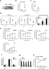
Crlr/Ramp1 signaling is essential for maintenance hematopoietic cells under stress conditions. (a) Expression levels of CGRP in BM stromal cells of WT mice with 10 Gy irradiation and without irradiation were determined by immunoblot analysis. BMMNCs from three mice for each group were analyzed. The full-length gels/blots are presented in Supplemental Fig. S4. (b) Expression levels of proCGRP mRNA were determined in BM stromal cells of WT mice treated with 10 Gy irradiation and without irradiation were determined by real-time PCR. ***p < 0.001 compared with control (Student’s t test). (c) The numbers of BMMNCs were compared between WT and Ramp1−/− mice at seven days after 5-FU injection. Data are shown as means ± SD of four mice. *p < 0.05, compared with WT mice (Student’s t test). (d) Under the same experiment in (c), the cell numbers and percentages of LSK or myeloid progenitors (MP) fractions were compared between WT and Ramp1−/− mice at seven days after 5-FU injection. The percentages of CMP, GMP, and MEP cells in the MP fraction were compared between the two groups. Data are shown as means ± SD of four mice. *p < 0.05, **p < 0.01, ***p < 0.001 compared with WT mice (Student’s t test). (e) Chimerism (percent donor-derived cells, CD45.2) from Ramp1−/− and WT mice in BMMNCs, myeloid cells, B lymphocytes, and T lymphocytes from the PB of lethally irradiated WT recipients (CD45.1) at each indicated time point after transplantation (1, 2, 3, and 4 months). Donor/recipient ratio: 50:50. Data are shown as means ± SD of four mice. *p < 0.05, **p < 0.01 compared with WT mice (Student’s t test). (f) The proportion of donor-derived myeloid, B lymphocytes, and T lymphocytes (Ramp1−/− and WT mice) among donor-derived WBCs in the PB of lethally irradiated WT recipients at four months after transplantation. Data are shown as means ± SD of four mice. (g) Chimerism (percent donor-derived cells, CD45.2) from Ramp1−/− and WT mice in BMMNCs, Lin+, Lin, MP, and LSK cells in BMMNCs from lethally irradiated WT recipients (CD45.1). Donor/recipient ratio: 50:50. Data are shown as means ± SD of four mice. *p < 0.05, **p < 0.01 compared with WT mice (Student’s t test). (h) Chimerism (percent donor-derived cells, CD45.2) from Ramp1−/− and WT mice in BMMNCs of lethally irradiated WT recipients (CD45.1) at each indicated time point after second transplantation (1, 2, 3, and 4 months). Donor/recipient ratio: 50:50. Data are shown as means ± SD of three mice. ***p < 0.001 compared with WT mice (Student’s t test).
Figure 3
Figure 3
CGRP enhances cell proliferation with suppression of ROS production and cell apoptosis in HSPCs under stress conditions. (a) Percentages of BrdU-positive cells in each BM cell fraction (LSK, Lin, BMMNCs, and myeloid cells) of the recipient mice (CD45.1) following transplantation of BM cells (CD45.2) from Ramp1−/− and WT mice were determined by flow cytometry. Data are shown as means ± SD of three mice. *p < 0.05, **p < 0.01 compared with WT mice (Student’s t test) (b) The homing ability of the transplanted donor BMMNCs (Ramp1−/− and WT) into the BM of recipient mice was determined by flow cytometry. Total numbers of donor-derived CD45.2+ BMMNCs in each recipient mouse are shown. (c) The ROS production in Lin cells from BMMNCs of the recipient mice transplanted with donor BM cells from Ramp1−/− or WT mice was determined by flow cytometry using redox-sensitive MitoTracker Orange CMH2TMROS fluorescence staining 16 hours after transplantation. Data are shown as means ± SD of three mice. *p < 0.05 compared with WT mice (Student’s t test). (d) The percentage of apoptotic cells in Lin cells from BMMNCs of the recipient mice transplanted with donor BM cells from Ramp1−/− or WT mice were determined by flow cytometry using annexin V staining after transplantation. Data are shown as means ± SD of three mice. *p < 0.05 compared with WT mice (Student’s t test). (e) The percentages and absolute numbers of LSK cells in the Lineage negative population from BMMNCs cultured alone or co-cultured with BM stromal cells in the presence or absence of CGRP, as determined by flow cytometery. Data are shown as means ± SD of three mice. *p < 0.05, **p < 0.01 compared with WT mice (Ordinary one-way ANOVA and Turkey’s multiple comparisons test).
Figure 4
Figure 4
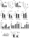
Long-term CGRP treatment affects hematopoiesis. (a) The colony-forming abilities of BMMNCs (left) and the percentage of Gr-1+CD11b+ cells in BMMNCs (right) from WT mice were determined by serial plating in methylcellulose culture with or without CGRP (100 nM). Data are shown as means ± SD of three mice. *p < 0.05, **p < 0.01 compared with WT mice (Student’s t test). (b) The colony-forming abilities of BMMNCs (left) and the percentage of Gr-1+CD11b+ cells in BMMNCs (right) from Ramp1−/− mice were determined by serial plating in methylcellulose culture with or without CGRP (100 nM). Data are shown as means ± SD of three mice. (c) The WBC, myeloid cells, RBC, and Plt counts in the PB of WT mice treated with CGRP or PBS for one and two weeks were determined. Data are shown as means ± SD of three mice. *p < 0.05 compared with PBS-treated mice (Student’s t test). (d) The percentages of differentiated white blood cells populations (myeloid cells, B cells, and T cells) in PB cells from WT mice treated with CGRP or PBS for two weeks were determined. Data are shown as means ± SD of three mice. *p < 0.05 compared with PBS-treated mice (Student’s t test). (e) The cell numbers (left) and percentages (right) of hematopoietic progenitor subpopulations (HSC, MPP, CMP, GMP, and MEP) in BMMNCs of WT mice treated with CGRP or PBS for two weeks were determined. Data are shown as means ± SD of three mice. *p < 0.05, **p < 0.01, ***p < 0.001, ****p < 0.0001 compared with PBS-treated mice (Student’s t test). (f) Expression levels of Crlr in BMMNCs of WT mice treated with CGRP or PBS for two weeks was determined by immunoblot analysis. BMMNCs from three mice for each group were analyzed. The full-length gels/blots are presented in Supplemental Fig. S4. (g) The colony forming abilities of BMMNCs of PBS-treated (PBS+) or CGRP- treated (CGRP+) mice for two weeks were determined in the in vitro cell culture with (CGRP) or without CGRP (Control). BMMNCs from three mice for each group were analyzed. ns; not significant, *p < 0.05, **p < 0.01 compared with PBS-treated mice (Ordinary one-way ANOVA and Dunnett’s multiple comparisons test).

Similar articles

Cited by

References

    1. Muller-Sieburg CE, Whitlock CA, Weissman IL. Isolation of two early B lymphocyte progenitors from mouse marrow: A committed Pre-Pre-B cell and a clonogenic Thy-1lo hematopoietic stem cell. Cell. 1986;44:653–662. doi: 10.1016/0092-8674(86)90274-6. - DOI - PubMed
    1. Kondo M, Weissman IL, Akashi K. Identification of Clonogenic Common Lymphoid Progenitors in Mouse Bone Marrow. Cell. 1997;91:661–672. doi: 10.1016/S0092-8674(00)80453-5. - DOI - PubMed
    1. Morrison SJ, Wandycz AM, Hemmati HD, Wright DE, Weissman IL. Identification of a lineage of multipotent hematopoietic progenitors. Development. 1997;124:1929–1939. - PubMed
    1. Akashi K, Traver D, Miyamoto T, Weissman IL. A clonogenic common myeloid progenitor that gives rise to all myeloid lineages. Nature. 2000;404:193–197. doi: 10.1038/35004599. - DOI - PubMed
    1. Manz MG, Miyamoto T, Akashi K, Weissman IL. Prospective isolation of human clonogenic common myeloid progenitors. Proceedings of the National Academy of Sciences. 2002;99:11872–11877. doi: 10.1073/pnas.172384399. - DOI - PMC - PubMed

Publication types

MeSH terms

Substances