

In vivo assessment of increased oxidation of branched-chain amino acids in glioblastoma
- PMID: 30674979
- PMCID: PMC6344513
- DOI: 10.1038/s41598-018-37390-0
In vivo assessment of increased oxidation of branched-chain amino acids in glioblastoma
Abstract
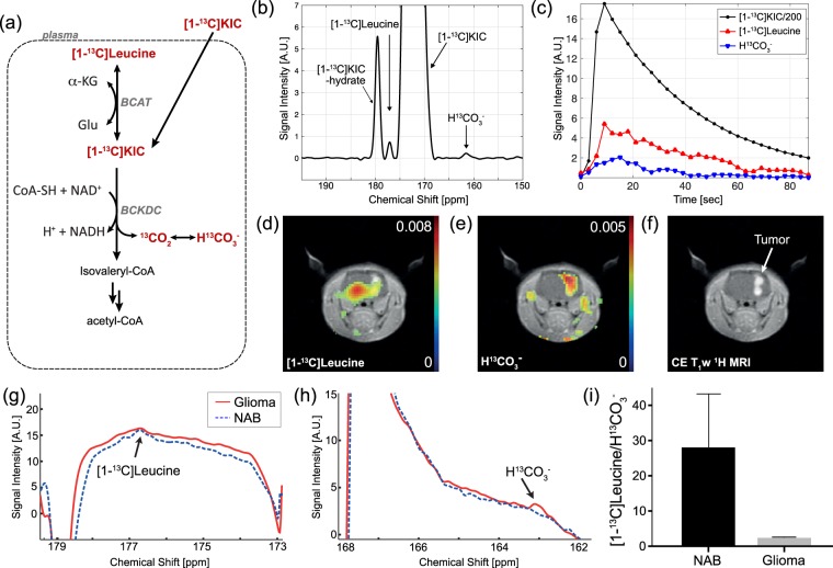
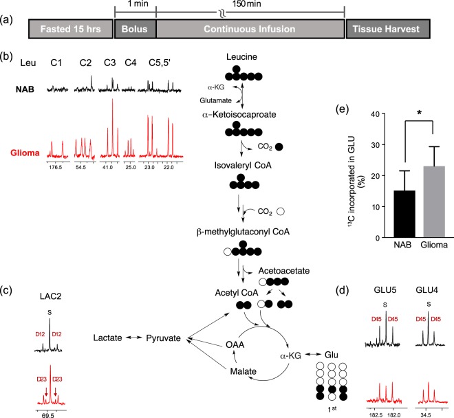
Altered branched-chain amino acids (BCAAs) metabolism is a distinctive feature of various cancers and plays an important role in sustaining tumor proliferation and aggressiveness. Despite the therapeutic and diagnostic potentials, the role of BCAA metabolism in cancer and the activities of associated enzymes remain unclear. Due to its pivotal role in BCAA metabolism and rapid cellular transport, hyperpolarized 13C-labeled α-ketoisocaproate (KIC), the α-keto acid corresponding to leucine, can assess both BCAA aminotransferase (BCAT) and branched-chain α-keto acid dehydrogenase complex (BCKDC) activities via production of [1-13C]leucine or 13CO2 (and thus H13CO3-), respectively. Here, we investigated BCAA metabolism of F98 rat glioma model in vivo using hyperpolarized 13C-KIC. In tumor regions, we observed a decrease in 13C-leucine production from injected hyperpolarized 13C-KIC via BCAT compared to the contralateral normal-appearing brain, and an increase in H13CO3-, a catabolic product of KIC through the mitochondrial BCKDC. A parallel ex vivo 13C NMR isotopomer analysis following steady-state infusion of [U-13C]leucine to glioma-bearing rats verified the increased oxidation of leucine in glioma tissue. Both the in vivo hyperpolarized KIC imaging and the leucine infusion study indicate that KIC catabolism is upregulated through BCAT/BCKDC and further oxidized via the citric acid cycle in F98 glioma.
Conflict of interest statement
The authors declare no competing interests.
Figures


Similar articles
-
Hyperpolarized α-keto[1-13C]isocaproate as a 13C magnetic resonance spectroscopic agent for profiling branched chain amino acid metabolism in tumors.2010 Jan 15 [updated 2010 May 19]. In: Molecular Imaging and Contrast Agent Database (MICAD) [Internet]. Bethesda (MD): National Center for Biotechnology Information (US); 2004–2013. 2010 Jan 15 [updated 2010 May 19]. In: Molecular Imaging and Contrast Agent Database (MICAD) [Internet]. Bethesda (MD): National Center for Biotechnology Information (US); 2004–2013. PMID: 20641995 Free Books & Documents. Review.
-
The feasibility of assessing branched-chain amino acid metabolism in cellular models of prostate cancer with hyperpolarized [1-(13)C]-ketoisocaproate.Magn Reson Imaging. 2014 Sep;32(7):791-5. doi: 10.1016/j.mri.2014.04.015. Epub 2014 Apr 28. Magn Reson Imaging. 2014. PMID: 24907854 Free PMC article.
-
Metabolic approach of absence seizures in a genetic model of absence epilepsy, the GAERS: study of the leucine-glutamate cycle.J Neurosci Res. 2001 Dec 1;66(5):923-30. doi: 10.1002/jnr.10086. J Neurosci Res. 2001. PMID: 11746420
-
Diabetes and branched-chain amino acids: What is the link?J Diabetes. 2018 May;10(5):350-352. doi: 10.1111/1753-0407.12645. Epub 2018 Feb 13. J Diabetes. 2018. PMID: 29369529
-
The Critical Role of the Branched Chain Amino Acids (BCAAs) Catabolism-Regulating Enzymes, Branched-Chain Aminotransferase (BCAT) and Branched-Chain α-Keto Acid Dehydrogenase (BCKD), in Human Pathophysiology.Int J Mol Sci. 2022 Apr 5;23(7):4022. doi: 10.3390/ijms23074022. Int J Mol Sci. 2022. PMID: 35409380 Free PMC article. Review.
Cited by
-
Enrichment of branched chain amino acid transaminase 1 correlates with multiple biological processes and contributes to poor survival of IDH1 wild-type gliomas.Aging (Albany NY). 2021 Jan 20;13(3):3645-3660. doi: 10.18632/aging.202328. Epub 2021 Jan 20. Aging (Albany NY). 2021. PMID: 33493139 Free PMC article.
-
Amino Acid Transporters in Glioblastoma: Implications for Diagnosis, Disease Monitoring, Therapeutic Targeting, and Drug Delivery.Mol Diagn Ther. 2025 Aug 22. doi: 10.1007/s40291-025-00810-9. Online ahead of print. Mol Diagn Ther. 2025. PMID: 40844759 Review.
-
Transporters at the Interface between Cytosolic and Mitochondrial Amino Acid Metabolism.Metabolites. 2021 Feb 16;11(2):112. doi: 10.3390/metabo11020112. Metabolites. 2021. PMID: 33669382 Free PMC article. Review.
-
Detecting biomarkers by dynamic nuclear polarization enhanced magnetic resonance.Natl Sci Rev. 2024 Jun 29;11(9):nwae228. doi: 10.1093/nsr/nwae228. eCollection 2024 Sep. Natl Sci Rev. 2024. PMID: 39144741 Free PMC article. Review.
-
Measuring the Metabolic Evolution of Glioblastoma throughout Tumor Development, Regression, and Recurrence with Hyperpolarized Magnetic Resonance.Cells. 2021 Oct 1;10(10):2621. doi: 10.3390/cells10102621. Cells. 2021. PMID: 34685601 Free PMC article.
References
-
- Warburg O. über den Stoffwechsel der Carcinomzelle. Klinische Wochenschrift. 1925;4:534–536. doi: 10.1007/BF01726151. - DOI
Publication types
MeSH terms
Substances
Grants and funding
LinkOut - more resources
Full Text Sources
