

Patho- physiological role of BDNF in fibrin clotting
- PMID: 30674980
- PMCID: PMC6344484
- DOI: 10.1038/s41598-018-37117-1
Patho- physiological role of BDNF in fibrin clotting
Abstract
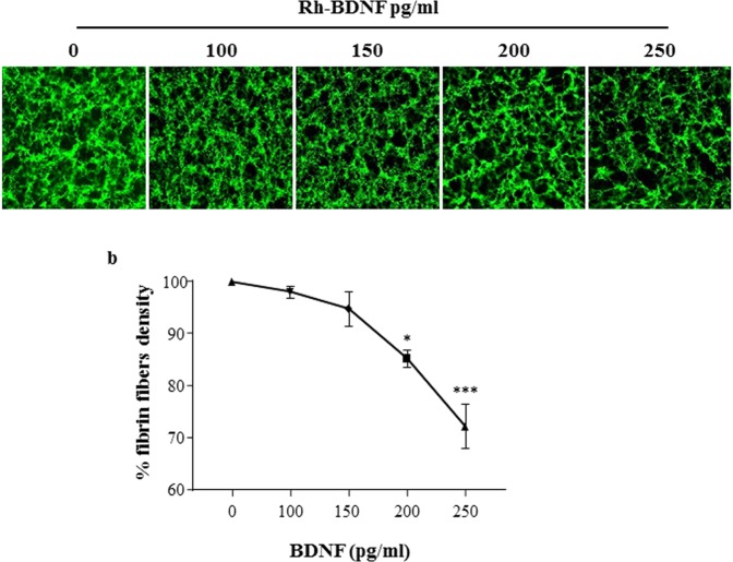
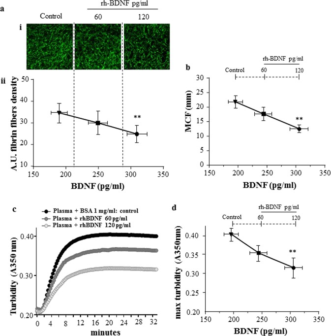
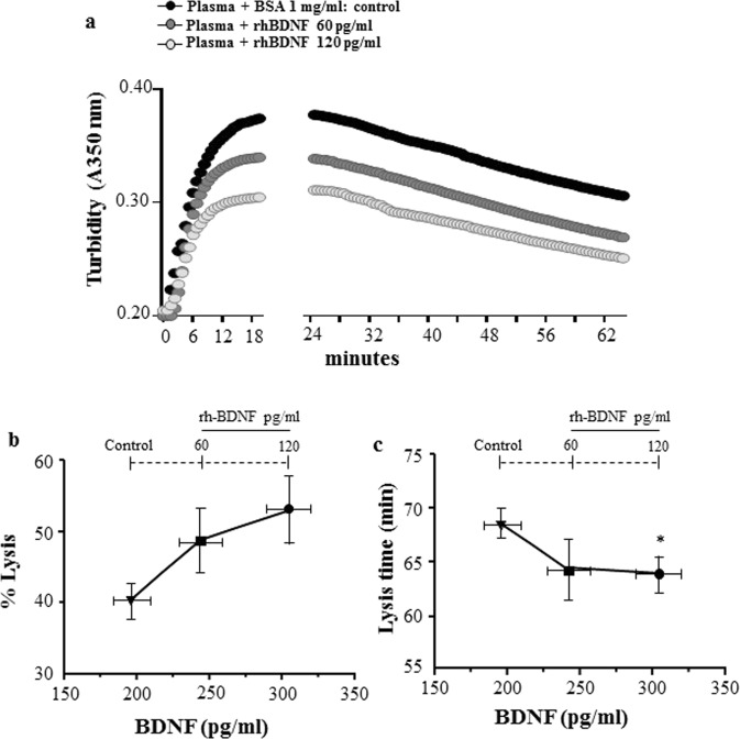
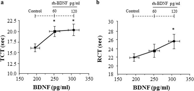
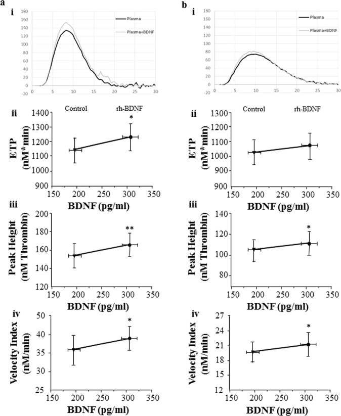
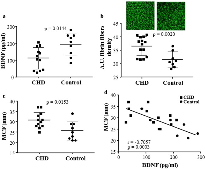
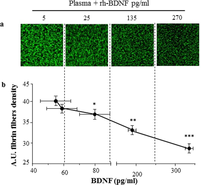
Circulating levels of Brain Derived Neurotrophic Factor (BDNF) are lower in coronary heart disease (CHD) than in healthy subjects and are associated with coronary events and mortality. However, the mechanism(s) underling this association is not fully understood. We hypothesize that BDNF may influence fibrin fiber structure and clot stability, favoring clot lysis and thrombus resolution. We showed that recombinant BDNF (rh-BDNF) influenced with clot formation in a concentration-dependent manner in both purified fibrinogen and plasma from healthy subjects. In particular, rh-BDNF reduced the density of fibrin fibers, the maximum clot firmness (MCF) and the maximum clot turbidity, and affected the lysis of clot. In addition, both thrombin and reptilase clotting time were prolonged by rh-BDNF, despite the amount of thrombin formed was greater. Intriguingly, CHD patients had lower levels of BDNF, greater fibrin fibers density, higher MCF than control subjects, and a negative correlation between BDNF and MCF was found. Of note, rh-BDNF markedly modified fibrin clot profile restoring physiological clot morphology in CHD plasma. In conclusion, we provide evidence that low levels of BDNF correlate with the formation of bigger thrombi (in vitro) and that this effect is mediated, at least partially, by the alteration of fibrin fibers formation.
Conflict of interest statement
The authors declare no competing interests.
Figures
Similar articles
-
Unfavorably Altered Fibrin Clot Properties in Patients with Eosinophilic Granulomatosis with Polyangiitis (Churg-Strauss Syndrome): Association with Thrombin Generation and Eosinophilia.PLoS One. 2015 Nov 5;10(11):e0142167. doi: 10.1371/journal.pone.0142167. eCollection 2015. PLoS One. 2015. PMID: 26540111 Free PMC article.
-
Fibrin clot structure - pro-fibrinolytic effect of oral contraceptives in apparently healthy women.Thromb Haemost. 2017 Apr 3;117(4):700-705. doi: 10.1160/TH16-10-0748. Epub 2017 Feb 2. Thromb Haemost. 2017. PMID: 28150855 Clinical Trial.
-
Prolonged duration of type 2 diabetes is associated with increased thrombin generation, prothrombotic fibrin clot phenotype and impaired fibrinolysis.Thromb Haemost. 2014 Apr 1;111(4):685-93. doi: 10.1160/TH13-07-0566. Epub 2013 Dec 5. Thromb Haemost. 2014. PMID: 24306139
-
Thrombin generation and fibrin clot structure.Blood Rev. 2007 May;21(3):131-42. doi: 10.1016/j.blre.2006.11.001. Epub 2007 Jan 8. Blood Rev. 2007. PMID: 17208341 Review.
-
Antithrombotic medications and their impact on fibrin clot structure and function.J Physiol Pharmacol. 2018 Aug;69(4). doi: 10.26402/jpp.2018.4.02. Epub 2018 Nov 7. J Physiol Pharmacol. 2018. PMID: 30415236 Review.
Cited by
-
Indole-3-Carbinol and Its Derivatives as Neuroprotective Modulators.Brain Sci. 2024 Jul 2;14(7):674. doi: 10.3390/brainsci14070674. Brain Sci. 2024. PMID: 39061415 Free PMC article. Review.
-
Brain-Derived Neurotrophic Factor (BDNF) Is Associated with Platelet Activity and Bleeding Tendency in Patients with Gaucher Disease.Int J Mol Sci. 2022 Nov 12;23(22):13982. doi: 10.3390/ijms232213982. Int J Mol Sci. 2022. PMID: 36430458 Free PMC article.
-
Molecular insights of exercise therapy in disease prevention and treatment.Signal Transduct Target Ther. 2024 May 29;9(1):138. doi: 10.1038/s41392-024-01841-0. Signal Transduct Target Ther. 2024. PMID: 38806473 Free PMC article. Review.
-
The multifaceted role of platelets in mediating brain function.Blood. 2022 Aug 25;140(8):815-827. doi: 10.1182/blood.2022015970. Blood. 2022. PMID: 35609283 Free PMC article. Review.
-
Preliminary results of the cross-sectional associations of sedentary behavior and physical activity with serum brain-derived neurotrophic factor in adults with coronary heart disease.Sci Rep. 2022 Nov 16;12(1):19685. doi: 10.1038/s41598-022-23706-8. Sci Rep. 2022. PMID: 36385629 Free PMC article.
