. 2019 Jan 23;9(1):382.
doi: 10.1038/s41598-018-37212-3.

An improved yeast surface display platform for the screening of nanobody immune libraries

Affiliations

An improved yeast surface display platform for the screening of nanobody immune libraries

Tomasz Uchański et al. Sci Rep. .

Abstract

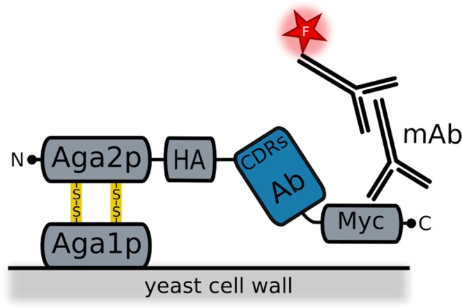
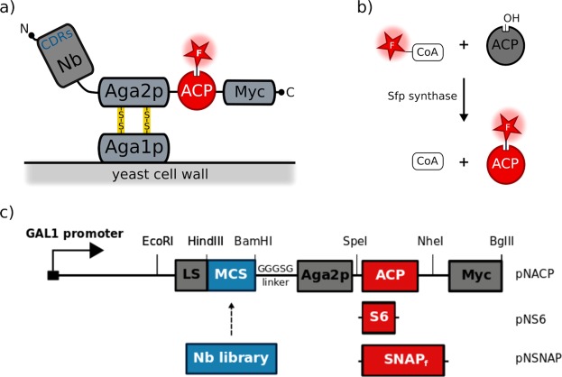
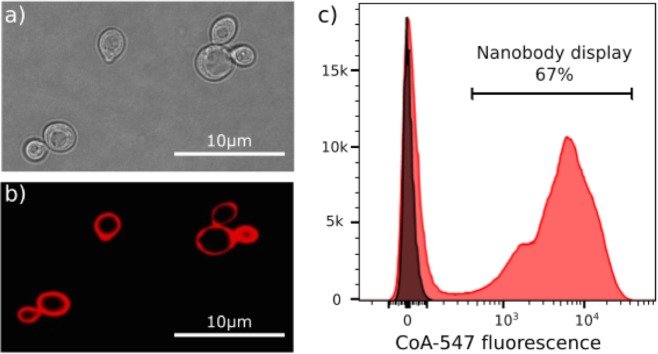
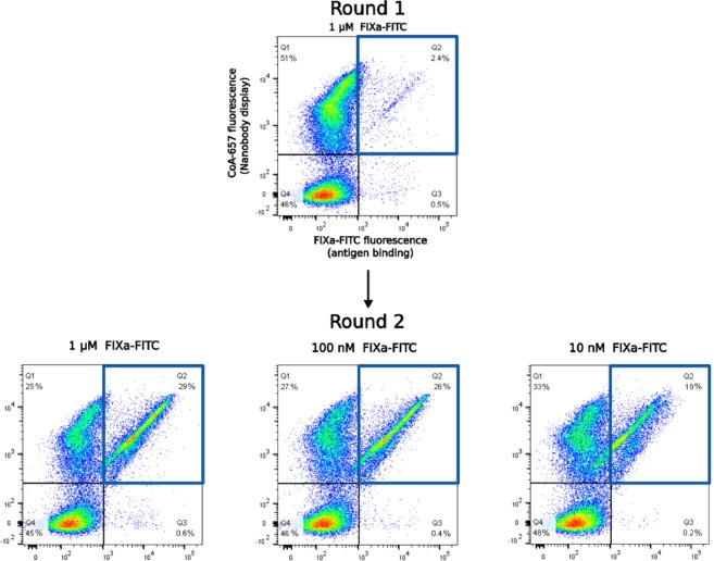
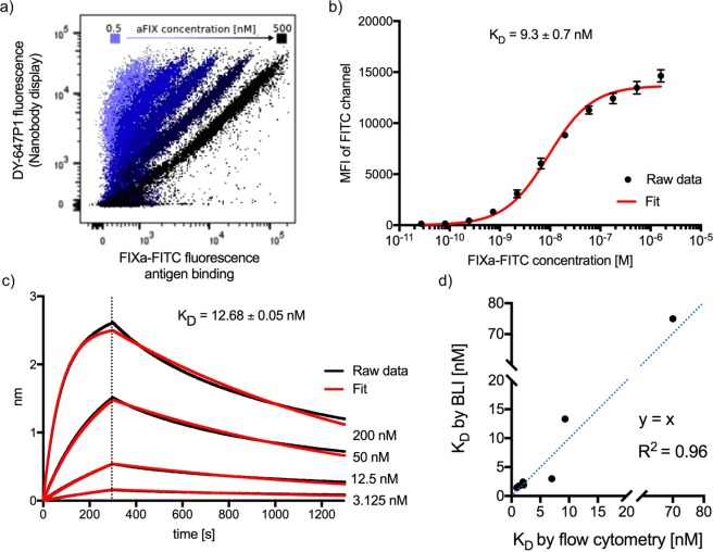
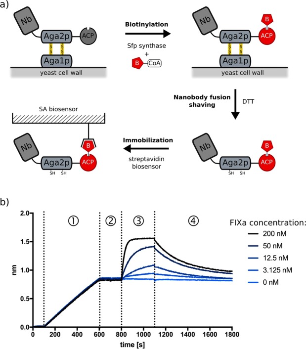
Fusions to the C-terminal end of the Aga2p mating adhesion of Saccharomyces cerevisiae have been used in many studies for the selection of affinity reagents by yeast display followed by flow cytometric analysis. Here we present an improved yeast display system for the screening of Nanobody immune libraries where we fused the Nanobody to the N-terminal end of Aga2p to avoid steric hindrance between the fused Nanobody and the antigen. Moreover, the display level of a cloned Nanobody on the surface of an individual yeast cell can be monitored through a covalent fluorophore that is attached in a single enzymatic step to an orthogonal acyl carrier protein (ACP). Additionally, the displayed Nanobody can be easily released from the yeast surface and immobilised on solid surfaces for rapid analysis. To prove the generic nature of this novel Nanobody discovery platform, we conveniently selected Nanobodies against three different antigens, including two membrane proteins.


Conflict of interest statement

The authors declare no competing interests.

Figures

Figure 1
Figure 1
Conventional yeast surface display system for the screening of antigen-binding scaffold libraries (Adapted from). Affinity reagents, including single-domain antibodies (blue) can be fused via its N-terminal end to the C-terminus of Aga2p. Surface expression can be detected by using fluorescently labelled antibodies that bind the Myc or HA tags.
Figure 2
Figure 2
Optimised yeast display system for the screening of Nanobody libraries by cell sorting. (a) We designed novel vectors for the extracellular display of fusion proteins consisting of a Nanobody followed by Aga2p and by ACP. The Nanobody is fused at its C-terminus to Aga2p, leaving the CDRs fully exposed for antigen binding. (b) Enzymes such as Sfp Synthase can be used to covalently attach CoA derivatives containing fluorophores or biotin to a unique serine residue of the C-terminal ACP tag. (c) In our vectors, Nanobody libraries can be cloned as fusion proteins under the transcriptional control of the GAL1 promoter. The fusion protein is secreted by using the appS4 leader sequence. ACP can be replaced by other tags that are amenable to covalent orthogonal labelling of the displayed fusion protein: S6 (pNS6 vector) or SNAPf (pNSNAP vector).
Figure 3
Figure 3
Orthogonal labelling of displayed Nanobodies on the surface of yeast. EBY100 yeast cells containing pNACP_Nb35 were grown and induced overnight in galactose-rich medium. The Nb35-Aga2p-ACP fusion was orthogonally labelled by incubating these cells with CoA-547 in the presence of Sfp synthase. Surface display of the fusion protein was analysed by light microscopy (a) and confocal microscopy (b,c) Histogram of the flow cytometric analysis of CoA-547 labelled cells expressing the fusion (pNACP_Nb35, red), compared to labelled yeast cells that do not display a Nb-Aga2p-ACP fusion (pNACP, grey). The fluorescence intensity of each cell was monitored at 582 nm upon excitation with a 561 nm yellow-green laser.
Figure 4
Figure 4
Consecutive rounds of selection of Nanobodies that bind FIXa by yeast display and two-dimensional flow cytometry. Each dot represents two fluorescent signals of a separate yeast cell of the (sub) library. The x-axis is a measure of the amount of the fluorescent antigen that is bound (FITC fluorescence) whereas the y-axis gives an indication of the Nanobody display level (DY-647P1 fluorescence). In this example, the yeast cells displaying FIXa FITC specific Nanobodies (gate Q2 represented as a blue square), were enriched from 2.4% in the first round of selection to 19% in the second round at the lowest antigen concentration (10 nM of FIXa-FITC).
Figure 5
Figure 5
The binding affinities of Nanobodies that are displayed on yeast can conveniently be estimated by flow cytometry. (a) Yeast cells displaying one particular FIXa-specific Nanobody (MP1031_B7) were cultured overnight and orthogonally labelled with CoA-647, then divided in six aliquots and incubated separately with different concentrations of FIXa-FITC (0.5, 2, 8, 32, 128, 500 nM) and analysed in two dimensions by flow cytometry. (b) An apparent affinity of displayed Nanobody MP1031_B7 for the fluorescent antigen was determined by plotting the FITC mean fluorescence intensity (MFI) of Nanobody displaying cells versus the FIXa-FITC concentration. An apparent KD (9.3 ± 0.7 nM) can conveniently be calculated by standard software like Prism 7 (GraphPad), using the simple one-site specific binding model. The data is shown as mean standard error of the mean (s.e.m.) from n = 3 independent experiments. (c) Association and dissociation isotherms of FIXa to Nanobody MP1031_B7. The Nb was immobilised on an Ni-NTA bio-sensor and the binding kinetics were monitored by bio‐layer interferometry (BLI) on OctetRED96 (ForteBio). The measured responses (red lines) were fitted to a monophasic 1:1 binding model (black lines). (d) Comparison of the apparent affinities of eight different Nanobodies as determined by flow cytometry (yeast displayed Nb-Aga2p-ACP fusion, y-axis) or BLI (subcloned and purified Nb, x-axis).
Figure 6
Figure 6
Orthogonally labelled Nanobodies can be functionally shaved from yeast cells and immobilised. (a) Nanobodies can be displayed on the yeast surface as Aga2p-ACP fusions and orthogonally biotinylated via ACP by adding a CoA-biotin derivative in the presence of Sfp synthase. Subsequent, this biotinylated Nanobody-Aga2p-ACP fusion protein can efficiently be shaved from the yeast surface by DTT and immobilised on streptavidin coated (SA) materials for other applications including BLI. (b) Binding isotherms (raw data) of the immobilisation of a biotinylated Nanobody-Aga2p-ACP fusion protein and the subsequent binding of its antigen to an SA-coated biosensor measured by BLI on OctetRED96. A 10 ml culture of yeast, displaying Nanobody MP1031_B7 as an Nb-Aga2p-ACP fusion was resuspended in with CoA-biotin and Sfp synthase and shaved by adding 2 mM DTT. A SA biosensor (ForteBio) was dipped into the supernatant (step 1) and equilibrated into buffer (step 2). Next, the biosensor was incubated with different FIXa concentrations to follow the association (step 3) and dissociation (step 4) of the antigen.

Similar articles

Cited by

References

    1. Bradbury ARM, Sidhu S, Dübel S, McCafferty J. Beyond natural antibodies: The power of in vitro display technologies. Nature Biotechnology. 2011;29:245–254. doi: 10.1038/nbt.1791. - DOI - PMC - PubMed
    1. Binz HK, et al. High-affinity binders selected from designed ankyrin repeat protein libraries. Nat. Biotechnol. 2004;22:575–582. doi: 10.1038/nbt962. - DOI - PubMed
    1. Koide A, Gilbreth RN, Esaki K, Tereshko V, Koide S. High-affinity single-domain binding proteins with a binary-code interface. Proc. Natl. Acad. Sci. 2007;104:6632–6637. doi: 10.1073/pnas.0700149104. - DOI - PMC - PubMed
    1. Muyldermans S. Nanobodies: Natural Single-Domain Antibodies. Annu. Rev. Biochem. 2013;82:775–797. doi: 10.1146/annurev-biochem-063011-092449. - DOI - PubMed
    1. Kubala MH, Kovtun O, Alexandrov K, Collins BM. Structural and thermodynamic analysis of the GFP:GFP-nanobody complex. Protein Sci. 2010;19:2389–2401. doi: 10.1002/pro.519. - DOI - PMC - PubMed

Publication types

MeSH terms

Substances