

Fifty Years of Ventricular Tachycardia in a Single Patient
- PMID: 30675072
- PMCID: PMC6342044
Fifty Years of Ventricular Tachycardia in a Single Patient
Abstract
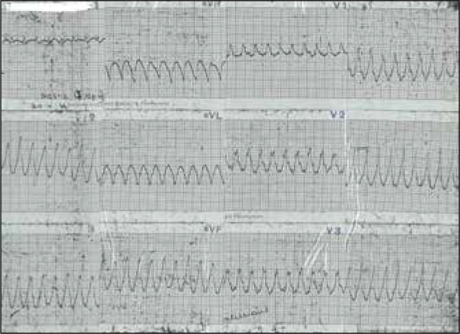
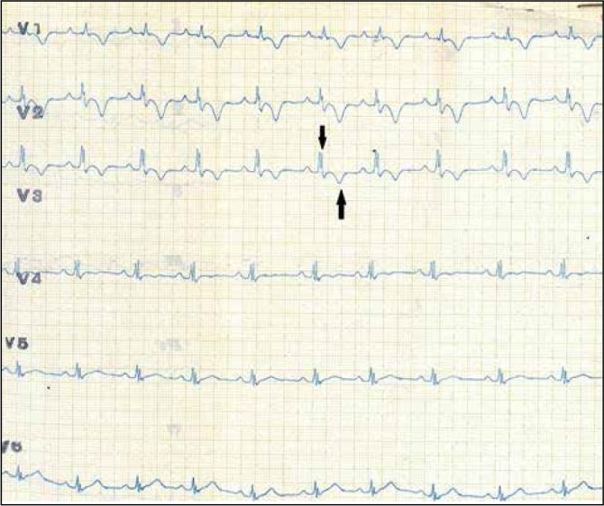
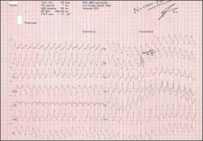
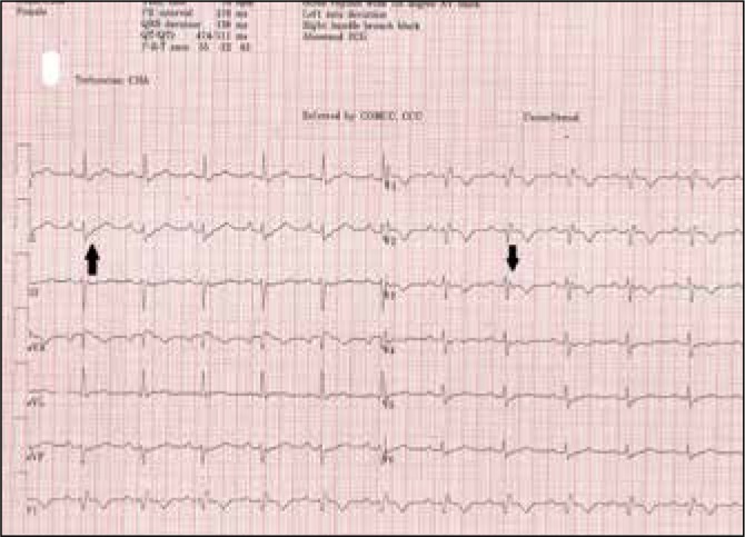
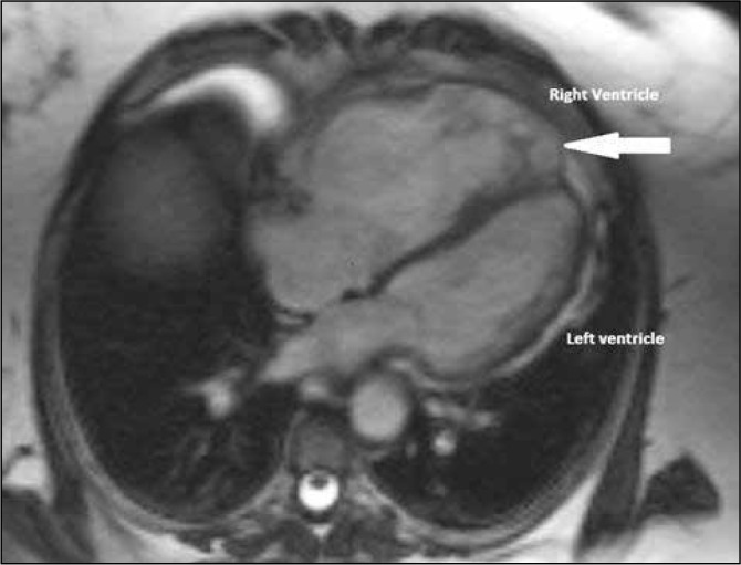
We report a patient who first presented during childhood in the early 1960's with several episodes of ventricular tachycardia (VT) and we describe her management which reflected the best medical knowledge at the time. She then presented more than 50 years later, again with VT, at which time a definitive diagnosis of the underlying cause was made. Her case illustrates the evolution in the understanding and management of VT over the past 50 years. This in turn reflects the clinical and technological advances in the management of cardiovascular disease over time.
Keywords: Arrhythmogenic Right Ventricular Cardiomyopathy; Dysplasia; Tachycardia; Ventricular.
Conflict of interest statement
Provenance: externally peer-reviewed.
Figures
Similar articles
-
Ventricular tachycardia in arrhythmogenic right ventricular dysplasia/cardiomyopathy: clinical presentation, risk stratification and results of long-term follow-up.Int J Cardiol. 2006 Mar 8;107(3):360-8. doi: 10.1016/j.ijcard.2005.03.049. Int J Cardiol. 2006. PMID: 16503259
-
Arrhythmogenic Right Ventricular Cardiomyopathy: An Exuberant Case Affecting Both Ventricles.Circ Cardiovasc Imaging. 2020 Sep;13(9):e010243. doi: 10.1161/CIRCIMAGING.119.010243. Epub 2020 Sep 2. Circ Cardiovasc Imaging. 2020. PMID: 32873069 No abstract available.
-
Epsilon Wave Masked by ST-Segment Elevation After Cardioversion From Sustained Ventricular Tachycardia: An Exceptional Manifestation of Arrhythmogenic Right Ventricular Dysplasia/Cardiomyopathy.J Emerg Med. 2022 Feb;62(2):254-259. doi: 10.1016/j.jemermed.2021.10.033. Epub 2022 Jan 17. J Emerg Med. 2022. PMID: 35058095
-
Arrhythmogenic right ventricular cardiomyopathy 2012: diagnostic challenges and treatment.J Cardiovasc Electrophysiol. 2012 Oct;23(10):1149-53. doi: 10.1111/j.1540-8167.2012.02412.x. Epub 2012 Aug 21. J Cardiovasc Electrophysiol. 2012. PMID: 22909229 Review.
-
Postoperative ventricular tachycardia in patients with congenital heart disease: diagnosis and management.Nat Clin Pract Cardiovasc Med. 2008 Aug;5(8):469-76. doi: 10.1038/ncpcardio1275. Epub 2008 Jul 1. Nat Clin Pract Cardiovasc Med. 2008. PMID: 18594548 Review.
Cited by
-
A case report of arrhythmogenic ventricular cardiomyopathy presenting with sustained ventricular tachycardia arising from the right and the left ventricles before structural changes are documented.Eur Heart J Case Rep. 2020 Jan 25;4(1):1-7. doi: 10.1093/ehjcr/ytz239. eCollection 2020 Feb. Eur Heart J Case Rep. 2020. PMID: 32128479 Free PMC article.
References
-
- Cox M, van der Zwaag Paul, van der Verf C, van der Smagt J, Noorman M, Bhuiyan ZA, et al. Arrhythmogenic right ventricular dysplasia/cardiomyopathy: pathogenic desmosome mutations in index-patients predict outcome of family screening: Dutch arrhythmogenic right ventricular dysplasia/cardiomyopathy genotype-phenotype follow-up study. Circulation. 2011;123(23):2690–700. - PubMed
-
- Corrado D, Buja G, Basso C, Thiene G. Clinical diagnosis and management strategies in arrhythmogenic right ventricular cardiomyopathy. J Electrocardiol. 2000;33((Suppl 1)):49–55. - PubMed
Publication types
MeSH terms
LinkOut - more resources
Full Text Sources
Miscellaneous