. 2018 Dec 24:2018:1310904.
doi: 10.1155/2018/1310904. eCollection 2018.

Efficient Nonviral Transfection of Human Bone Marrow Mesenchymal Stromal Cells Shown Using Placental Growth Factor Overexpression

Affiliations

Efficient Nonviral Transfection of Human Bone Marrow Mesenchymal Stromal Cells Shown Using Placental Growth Factor Overexpression

Winston Y Cheung et al. Stem Cells Int. .

Abstract

Background: Human mesenchymal stromal/stem cells (hMSCs) hold great therapeutic potential due to their immunomodulatory and tissue regenerative properties. Enhancement of biological features of hMSCs by transfection has become a focus of investigation for cell- and gene-based therapies. However, many of the current transient transfection methods result in either low transfection efficiency or high cytotoxicity.

Methods: In order to find a transfection method that would address the current issues of low transfection efficiency and high cytotoxicity, 6 commercially available cationic lipid and polymer reagents were tested on human bone marrow-derived MSCs (hBM-MSCs) using GFP as a reporter gene. One transfection method using TransIT-2020 was selected and tested with an emphasis on cell quality (viability, identity, and yield), as well as efficacy with a human placental growth factor (PlGF) plasmid.

Results: TransIT-2020 yielded the highest fluorescence signal per cell out of the methods that did not decrease cell recovery. Transfecting GFP to 5 hBM-MSC donors using TransIT-2020 yielded 24-36% GFP-expressing cells with a viability of 85-96%. hBM-MSC identity was unaffected as CD90, CD105, and CD73 markers were retained (>95%+) after transfection. When this method was applied to PlGF expression, there was up to a 220-fold increase in secretion. Both growth and secretion of PlGF in overexpressing hBM-MSC were sustained over 7 days, confirming the sustainability and applicability of the TransIT-2020 transfection system.

Discussion: We report a simple and efficient method for transient transfection that has not been reported for hBM-MSCs, encompassing high levels of plasmid expression without significant changes to fundamental hBM-MSC characteristics.


Figures

Figure 1
Figure 1
A multivariate approach to test the transfection efficiency of 6 commercially available cationic lipids and polymers using hBM-MSC #15. (a) Table of conditions for the 96-well multivariate transfection screening experiment with an untransfected control (UT) where no transfection reagent or DNA is added. (b) Representative 4X fluorescent overlay images of GFP+ cells (green) and Hoechst nuclei staining (blue) for the 6 commercially available cationic lipids and polymers assayed on hBM-MSCs. Images were taken after 24 hours at 2 different reagent/DNA (R/DNA) ratios per condition (1 and 2). Scale bars represent 1000 μm. (c) GFP fluorescence was quantified using a plate reader at 24 hours using 2 different R/DNA ratios for all 6 transfection conditions, along with their respective transfection agent control. (d) Total cell recovery quantification of all transfection conditions assayed on hBM-MSCs after 24 hours at 2 different R/DNA ratios and their respective transfection agent control. Cell recovery is expressed as a percentage of total cells measured in an experimental condition divided by total cells measured in the untransfected control. Results are from 4 independent experiments using 3 technical replicates with bars representing means ± S.E.M. Statistical significance was obtained using a one-tailed t-test for (c) and a one-way ANOVA with a Dunnet post hoc analysis for (d). p < 0.05 and ∗∗ p < 0.01.
Figure 2
Figure 2
Effect of donor variability on hBM-MSC cytotoxicity, viability, surface marker profiling, and efficiency after transfection. (a) Representative 20X fluorescent images of GFP-transfected cells using TransIT-2020 where green represents GFP+ cells. Transfection was done 24 hours prior on 5 different hBM-MSC cultures (12RB, 37RB, 48RB, 56RB, and 85RB). Scale bars represent 200 μm. (b) Quantification by flow cytometry of percent GFP+ cells of 5 hBM-MSC donors. (c) Percentage of viable cells quantified by flow cytometry using SYTOX Orange. (d) Percentage of cytotoxicity in the cells following transfection via quantification of lactate dehydrogenase (LDH). (e) Percentage of cells positive for hBM-MSC CD73, CD90, and CD105 surface markers after transfection. Results from 4 (a, b, c, and e) or 3 (d) independent experiments with technical duplicates, where error bars represent S.E.M. Statistical significance was obtained using multiple t-tests followed by a Holm-Sidak correction. p < 0.05, ∗∗ p < 0.01, and ∗∗∗ p < 0.001.
Figure 3
Figure 3
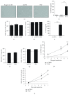
Sustained hBM-MSC growth and secretion of placental growth factor after TransIT-2020 transfection. (a) Representative 20X phase contrast images showing cell morphology of hBM-MSCs with reagent only control (R) and the 4h-OM condition with either the pCMV6 empty control or pCMV6-PlGF vector. Scale bars represent 200 μm. (b) Quantification of PlGF secretion per well in a 6-well plate (pg/ml) in the hBM-MSC cell-conditioned media 24 hours after transfection by ELISA. (c) Amount of viable cells present 24 hours after transfection expressed as a percentage of initial seeding density in a 6-well plate. (d) Percentage of viable cells after transfection in a 6-well plate. (e) Quantification of PlGF secretion (pg/ml) per T75 flask. (f) Amount of viable cells present 24 hours after transfection in a T75. (g) Percentage of viable cells after transfection in a T75. (h) Secretion of PlGF (pg/ml) throughout the 7 days after transfection. (i) Amount of cell growth up to 7 days after transfection, expressed as a percentage of viable cells measured divided by the initial seeding of 37,500 cells. Results from 3 (a–d) or 4 (e–i) independent experiments with technical duplicates. Error bars represent S.E.M. Statistical significance was obtained using a one-tailed t-test for (b) and a two-way ANOVA with a Bonferroni post hoc analysis for (f–g). p < 0.05 and ∗∗ p < 0.01.

Similar articles

Cited by

References

    1. Kode J. A., Mukherjee S., Joglekar M. V., Hardikar A. A. Mesenchymal stem cells: immunobiology and role in immunomodulation and tissue regeneration. Cytotherapy. 2009;11(4):377–391. doi: 10.1080/14653240903080367. - DOI - PubMed
    1. Squillaro T., Peluso G., Galderisi U. Clinical trials with mesenchymal stem cells: an update. Cell Transplantation. 2016;25(5):829–848. doi: 10.3727/096368915X689622. - DOI - PubMed
    1. Mendicino M., Bailey A. M., Wonnacott K., Puri R. K., Bauer S. R. MSC-based product characterization for clinical trials: an FDA perspective. Cell Stem Cell. 2014;14(2):141–145. doi: 10.1016/j.stem.2014.01.013. - DOI - PubMed
    1. Baksh D., Song L., Tuan R. S. Adult mesenchymal stem cells: characterization, differentiation, and application in cell and gene therapy. Journal of Cellular and Molecular Medicine. 2004;8(3):301–316. doi: 10.1111/j.1582-4934.2004.tb00320.x. - DOI - PMC - PubMed
    1. Mangi A. A., Noiseux N., Kong D., et al. Mesenchymal stem cells modified with Akt prevent remodeling and restore performance of infarcted hearts. Nature Medicine. 2003;9(9):1195–1201. doi: 10.1038/nm912. - DOI - PubMed