. 2019 Jan 19:10:4.
doi: 10.1186/s13100-019-0146-7. eCollection 2019.

Features of a novel protein, rusticalin, from the ascidian Styela rustica reveal ancestral horizontal gene transfer event

Affiliations

Features of a novel protein, rusticalin, from the ascidian Styela rustica reveal ancestral horizontal gene transfer event

Maria A Daugavet et al. Mob DNA. .

Abstract

Background: The transfer of genetic material from non-parent organisms is called horizontal gene transfer (HGT). One of the most conclusive cases of HGT in metazoans was previously described for the cellulose synthase gene in ascidians.

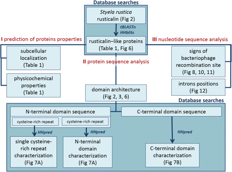
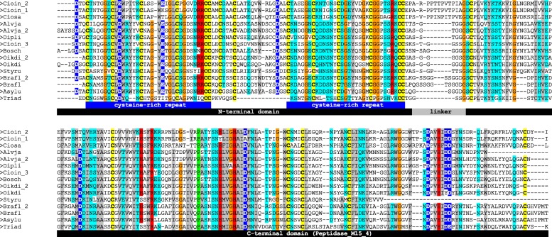
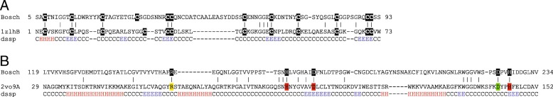
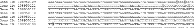
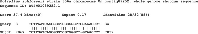
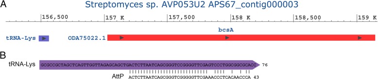
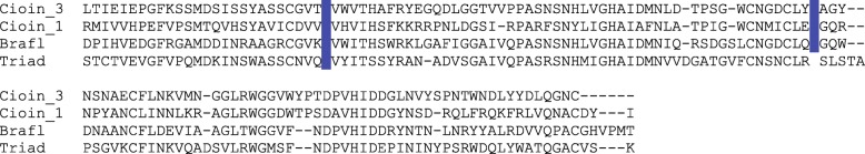
Results: In this study we identified a new protein, rusticalin, from the ascidian Styela rustica and presented evidence for its likely origin by HGT. Discernible homologues of rusticalin were found in placozoans, coral, and basal Chordates. Rusticalin was predicted to consist of two distinct regions, an N-terminal domain and a C-terminal domain. The N-terminal domain comprises two cysteine-rich repeats and shows remote similarity to the tick carboxypeptidase inhibitor. The C-terminal domain shares significant sequence similarity with bacterial MD peptidases and bacteriophage A500 L-alanyl-D-glutamate peptidase. A possible transfer of the C-terminal domain by bacteriophage was confirmed by an analysis of noncoding sequences of C. intestinalis rusticalin-like gene, which was found to contain a sequence similar to the bacteriophage A500 recombination site. Moreover, a sequence similar to the bacteriophage recombination site was found to be adjacent to the cellulose synthase catalytic subunit gene in the genome of Streptomices sp., the donor of ascidian cellulose synthase.

Conclusions: The C-terminal domain of rusticalin and rusticalin-like proteins is likely to be horizontally transferred by the bacteriophage A500. A common mechanism involving bacteriophage mediated gene transfer can be proposed for at least two HGT events in ascidians.

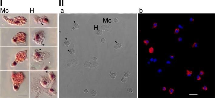
Keywords: Ascidians; Bacteriophage; Hemocytes; Horizontal gene transfer; L-alanyl-D-glutamate peptidase; Trichoplax; tRNA.


Conflict of interest statement

Not applicable.Not applicable.The authors declare that they have no competing interests.Springer Nature remains neutral with regard to jurisdictional claims in published maps and institutional affiliations.

Figures

Fig. 1
Fig. 1
SDS-PAGE of blood cells proteins of ascidian Styela rustica. Lane T: whole blood cells; lane 1: the upper fraction of blood cells from discontinuous percoll gradient (above 35% percoll); lane 2: the intermediate fraction (between 35 and 45% percoll); lane 3: morula cells fraction from discontinuous percoll gradient (between 45 and 60% percoll); lane 4: whole blood cells proteins transferred to PVDF membrane and stained with Ponceau S. The major band of the upper (hyalinocytes) fraction of 23 kDa (black arrow) subjected to further matrix-assisted laser desorption/ionization tandem mass spectrometry MALDI MS/MS analysis. M – Molecular weight markers in kDa
Fig. 2
Fig. 2
Deduced amino acid sequence of rusticalin. The predicted signal peptide is printed in red, the internal repeats detected with REPRO and RADAR are underlined, with cysteines shaded yellow, and a stretch of residues with relative solvent accessibility > 50% shaded black. Peptide sequence detected by de novo sequencing is printed in blue
Fig. 3
Fig. 3
Prediction of disorder. Disordered regions predicted using Disopred3 (red line), SPOT (blue line) and protein backbone dynamics predicted using DynaMine (green line) for Styela rustica rusticalin
Fig. 4
Fig. 4
FISH detection of rusticalin mRNA in the blood cells. I Hyalinocytes (H) and morula cells (Mc), hematoxylin and eosin staining, DIC microscopy. Note hyalinocytes containing numerous small spherical granules (arrows). Scale bar, 5 μm. II Confocal sections showing the distribution of transcripts (b) within the cells with the morphology of hyalinocytes (a). Scale bar, 10 μm. The hybrids with Probe 2 were detected with streptavidin-Alexa594 (red pseudo color). 4′-6-Diamidino-2-phenylindole (DAPI) was used as a general DNA dye (blue pseudo color)
Fig. 5
Fig. 5
Workflow of rusticalin sequence analysis
Fig. 6
Fig. 6
Multiple sequence alignment of rusticalin-like proteins. Predicted signal peptides were trimmed out before alignment. Note the perfect match and conservation of cysteine residues highlighted in yellow. N- and C-terminal domains of rusticalin-like proteins are shown in black. Note the lack of 40 C-terminal residues in rusticalin. The tentative linker region between N- and C-terminal domains is shown in gray. Two cysteine-rich repeats inside the N-terminal domain are shown in blue. Amino acid residues above 85% identity threshold are colored according to their physicochemical properties. Styru – Styela rustica, Bosch – Botryllus schlosseri, Cioin – Ciona intestinalis, Ciosa – Ciona savignyi, Dipli – Diplosoma listerianum, Oikdi – Oikopleura dioica, Brafl – Branchiostoma floridae, Alvja – Alveopora japonica, Triad – Trichoplax adhaerens
Fig. 7
Fig. 7
Sequence-structure alignment of N- and C-terminal domains of Botryllus schlosseri rusticalin sequence. a Alignment of the N-terminal domain with β-defensin-like fold of tick carboxypeptidase inhibitor (PDB ID 1ZLH). Conserved cysteine residues are highlighted in black. The sequence identity is 26%. b Alignment of C-terminal domain with Hedgehog/DD-peptidase fold of bacteriophage A500 L-alanyl-D-glutamate peptidase (PDB ID 2VO9). Amino acid residues involved in Zn2+ binding by 2VO9 are highlighted in red, catalytic and substrate-binding residues are highlighted in green and yellow, respectively. The sequence identity is 21%. ss_dssp – template secondary structure as determined by DSSP. The secondary structure is labeled ‘H’ for α-helix, ‘E’ for β-strand, and ‘C’ for coil
Fig. 8
Fig. 8
Rusticalin-like gene from Ciona intestinalis contains bacteriophage A500 recombination site inside the non-coding region. a Position and antisense orientation of seven tRNA-Arg genes inside the non-coding regions of C. intestinalis rusticalin-like gene (Gene ID: 100185212). The first tRNA gene is situated upstream of the protein coding sequence, four of tRNA genes are inside the second intron and two are inside the third intron neighboring the C-terminal domain coding region. b Alignment of bacteriophage A500 recombination site (AttP) with Ciona intestinalis tRNA gene (Gene ID: 108950122) situated inside the third intron. The sequence identity and score are, respectively, 65.8% and 73 (calculated by EMBOSS Matcher)
Fig. 9
Fig. 9
Alignment of seven Ciona intestinalis tRNA genes neighboring rusticalin-like gene coding sequence. Asterisks indicate conservative positions. Three of the tRNA genes differ from the other tRNA genes at four nucleotide positions (shaded grey). The sequence identity is 95–100%
Fig. 10
Fig. 10
Result of nucleotide BLAST of bacteriophage A500 recombination site (AttP) against Tunicata genome sequences. Alignment of AttP site with Botryllus schlosseri genome sequence - contig89252. The sequence identity is 88%, E-value: 0.17
Fig. 11
Fig. 11
Donor of ascidian cellulose synthase Streptomyces sp. contains bacteriophage recombination site adjacent to cellulose synthase gene. a Streptomyces sp. cellulose synthase catalytic subunit gene (bcsA) is adjacent to tRNA-lys gene. b Alignment of bacteriophage A500 recombination site (AttP) with the tRNA-Lys gene (APS67_000733). The sequence identity and score are, respectively, 91.2% and 143 (calculated by EMBOSS Matcher)
Fig. 12
Fig. 12
Alignment of C-terminal domains of rusticalin-like proteins with designated positions of introns. Blue vertical bars represent the positions of introns in the corresponding DNA sequence of: Cioin_3 – Ciona intestinalis XP_002122335.1; Cioin_1 – Ciona intestinalis XP_002128942.1; Brafl – Branchiostoma floridae XP_002588042.1; Triad – Trichoplax adhaerens XP_002117795.1

Similar articles

Cited by

References

    1. Delsuc F, Brinkmann H, Chourrout D, Philippe H. Tunicates and not cephalochordates are the closest living relatives of vertebrates. Nature. 2006;439:965. doi: 10.1038/nature04336. - DOI - PubMed
    1. Daele Y, Revol J, Gaill F, Goffinet G. Characterization and supramolecular architecture of the cellulose-protein fibrils in the tunic of the sea peach (Halocynthia papillosa, Ascidiacea, Urochordata) Biol Cell. 1992;76:87–96. doi: 10.1016/0248-4900(92)90198-A. - DOI
    1. Shenkar N, Swalla BJ. Global diversity of ascidiacea. PLoS One. 2011;6(6):e20657. doi: 10.1371/journal.pone.0020657. - DOI - PMC - PubMed
    1. Schreiber L, Kjeldsen KU, Funch P, Jensen J, Obst M, López-Legentil S, et al. Endozoicomonas are specific, facultative symbionts of sea squirts. Front Microbiol. 2016;7:1042. doi: 10.3389/fmicb.2016.01042. - DOI - PMC - PubMed
    1. Nakashima K, Yamada L, Satou Y, Azuma J, Satoh N. The evolutionary origin of animal cellulose synthase. Dev Genes Evol. 2004;214(2):81–88. doi: 10.1007/s00427-003-0379-8. - DOI - PubMed

LinkOut - more resources