. 2019 Jan;5(1):13-19.
doi: 10.1159/000487949. Epub 2018 Apr 17.

Insights from a Case of Vitreoretinal Lymphoma

Affiliations

Insights from a Case of Vitreoretinal Lymphoma

Bertil E Damato et al. Ocul Oncol Pathol. 2019 Jan.

Abstract

Purpose/background: The aim of this study was to report a patient with vitreoretinal lymphoma with clinical features providing hypothesis-generating insights into the pathophysiology of this disease.

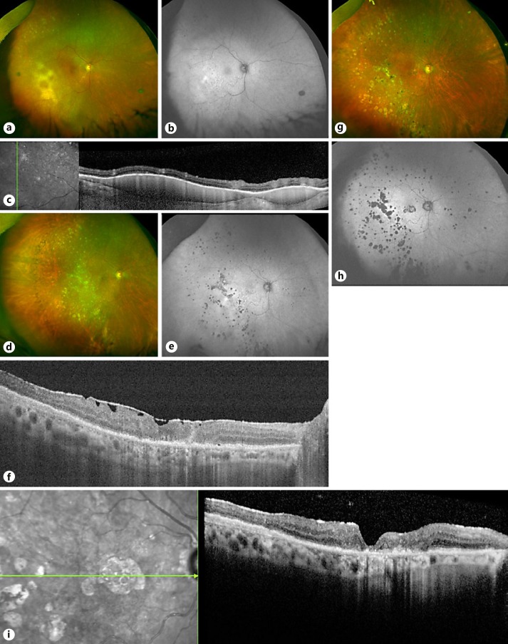
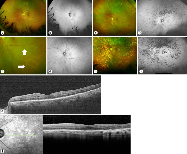
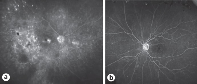
Methods: Clinical history and imaging studies (i.e., fundus photography, optical coherence tomography, fundus autofluorescence, and fluorescein angiography) were documented.

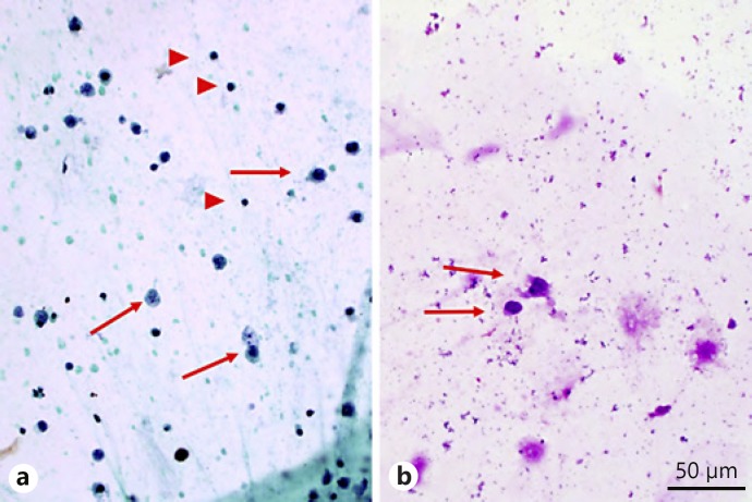
Results: A 71-year-old woman presented with a 2-month history of blurred vision in the right eye and bilateral vitreous infiltrates unresponsive to topical and systemic steroids. Vitreous biopsy of the left eye was diagnostic for lymphoma. Bulky subretinal deposits in the right eye responded to systemic therapy. The left fundus showed diffuse hypoautofluorescence and punctate, hyperfluorescent sub-retinal pigment epithelial tumor deposits, which resolved leaving hypoautofluorescent atrophic retinal pigment epithelium (RPE) scars, except inferotemporally, where retinal vasculopathy had occurred.

Conclusions: The clinical features suggest that occlusion of the inferotemporal retinal arteriole prevented sub-RPE lymphomatous deposits and subsequent RPE atrophy in this area of vascular nonperfusion. This suggests that "primary" vitreoretinal lymphoma is secondary to hematogenous spread from systemic loci. This finding, together with the ocular tumor control achieved entirely by systemic therapy, indicates scope for studies investigating systemic treatment protocols, especially those including immune-modulatory agents.

Keywords: Lymphoma; Retinal disease.


Figures

Fig. 1.
Fig. 1.
Cytology from vitreous biopsy of the left eye with arrows showing large atypical cells and arrowheads showing small mature lymphocytes for size comparison. a Aspirate smear, alcohol-fixed Papanicolau stain. Magnification ×400. b Aspirate smear, air-dried May-Grünwald-Giemsa stain. Magnification ×400. Courtesy of Melike Pekmezci.
Fig. 2.
Fig. 2.
Imaging of the right eye over time. a September 2014: color photograph showing pale, yellow sub-RPE deposits inferotemporally and sheathing of an inferior macular arteriole. b Corresponding autofluorescence image, showing hyperautofluorescence in the area of infiltration, with hyperfluorescent spots elsewhere indicating active lesions and hypofluorescent spots indicating atrophic RPE scars. c OCT performed on the same date showing sub-RPE infiltrates in the temporal macula. d Color photograph of the right fundus from March 2015 showing resolution of the large sub-RPE deposits with only systemic therapy. e Corresponding fundus autofluorescence image showing hyper- and hypofluorescent spots indicating active lesions and atrophic RPE scars, respectively. f OCT of the macula on the same date showing an ERM. g Col or photograph of the right fundus, taken in May 2017, showing atrophic RPE lesions, mostly temporally. h Corresponding fundus autofluorescence image showing hypoautofluorescence scars. i OCT of the macula on the same date showing RPE atrophy and a mild ERM. RPE, retinal pigment epithelium; OCT, optical coherence tomography; ERM, epiretinal membrane.
Fig. 3.
Fig. 3.
Imaging of the left eye over time. a Color photograph in September 2014, prior to treatment, showing only faint microflecks. b Corresponding fundus autofluorescence image showing diffuse hypoautofluorescence inferonasally and perhaps superiorly and temporally. c Color photograph from November 2015, showing sheathing of inferotemporal arterioles (white arrows). d Corresponding fundus autofluorescence image showing diffuse hypoautofluorescence, except inferotemporally. e OCT of the left eye in November 2015 showing bumpy appearance of the RPE layer. f Color photograph from March 2016 showing increasing RPE atrophy, especially superiorly. g Corresponding fundus autofluorescence image showing active hyperautofluorescent infiltrates and hypoautofluorescent atrophic RPE scars. h Fundus photograph from May 2017 showing extensive RPE atrophy that spares the inferotemporal retina. i Corresponding autofluorescence image, which shows sparing of the inferotemporal retina from atrophic RPE lesions. This is the area of prior vascular occlusion. j OCT on the same date showing a mild ERM as well as extensive loss of the RPE layer corresponding to atrophy seen in color and autofluorescence imaging. RPE, retinal pigment epithelium; OCT, optical coherence tomography; ERM, epiretinal membrane.
Fig. 4.
Fig. 4.
Fluorescein angiography prior to treatment in September 2014. a Late-frame image of the right fundus showing hypofluorescence temporal to the macula at the site of the largest sub-RPE lymphomatous infiltrate, with macular edema as well as focal lesions elsewhere, with fuzzy spots indicating active lesions and discrete spots indicating RPE scars. b Late-frame image of the left fundus showing only a few areas of hyperfluorescence nasally, with no obvious retinal vascular abnormalities. RPE, retinal pigment epithelium.

References

    1. Araujo I, Coupland SE. Primary vitreoretinal lymphoma - a review. Asia Pac J Ophthalmol. 2017;6:283–289. - PubMed
    1. Reichstein D. Primary vitreoretinal lymphoma: an update on pathogenesis, diagnosis and treatment. Curr Opin Ophthalmol. 2016;27:177–184. - PubMed
    1. Witmer MT. Primary vitreoretinal lymphoma: management of isolated ocular disease. Cancer Control. 2016;23:110–116. - PubMed
    1. Teckie S, Yahalom J. Primary intraocular lymphoma: treatment outcomes with ocular radiation therapy alone. Leuk Lymphoma. 2014;55:795–801. - PubMed
    1. Frenkel S, Hendler K, Siegal T, Shalom E, Pe'er J. Intravitreal methotrexate for treating vitreoretinal lymphoma: 10 years of experience. Br J Ophthalmol. 2008;92:383–388. - PubMed