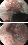
Narrow-Band Imaging: Clinical Application in Gastrointestinal Endoscopy
- PMID: 30675503
- PMCID: PMC6341367
- DOI: 10.1159/000487470
Narrow-Band Imaging: Clinical Application in Gastrointestinal Endoscopy
Abstract
Narrow-band imaging is an advanced imaging system that applies optic digital methods to enhance endoscopic images and improves visualization of the mucosal surface architecture and microvascular pattern. Narrow-band imaging use has been suggested to be an important adjunctive tool to white-light endoscopy to improve the detection of lesions in the digestive tract. Importantly, it also allows the distinction between benign and malignant lesions, targeting biopsies, prediction of the risk of invasive cancer, delimitation of resection margins, and identification of residual neoplasia in a scar. Thus, in expert hands it is a useful tool that enables the physician to decide on the best treatment (endoscopic or surgical) and management. Current evidence suggests that it should be used routinely for patients at increased risk for digestive neoplastic lesions and could become the standard of care in the near future, at least in referral centers. However, adequate training programs to promote the implementation of narrow-band imaging in daily clinical practice are needed. In this review, we summarize the current scientific evidence on the clinical usefulness of narrow-band imaging in the diagnosis and characterization of digestive tract lesions/cancers and describe the available classification systems.
O sistema de iluminação narrow-band imaging é um sistema de imagem avançada que utiliza ferramentas digitais óticas para realçar imagens endoscópicas e melhorar a observação da superfície e do padrão microvascular da mucosa. O narrow-band imaging tem demonstrado ser um importante adjuvante à endoscopia com luz branca, melhorando a deteção de lesões no tubo digestivo. Tam-bém, possibilita a distinção entre lesões benignas e mali-gnas, guia as biópsias para zonas suspeitas, prediz o risco de cancro invasivo, delimita as margens de ressecção e identifica lesões residuais em cicatrizes. Portanto, em mãos experientes, é uma ferramenta útil que permite ao médico decidir o melhor tratamento (endoscópico ou cirúrgico) e orientação. A evidência atual sugere que esta técnica deve ser utilizada por rotina em doentes com risco aumentado para lesões neoplásicas do tubo digestivo e poderá tornar-se o método de escolha num futuro próxi-mo, pelo menos nos centros de referência. Contudo, são necessários programas de treino adequados para pro-mover a utilização do narrow-band imaging na prática clinica diária. Nesta revisão, resumimos a evidência científi-ca disponível acerca da utilidade do narrow-band imaging no diagnóstico e caracterização das lesões do tubo di-gestivo e descrevem-se os sistemas de classificação dis-poníveis.
Keywords: Barrett esophagus; Chromoendoscopy; Colonic polyps; Early gastric cancer; Endoscopic classifications; Gastric intestinal metaplasia; Narrow-band imaging; Squamous cell carcinoma.
Figures


Similar articles
-
Usefulness of Narrow-Band Imaging in Endoscopic Submucosal Dissection of the Stomach.Clin Endosc. 2018 Oct;51(6):527-533. doi: 10.5946/ce.2018.186. Epub 2018 Nov 19. Clin Endosc. 2018. PMID: 30453446 Free PMC article.
-
AGA Clinical Practice Update on Endoscopic Treatment of Barrett's Esophagus With Dysplasia and/or Early Cancer: Expert Review.Gastroenterology. 2020 Feb;158(3):760-769. doi: 10.1053/j.gastro.2019.09.051. Epub 2019 Nov 12. Gastroenterology. 2020. PMID: 31730766 Review.
-
Narrow-band imaging for the head and neck region and the upper gastrointestinal tract.Jpn J Clin Oncol. 2013 May;43(5):458-65. doi: 10.1093/jjco/hyt042. Jpn J Clin Oncol. 2013. PMID: 23630386 Review.
-
Endoluminal Diagnosis of Early Gastric Cancer and Its Precursors: Bridging the Gap Between Endoscopy and Pathology.Adv Exp Med Biol. 2016;908:293-316. doi: 10.1007/978-3-319-41388-4_14. Adv Exp Med Biol. 2016. PMID: 27573777 Review.
-
Diagnosis and Management of Low-Grade Dysplasia in Barrett's Esophagus: Expert Review From the Clinical Practice Updates Committee of the American Gastroenterological Association.Gastroenterology. 2016 Nov;151(5):822-835. doi: 10.1053/j.gastro.2016.09.040. Epub 2016 Oct 1. Gastroenterology. 2016. PMID: 27702561 Review.
Cited by
-
Next-Generation Endoscopy in Inflammatory Bowel Disease.Diagnostics (Basel). 2023 Jul 31;13(15):2547. doi: 10.3390/diagnostics13152547. Diagnostics (Basel). 2023. PMID: 37568910 Free PMC article. Review.
-
Diagnostic value of artificial intelligence-assisted endoscopy for chronic atrophic gastritis: a systematic review and meta-analysis.Front Med (Lausanne). 2023 May 2;10:1134980. doi: 10.3389/fmed.2023.1134980. eCollection 2023. Front Med (Lausanne). 2023. PMID: 37200961 Free PMC article.
-
Future of image enhanced endoscopy of esophageal adenocarcinoma.Clin Endosc. 2025 Jul;58(4):503-513. doi: 10.5946/ce.2024.324. Epub 2025 May 20. Clin Endosc. 2025. PMID: 40394929 Free PMC article. Review.
-
Artificial Intelligence in Colorectal Cancer Screening, Diagnosis and Treatment. A New Era.Curr Oncol. 2021 Apr 23;28(3):1581-1607. doi: 10.3390/curroncol28030149. Curr Oncol. 2021. PMID: 33922402 Free PMC article. Review.
-
Crush cytology: an expeditious diagnostic tool for gastrointestinal tract malignancy.Endosc Int Open. 2021 May;9(5):E735-E740. doi: 10.1055/a-1388-6479. Epub 2021 Apr 22. Endosc Int Open. 2021. PMID: 33937515 Free PMC article.
References
-
- East JE, Vleugels JL, Roelandt P, Bhandari P, Bisschops R, Dekker E, Hassan C, et al. Advanced endoscopic imaging: European Society of Gastrointestinal Endoscopy (ESGE) Technology Review. Endoscopy. 2016;48:1029–1045. - PubMed
-
- Kamiński MF, Hassan C, Bisschops R, Pohl J, Pellisé M, Dekker E, Ignjatovic-Wilson A, et al. Advanced imaging for detection and differentiation of colorectal neoplasia: European Society of Gastrointestinal Endoscopy (ESGE) Guideline. Endoscopy. 2014;46:435–449. - PubMed
-
- ASGE Technology Committee, Manfredi MA, Abu Dayyeh BK, Bhat YM, Chauhan SS, Gottlieb KT, Hwang JH, et al. Electronic chromoendoscopy. Gastrointest Endosc. 2015;81:249–261. - PubMed
-
- Kudo S, Rubio CA, Teixeira CR, Kashida H, Kogure E. Pit pattern in colorectal neoplasia: endoscopic magnifying view. Endoscopy. 2001;33:367–373. - PubMed
-
- Sano Y, Horimatsu T, Fu KI, Katagiri A, Muto M, Ishikawa H. Magnifying observation of microvascular architecture of colorectal lesions using a narrow-band imaging system. Dig Endosc. 2006;18:S44–S51.
Publication types
LinkOut - more resources
Full Text Sources

