Review
. 2018 Mar 1;2(1):74-83.
doi: 10.1016/j.jses.2017.11.006. eCollection 2018 Mar.

Subscapularis tears: hidden and forgotten no more

Affiliations
Review

Subscapularis tears: hidden and forgotten no more

Julia Lee et al. JSES Open Access. .

Abstract

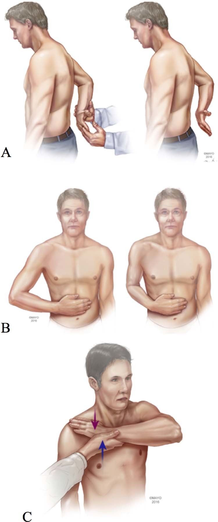
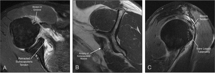
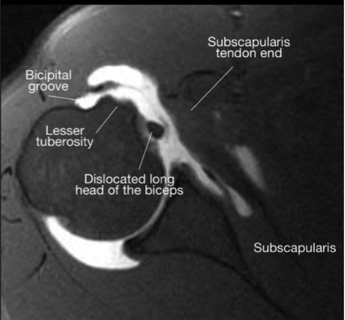
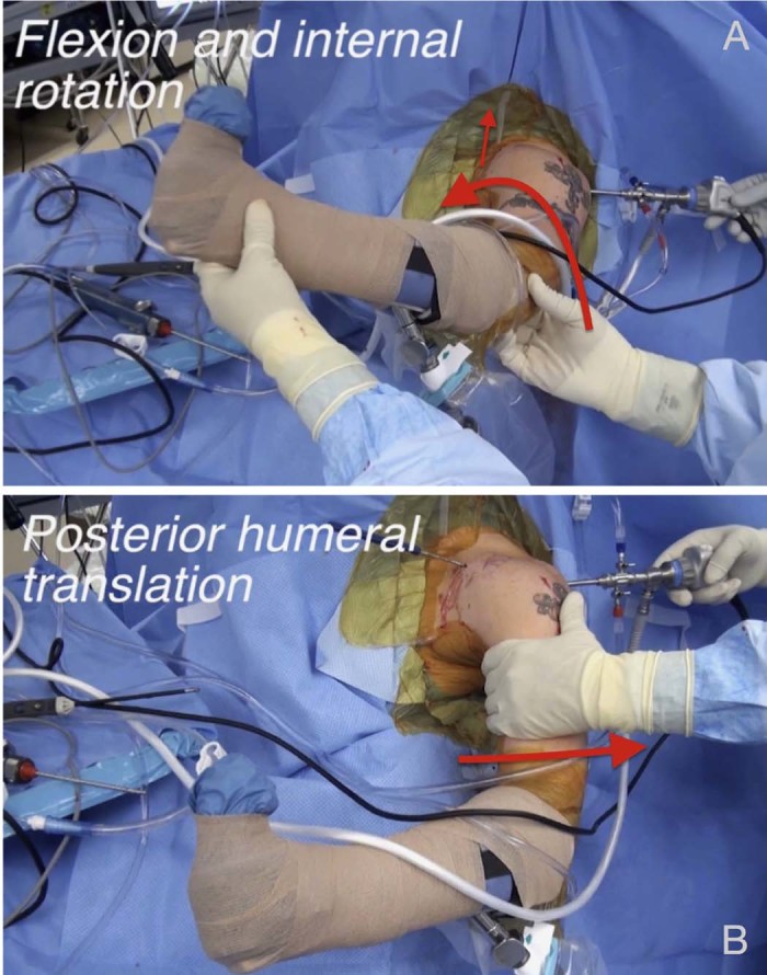
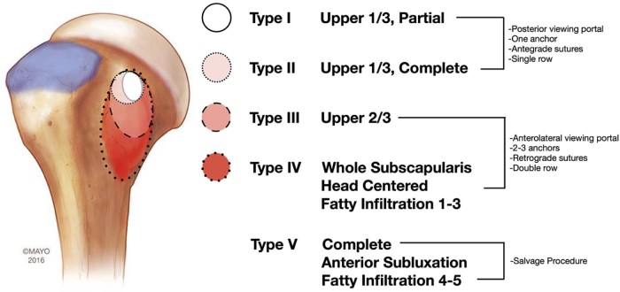
The subscapularis tendon, at one point, was thought of as the forgotten tendon, with "hidden lesions" that referred to partial tears of this tendon. Better understanding of anatomy and biomechanics combined with improved imaging technology and the widespread use of arthroscopy has led to a higher rate of subscapularis tear diagnoses and repairs. The bulk mass of the subscapularis muscle is more than that of all 3 other rotator cuff muscles combined. It functions as the internal rotator of the shoulder as the stout, rolled border of its tendon inserts onto the superior portion of the lesser tuberosity. A thorough history combined with specific physical examination maneuvers (including the bear hug, lift-off, and belly-press tests) is critical for accurate diagnosis. A systematic approach to advanced shoulder imaging also improves diagnostic capacity. Once identified, most subscapularis tendon tears can be successfully repaired arthroscopically. The Lafosse classification is useful as part of a treatment algorithm. Type I and II tears may be addressed while viewing from the standard posterior glenohumeral portal; larger Lafosse type III and IV tears are best repaired with anterior visualization at the subacromial or subdeltoid space. Tendon mobilization for larger tears is critical for adequate repair. In Lafosse type V tears, in which there is glenohumeral imbalance, tendon transfers and reverse replacement are commonly considered salvage options.

Keywords: Arthroscopic rotator cuff repair; Irreparable subscapularis; Rotator cuff imaging; Rotator cuff tear; Shoulder tendon transfer; Subscapularis physical examination; Subscapularis tear.


Figures

Figure 1
Figure 1
Subscapularis physical examination maneuvers. (A) Lift-off test. (B) Belly-press test. (C) Bear hug test.
Figure 2
Figure 2
Applying the 4-step magnetic resonance imaging evaluation by Adams et al. (A) Axial image showing the biceps tendon in the groove with an obvious retracted tear of the subscapularis tendon. (B) Sagittal image showing grade 2 fatty infiltration of the subscapularis muscle. (C) Sagittal image showing a bare lesser tuberosity.
Figure 3
Figure 3
Medial dislocation of the long head of the biceps tendon.
Figure 4
Figure 4
Manipulation methods for better arthroscopic subscapularis visualization. (A) Flexion and internal rotation of affected extremity. (B) “Posterior lever push,” whereby a posteriorly directed force is placed on the proximal humerus.
Figure 5
Figure 5
Lafosse classification of subscapularis tears and treatment algorithm.
Figure 6
Figure 6
Intraoperative images of a subscapularis tear. (A) Traction on the subscapularis reveals the poor-quality tendon associated with a tear. (B) Tear in continuity seen in subscapularis tendon. (C) Intraoperative fixation of tendon with retrograde passing of sutures for horizontal mattress suture fixation. (D) Final picture of re-established rolled border.
Figure 7
Figure 7
Arthroscopic fixation of type I and type II tears. (A) Type I tear. (B) Fixation of type I tear. (C) Type II tear. (D) Fixation of type II tear.
Figure 8
Figure 8
Arthroscopic fixation of type III and type IV tears. (A-C) Type III tear. Note the disruption of the superior tendinous portion in (A) and continuity of the muscle portion in (B). (C) Fixation of type III tear. (D-F) Type IV tear. (D) Before mobilization of tendon. (E) After mobilization of tendon; note how far back the tendon is retracted. (F) Suture fixation of the tendon with multiple anchors.

References

    1. Adams C.R., Brady P.C., Koo S.S., Narbona P., Arrigoni P., Karnes G.J. A systematic approach for diagnosing subscapularis tendon tears with preoperative magnetic resonance imaging scans. Arthroscopy. 2012;28:1592–1600. doi: 10.1016/j.arthro.2012.04.142. - DOI - PubMed
    1. Adams C.R., Schoolfield J.D., Burkhart S.S. The results of arthroscopic subscapularis tendon repairs. Arthroscopy. 2008;24:1381–1389. doi: 10.1016/j.arthro.2008.08.004. - DOI - PubMed
    1. Adams C.R., Schoolfield J.D., Burkhart S.S. Accuracy of preoperative magnetic resonance imaging in predicting a subscapularis tendon tear based on arthroscopy. Arthroscopy. 2010;26:1427–1433. doi: 10.1016/j.arthro.2010.02.028. - DOI - PubMed
    1. Alcid J.G., Powell S.E., Tibone J.E. Revision anterior capsular shoulder stabilization using hamstring tendon autograft and tibialis tendon allograft reinforcement: minimum two-year follow-up. J Shoulder Elbow Surg. 2007;16:268–272. doi: 10.1016/j.jse.2006.07.008. - DOI - PubMed
    1. Arai R., Sugaya H., Mochizuki T., Nimura A., Moriishi J., Akita K. Subscapularis tendon tear: an anatomic and clinical investigation. Arthroscopy. 2008;24:997–1004. doi: 10.1016/j.arthro.2008.04.076. - DOI - PubMed

LinkOut - more resources