. 2019 May 1;316(5):L872-L885.
doi: 10.1152/ajplung.00254.2018. Epub 2019 Jan 24.

The Tcf21 lineage constitutes the lung lipofibroblast population

Affiliations

The Tcf21 lineage constitutes the lung lipofibroblast population

Juwon Park et al. Am J Physiol Lung Cell Mol Physiol. .

Abstract

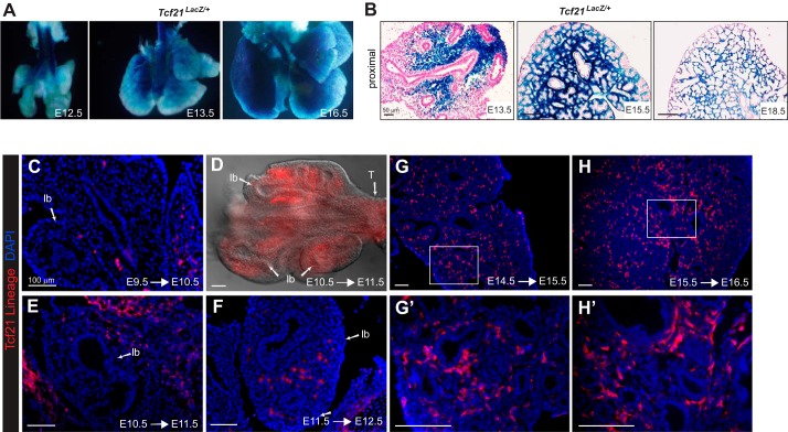
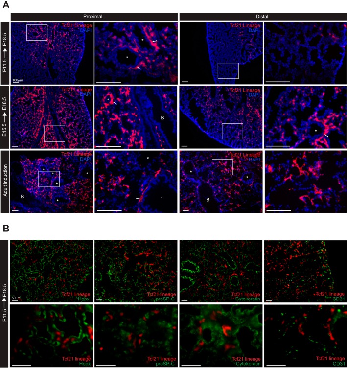
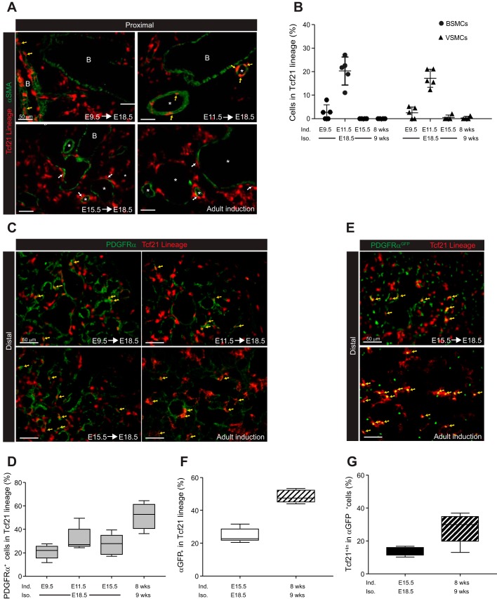
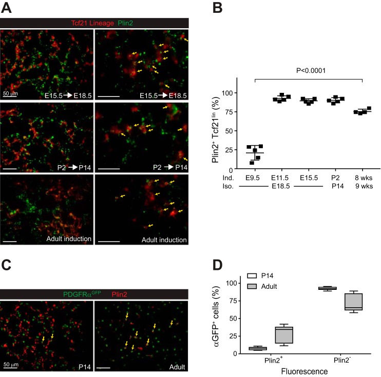
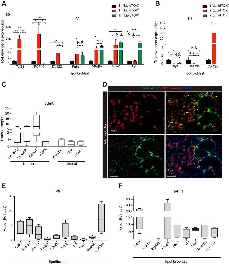
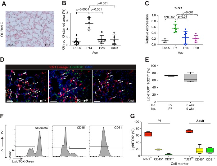
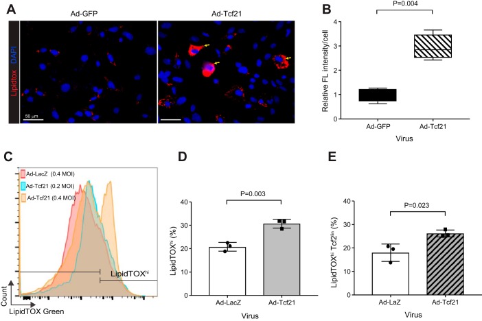
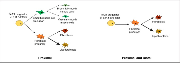
Transcription factor 21 (Tcf21) is a basic helix-loop-helix transcription factor required for mesenchymal development in several organs. Others have demonstrated that Tcf21 is expressed in embryonic lung mesenchyme and that loss of Tcf21 results in a pulmonary hypoplasia phenotype. Although recent single-cell transcriptome analysis has described multiple mesenchymal cell types in the lung, few have characterized the Tcf21 expressing population. To explore the Tcf21 mesenchymal lineage, we traced Tcf21-expressing cells during embryogenesis and in the adult. Our results showed that Tcf21 progenitor cells at embryonic day (E)11.5 generated a subpopulation of fibroblasts and lipofibroblasts and a limited number of smooth muscle cells. After E15.5, Tcf21 progenitor cells exclusively become lipofibroblasts and interstitial fibroblasts. Lipid metabolism genes were highly expressed in perinatal and adult Tcf21 lineage cells. Overexpression of Tcf21 in primary neonatal lung fibroblasts led to increases in intracellular neutral lipids, suggesting a regulatory role for Tcf21 in lipofibroblast function. Collectively, our results reveal that Tcf21 expression after E15.5 delineates the lipofibroblast and a population of interstitial fibroblasts. The Tcf21 inducible Cre mouse line provides a novel method for identifying and manipulating the lipofibroblast.

Keywords: fibroblast; lipofibroblast; lung; perilipin 2; transcription factor 21.


Conflict of interest statement

No conflicts of interest, financial or otherwise, are declared by the authors.

Figures

Fig. 1.
Fig. 1.
Transcription factor 21 (Tcf21) expression in embryonic lung. A: whole mount images of Tcf21LacZ/+ reporter embryonic lungs. B: histologic sections of Tcf21LacZ/+ reporter lungs counterstained with nuclear fast red. Representative images are top left lobe at embryonic day (E)13.5, E15.5, and E18.5; n = 3–5. Scale bars, 50 μm. C–H: Cre-mediated tdTomato expression in embryonic lungs after 1-day labeling. tdTomato expression was visualized using DsRed antibody. Boxed regions in G and H are magnified in G' and H'. D: whole mount fluorescence image of E11.5 lung; lb, lung bud. lb, lung bud; T, trachea. Scale bars, 100 μm.
Fig. 2.
Fig. 2.
Cre-mediated transcription factor 21 (Tcf21) expression pattern in embryonic and adult lung. A: representative images of Tcf21 lineage-tagged, tdTomato cells in proximal and distal embryonic and adult lungs from Tcf21mCrem/+;R26RtdT/tdT mice (R26R, Rosa 26 reporter). Thin white boxed regions indicate regions magnified on the right. Lungs were isolated at embryonic day (E)18.5 after a single dose of tamoxifen administration at E11.5 and E15.5. Adult lungs were isolated 3 mo after a single dose of tamoxifen administration. White arrows point to tdTomato+ cells found in regions surrounding vessel; n = 5–6. B, bronchiole; *, pulmonary vessels. Scale bars, 100 μm. B: representative images of E18.5 lungs stained for Hopx [HOP homeobox; alveolar type 1 (ATI) cells], proSP-C [prosurfactant protein C; (ATII) cells], cytokeratin (epithelial cells), and CD31 (endothelial cells). Tamoxifen induction at E11.5; n = 3. Scale bars, 50 μm.
Fig. 3.
Fig. 3.
Identification of smooth muscle cells and platelet-derived growth factor receptor-α (PDGFRα)-expressing cells derived from transcription factor 21 (Tcf21) progenitors. A: representative images of α-smooth muscle actin (α-SMA) staining in embryonic day (E)18.5 and adult lungs of Tcf21mCrem/+;R26RtdT/tdT mice treated with tamoxifen at indicated time points. Adult lungs were isolated 1 wk after a single tamoxifen induction. Yellow arrows indicate cells with coexpressed tdTomato and α-SMA; white arrows indicate tdTomato+ cells surrounding a bronchiole or vessel; n = 4–5. Scale bars, 50 μm. B: quantification of α-SMA+ cells in Tcf21 lineage; n = 4–5 with 6–10 fields of view (FOV). BSMCs, bronchial smooth muscle cells; VSMCs, vascular smooth muscle cells. Data are shown as scatter dot plot with means ± SD. C: representative images of PDGFRα immunohistochemistry. Yellow arrows indicate cells with overlapping tdTomato and PDGFRα; n = 4–5. Scale bars, 50 μm. D: quantification of PDGFRα+ cells in Tcf21 lineage; n = 4–5, 10 FOV were analyzed. E: representative images of distal E18.5 and adult lungs from Tcf21mCrem/+;R26RtdT/tdT;PDGFRαGFP/+ mice. Yellow arrows indicate cells with overlapping fluorescence. Note that PDGFRαGFP cells contain a nuclear localized green fluorescent protein (GFP); n = 5. Scale bars, 50 μm. F and G: quantification of fluorescence overlap shown in E; n = 5, 10 FOV were analyzed. Data are shown as box and whisker plot with whiskers indicating max/min values.
Fig. 4.
Fig. 4.
Transcription factor 21 (Tcf21) lineage cells become lipofibroblasts. A: representative images of perilipin 2 (Plin2)-stained lungs from Tcf21mCrem/+;R26RtdT/tdT mice treated with tamoxifen at indicated time points. Yellow arrows indicate cells with overlapping fluorescence; n = 4–5. Scale bars, 50 μm. Adult lungs were isolated 1 wk after a single tamoxifen induction. B: quantification of Plin2+ cells in Tcf21 lineage; n = 4–5, 6–10 fields of view (FOV), one-way ANOVA. Adult lungs were isolated 1 wk after a single tamoxifen induction. E, embryonic day; P, postnatal day; Ind, tamoxifen induction; Iso, lung isolation. C: representative images of Plin2-stained lungs from P14 and adult platelet-derived growth factor receptor-α (PDGFRα)GFP/+ mice. GFP, green fluorescent protein. Yellow arrows indicate cells with overlapping red and green fluorescence; n = 5. Scale bars, 50 μm. D: quantification of Plin2+ cells in PDGFRαGFP+ cells; n = 5, 10 FOV. Data are shown as box and whisker plot with whiskers indicating max/min values.
Fig. 5.
Fig. 5.
Gene expression in perinatal and adult lipofibroblasts. A and B: expression of putative lipofibroblast genes in lipofibroblasts and other lipid droplet-containing cells in the lung. Postnatal day (P)7 lungs from 3 tamoxifen-treated mice were pooled and sorted. Lineage (lin) with antibodies for CD45, CD31, and CD326 (epithelial cell adhesion molecule, EpCAM) that were positive (lin;LipidTOX+) or negative (lin;LipidTOX) for LipidTOX and lin+ that were positive for LipidTOX (lin+LipidTOX+); n = 3 replicates of pooled samples. FGF10, fibroblast growth factor 10; Zfp423, zinc finger protein-423; Fabp4, fatty acid-binding protein-4; PPARγ, peroxisome proliferator-activated receptor-γ; Plin2, perilipin 2; Lpl, lipoprotein lipase; Thy1, thymus cell antigen 1; Col13a1, collagen type XIIIα1. C: expression of lung fibroblast and epithelial genes in adult transcription factor 21 (Tcf21) lineage. PDGFRα, platelet-derived growth factor receptor-α; Col1a1, collagen type 1α1; α-SMA, α-smooth muscle actin; Sftpc, surfactant protein C; Nkx2.1, NK2 homeobox 1. D: representative images of Col1a1 reporter in adult lungs from Tcf21mCrem/+;R26RtdT/tdT;Col1a1-GFP mice. GFP, green fluorescent protein. Adult lungs were isolated 3 mo after 3 doses of tamoxifen induction. Yellow arrows indicate fluorescence overlap; n = 4. Scale bars, 100 μm. E and F: expression of putative lipofibroblast genes in P2 induced P9 isolated Tcf21 Ribotag lungs (E) and adult induced Tcf21 Ribotag lungs (F). Expression levels were normalized to that of GADPH and represented as a ratio of gene expression in the Tcf21 lineage (IP) to total lung RNA (input). Lungs were isolated after a single tamoxifen induction for P9 mice or 2–3 doses of tamoxifen induction for adult mice. Data are shown as box and whisker plot with whiskers indicating max/min values (n = 4). NS, not significant; *P < 0.05; **P < 0.01; ***P < 0.001.
Fig. 6.
Fig. 6.
Neutral lipid-containing cells in the lung. A: representative image of Oil red O-stained postnatal day (P)14 lung section; n = 5. B: quantification of Oil red O-stained area in lungs at stages indicated. E, embryonic day. Data are shown as scatter dot plot with means ± SD. Ten fields of view (FOV) from lungs were analyzed, unpaired t-test. C: expression of transcription factor 21 (Tcf21) in lungs at stages indicated. Data were normalized to expression of GADPH and are shown as scatter dot plot with means ± SD, unpaired t-test. D: representative images of LipidTOX green-stained postnatal and adult lungs from Tcf21mCrem/+;R26RtdT/tdT mice. White arrows indicate LipidTOX+ Tcf21 lineage. Adult lungs were isolated 1 wk after a single tamoxifen induction; n = 4. Scale bars, 50 μm. E: quantification of LipidTOX+ cells in Tcf21 lineage at indicated time points. Data are shown as box and whisker plot with whiskers indicating max/min values; n = 4. Seven to ten FOV from lungs were analyzed. Ind, induced; Iso, isolated. F: representative flow cytometry histograms of LipidTOX fluorescence intensity within tdTomato, CD45, or CD31 gates at P7 lungs. Tamoxifen induction at P2; n = 5. G: quantification of percent LipidTOX+ cells in P7 and adult lungs, gated on tdTomato+, CD45+, or CD31+. Adult lungs were isolated 2 wk after 3 doses of tamoxifen induction. Data are shown as box and whisker plot with whiskers indicating max/min values (n = 4–5).
Fig. 7.
Fig. 7.
Increased neutral lipid content in lung fibroblasts after transcription factor 21 (Tcf21) overexpression. A: representative images of lipid accumulation in wild-type postnatal day (P)7 primary lung fibroblasts after adenoviral GFP [Ad-GFP, 0.4 multiplicity of infection (MOI)] or adenoviral Tcf21 (Ad-Tcf21, 0.4 MOI) transduction. Yellow arrows indicate cells with increased neutral lipids; n = 4. B: quantification of fluorescent intensity per cell. Images and data analysis were performed 4 days after adenoviral transduction of wild-type P7 primary lung fibroblasts; n = 4, paired t-test. Data are shown as box and whisker plot with whiskers indicating max/min values. C: flow cytometry histograms of neutral lipid staining in P7 primary lung fibroblasts after transduction with adenoviral Tcf21 at MOI of 0.2 and 0.4 or adenoviral LacZ at a MOI of 0.4 as control. D and E: quantification of LipidTOXhi cells in P7 lung fibroblasts after Tcf21 overexpression. D: quantification of percent LipidTOXhi cells in P7 fibroblast after Ad-βgal (0.4 MOI) or Tcf21 (0.4 MOI) transduction. E: quantification of percent LipidTOXhi cells in P7 Tcf21 lineage. Tcf21 lineage cells were labeled at P1; n = 3, unpaired t-test.
Fig. 8.
Fig. 8.
Summary of fibroblast subtypes derived from transcription factor 21 (Tcf21) progenitors. Ad-GFP, adenoviral green fluorescent protein. Tcf21 lineage tracing results of the present study demonstrate that Tcf21 progenitors at embryonic day (E)11.5 to E13.5 generate smooth muscle cells, interstitial fibroblasts, and lipofibroblasts. After E14.5, Tcf21 progenitors give rise exclusively to lipofibroblasts and a subpopulation of fibroblasts.

Comment in

  • The lipofibroblast: more than a lipid-storage depot.
    McGowan SE. McGowan SE. Am J Physiol Lung Cell Mol Physiol. 2019 May 1;316(5):L869-L871. doi: 10.1152/ajplung.00109.2019. Epub 2019 Mar 6. Am J Physiol Lung Cell Mol Physiol. 2019. PMID: 30840482 No abstract available.

Similar articles

Cited by

References

    1. Acharya A, Baek ST, Banfi S, Eskiocak B, Tallquist MD. Efficient inducible Cre-mediated recombination in Tcf21 cell lineages in the heart and kidney. Genesis 49: 870–877, 2011. doi:10.1002/dvg.20750. - DOI - PMC - PubMed
    1. Acharya A, Baek ST, Huang G, Eskiocak B, Goetsch S, Sung CY, Banfi S, Sauer MF, Olsen GS, Duffield JS, Olson EN, Tallquist MD. The bHLH transcription factor Tcf21 is required for lineage-specific EMT of cardiac fibroblast progenitors. Development 139: 2139–2149, 2012. doi:10.1242/dev.079970. - DOI - PMC - PubMed
    1. Al Alam D, El Agha E, Sakurai R, Kheirollahi V, Moiseenko A, Danopoulos S, Shrestha A, Schmoldt C, Quantius J, Herold S, Chao CM, Tiozzo C, De Langhe S, Plikus MV, Thornton M, Grubbs B, Minoo P, Rehan VK, Bellusci S. Evidence for the involvement of fibroblast growth factor 10 in lipofibroblast formation during embryonic lung development. Development 142: 4139–4150, 2015. doi:10.1242/dev.109173. - DOI - PMC - PubMed
    1. Awonusonu F, Srinivasan S, Strange J, Al-Jumaily W, Bruce MC. Developmental shift in the relative percentages of lung fibroblast subsets: role of apoptosis postseptation. Am J Physiol 277: L848–L859, 1999. doi:10.1152/ajplung.1999.277.4.L848. - DOI - PubMed
    1. Barkauskas CE, Cronce MJ, Rackley CR, Bowie EJ, Keene DR, Stripp BR, Randell SH, Noble PW, Hogan BL. Type 2 alveolar cells are stem cells in adult lung. J Clin Invest 123: 3025–3036, 2013. doi:10.1172/JCI68782. - DOI - PMC - PubMed

Publication types

MeSH terms

Substances

LinkOut - more resources