

The effects of iclaprim on exotoxin production in methicillin-resistant and vancomycin-intermediate Staphylococcus aureus
- PMID: 30676310
- PMCID: PMC6580997
- DOI: 10.1099/jmm.0.000929
The effects of iclaprim on exotoxin production in methicillin-resistant and vancomycin-intermediate Staphylococcus aureus
Abstract
Purpose: Extracellular protein toxins contribute to the pathogenesis of Staphylococcus aureus infections. The present study compared the effects of iclaprim and trimethoprim - two folic acid synthesis inhibitors - with nafcillin and vancomycin on production of Panton-Valentine leukocidin (PVL), alpha haemolysin (AH) and toxic-shock syndrome toxin I (TSST-1) in methicillin-resistant and vancomycin-intermediate S. aureus (MRSA and VISA, respectively).
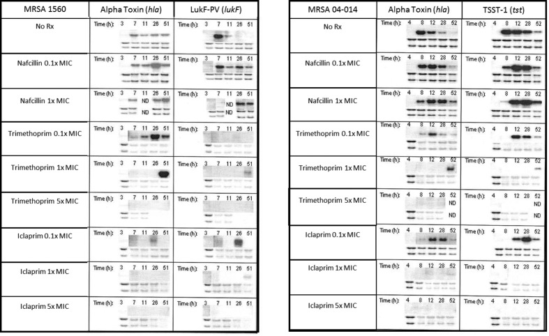
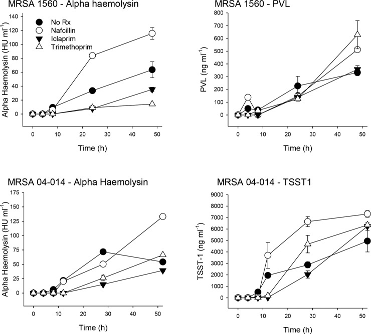
Methodology: Northern blotting and RT-PCR were used to assess gene transcription; toxin-specific bioassays were used to measure protein toxin production.
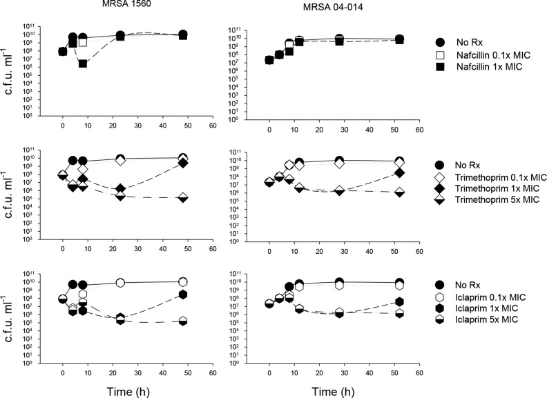
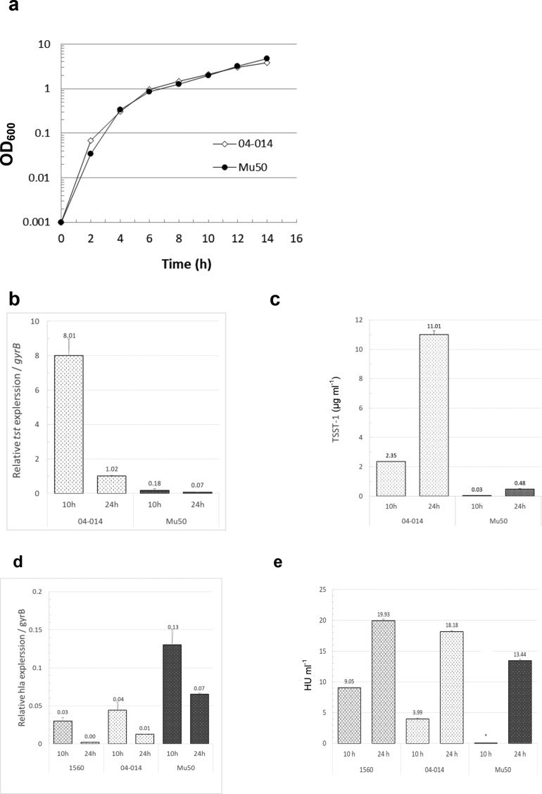
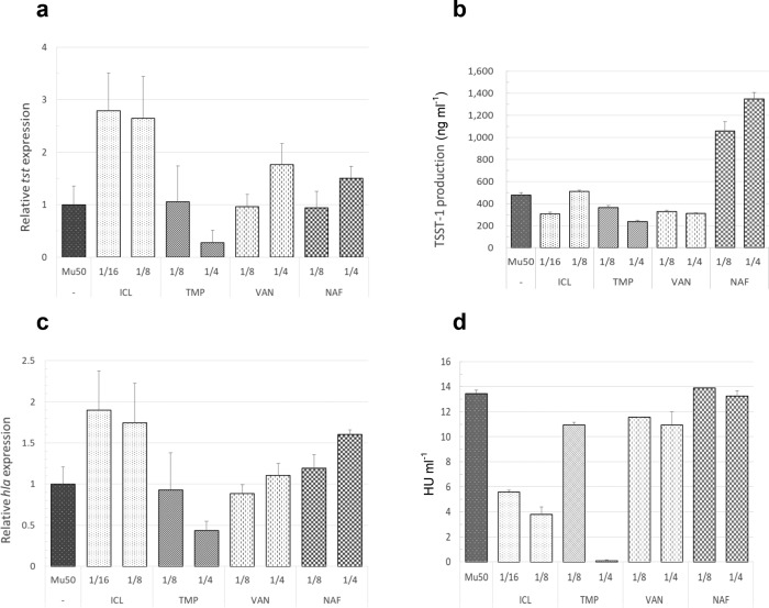
Results: As shown previously, sub-inhibitory concentrations (sub-MIC) of nafcillin increased and prolonged MRSA toxin gene transcription and enhanced PVL, TSST-1 and AH production. Sub-inhibitory doses of iclaprim and trimethoprim delayed maximal AH gene (hla) transcription and suppressed AH production; both drugs delayed, but neither reduced, maximal TSST-1 production. Trimethoprim significantly increased lukF-PV expression and PVL production compared to both untreated and iclaprim-treated cultures. Higher concentrations of iclaprim and trimethoprim markedly suppressed MRSA growth, mRNA synthesis and toxin production. In VISA, iclaprim, vancomycin and nafcillin variably increased tst and hla expression, but only nafcillin increased toxin production. Despite its ability to increase hla expression, iclaprim was the most potent inhibitor of AH production.
Conclusions: We conclude that, due to its ability to suppress toxin production, iclaprim should be effective against severe staphylococcal infections caused by toxin-producing MRSA and VISA strains, especially given its ability to concentrate at sites of infection such as skin and skin structures and the lung.
Keywords: Iclaprim; MRSA; Panton–Valentine leukocidin; Staphylococcus aureus; TSST-1; VISA; alpha haemolysin.
Conflict of interest statement
A.E.B., S.G., E.K. and D.L.S. have no conflict of interest to declare; D.B.H. is an employee of Motif BioSciences.
Figures
Similar articles
-
Subinhibitory concentrations of tedizolid potently inhibit extracellular toxin production by methicillin-sensitive and methicillin-resistant Staphylococcus aureus.J Med Microbiol. 2019 Feb;68(2):255-262. doi: 10.1099/jmm.0.000905. Epub 2018 Dec 17. J Med Microbiol. 2019. PMID: 30556803 Free PMC article.
-
Simulated antibiotic exposures in an in vitro hollow-fiber infection model influence toxin gene expression and production in community-associated methicillin-resistant Staphylococcus aureus strain MW2.Antimicrob Agents Chemother. 2012 Jan;56(1):140-7. doi: 10.1128/AAC.05113-11. Epub 2011 Nov 7. Antimicrob Agents Chemother. 2012. PMID: 22064533 Free PMC article.
-
Impact of antibiotics on expression of virulence-associated exotoxin genes in methicillin-sensitive and methicillin-resistant Staphylococcus aureus.J Infect Dis. 2007 Jan 15;195(2):202-11. doi: 10.1086/510396. Epub 2006 Dec 18. J Infect Dis. 2007. PMID: 17191165
-
Iclaprim: a differentiated option for the treatment of skin and skin structure infections.Expert Rev Anti Infect Ther. 2018 Nov;16(11):793-803. doi: 10.1080/14787210.2018.1536545. Epub 2018 Oct 23. Expert Rev Anti Infect Ther. 2018. PMID: 30317894 Review.
-
Review on Panton Valentine leukocidin toxin carriage among Staphylococcus aureus.J Nepal Health Res Counc. 2013 Sep;11(25):305-12. J Nepal Health Res Counc. 2013. PMID: 24908537 Review.
Cited by
-
Toxic Shock Syndrome Toxin-1 (TSST-1) in Staphylococcus aureus: Prevalence, Molecular Mechanisms, and Public Health Implications.Toxins (Basel). 2025 Jun 24;17(7):323. doi: 10.3390/toxins17070323. Toxins (Basel). 2025. PMID: 40711134 Free PMC article. Review.
-
Evaluation of in vitro activity of iclaprim in combination with other antimicrobials against pulmonary pathogens: a pilot study.Access Microbiol. 2019 May 20;1(3):e000027. doi: 10.1099/acmi.0.000027. eCollection 2019. Access Microbiol. 2019. PMID: 32974519 Free PMC article.
-
Iclaprim reduces the incidence and severity of Staphylococcus aureus-induced septic arthritis in a murine model.Access Microbiol. 2019 Aug 20;1(7):e000052. doi: 10.1099/acmi.0.000052. eCollection 2019. Access Microbiol. 2019. PMID: 32974543 Free PMC article.
-
Anti-virulence potential of iclaprim, a novel folic acid synthesis inhibitor, against Staphylococcus aureus.Appl Microbiol Biotechnol. 2024 Aug 5;108(1):432. doi: 10.1007/s00253-024-13268-2. Appl Microbiol Biotechnol. 2024. PMID: 39102054 Free PMC article.
-
Virulence alterations in staphylococcus aureus upon treatment with the sub-inhibitory concentrations of antibiotics.J Adv Res. 2021 Jan 23;31:165-175. doi: 10.1016/j.jare.2021.01.008. eCollection 2021 Jul. J Adv Res. 2021. PMID: 34194840 Free PMC article. Review.
References
-
- Hartman PG. Molecular aspects and mechanism of action of dihydrofolate reductase inhibitors. J Chemother. 1993;5:369–376. - PubMed
-
- Entenza JM, Haldimann A, Giddey M, Lociuro S, Hawser S, et al. Efficacy of iclaprim against wild-type and thymidine kinase-deficient methicillin-resistant Staphylococcus aureus isolates in an in vitro fibrin clot model. Antimicrob Agents Chemother. 2009;53:3635–3641. doi: 10.1128/AAC.00325-09. - DOI - PMC - PubMed
-
- Hamilton SM, Bryant AE, Carroll KC, Lockary V, Ma Y, et al. In vitro production of Panton-Valentine Leukocidin (PVL) among strains of methicillin-resistant Staphylococcus aureus causing diverse infections. Clin Infect Dis. 2007;45:1550–1558. - PubMed
MeSH terms
Substances
Grants and funding
LinkOut - more resources
Full Text Sources
Medical
Research Materials
Miscellaneous