. 2019 Mar 7;4(5):e125068.
doi: 10.1172/jci.insight.125068.

Piezo1 incorporates mechanical force signals into the genetic program that governs lymphatic valve development and maintenance

Affiliations

Piezo1 incorporates mechanical force signals into the genetic program that governs lymphatic valve development and maintenance

Dongwon Choi et al. JCI Insight. .

Abstract

The lymphatic system plays crucial roles in tissue homeostasis, lipid absorption, and immune cell trafficking. Although lymphatic valves ensure unidirectional lymph flows, the flow itself controls lymphatic valve formation. Here, we demonstrate that a mechanically activated ion channel Piezo1 senses oscillating shear stress (OSS) and incorporates the signal into the genetic program controlling lymphatic valve development and maintenance. Time-controlled deletion of Piezo1 using a pan-endothelial Cre driver (Cdh5[PAC]-CreERT2) or lymphatic-specific Cre driver (Prox1-CreERT2) equally inhibited lymphatic valve formation in newborn mice. Furthermore, Piezo1 deletion in adult lymphatics caused substantial lymphatic valve degeneration. Piezo1 knockdown in cultured lymphatic endothelial cells (LECs) largely abrogated the OSS-induced upregulation of the lymphatic valve signature genes. Conversely, ectopic Piezo1 overexpression upregulated the lymphatic valve genes in the absence of OSS. Remarkably, activation of Piezo1 using chemical agonist Yoda1 not only accelerated lymphatic valve formation in animals, but also triggered upregulation of some lymphatic valve genes in cultured LECs without exposure to OSS. In summary, our studies together demonstrate that Piezo1 is the force sensor in the mechanotransduction pathway controlling lymphatic valve development and maintenance, and Piezo1 activation is a potentially novel therapeutic strategy for congenital and surgery-associated lymphedema.

Keywords: Lymph; Vascular Biology; endothelial cells.


Conflict of interest statement

Conflict of interest: The authors have declared that no conflict of interest exists.

Figures

Figure 1
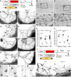
Figure 1. Piezo1 is essential for lymphatic valve development.
(AS) Effect of lymphatic deletion of Piezo1 on lymphatic valve development. (A) Experimental design. Control pups (CTR) harbor Prox1-tdTomato and Prox1-CreERT2, but lack the Piezo1fl/fl allele, while lymphatic Piezo1-KO pups (Piezo1ΔLEC) have all Prox1-tdTomato, Prox1-CreERT2, and Piezo1fl/fl. Tamoxifen (75 mg/kg) was s.c. injected into pups at P1, and tissues were harvested and analyzed at P7. (BI) Mesenteric lymphatics in the (BE) jejunum or (FI) colon are shown in the (B, D, F, and H) CTR or (C, E, G, and I) lymphatic Piezo1-KO pups. High-magnification images of mesenteric lymphatic valves of (J and K) CTR and (L and M) Piezo1-KO pups are also shown. (NQ) Dermal lymphatic valve development was impaired in the tail skin of the lymphatic Piezo1-KO pups. (R and S) Box-and-whisker plots showing lymphatic valve number (R) per lymphatic vessel length or (S) per branching point in the jejunum, colon, and tail. The box plots depict the minimum and maximum values (whiskers), the upper and lower quartiles, and the median. The length of the box represents the interquartile range. (TV) Impact of endothelial deletion of Piezo1 using a Cdh5(PAC)-CreERT2 driver of lymphatic valve development. (T) Experimental design: Peizo1 deletion was induced in pups [Prox1-tdTomato, Cdh5(PAC)-CreERT2, and/or Piezo1fl/fl] at P3 by i.p. tamoxifen injection (75 mg/kg), and the mesenteric lymphatic valves in the jejunum were analyzed at P10. (U and V) Endothelial Piezo1 deletion significantly inhibited lymphatic valve formation. All images of lymphatic vessels and valves were captured based on the tdTomato signal and shown in inverted grayscales. Boxed areas were enlarged in panels below, respectively. Arrows mark matured Prox1hi lymphatic valves. Scale bars: 1 mm (B, C, F, and G); 500 μm (D, E, H, and I); 50 μm (JM); 200 μm (NQ); and 100 μm (U and V). ***P < 0.001, unpaired, 2-tailed t test compared with the valve of the controls. More than 5 pups were used for each group. A representative of >10 images was chosen for each panel.
Figure 2
Figure 2. Piezo1 deletion in lymphatic valves by low-dose tamoxifen.
(AD) Dermal lymphatics in the ear of Piezo1-tdTomato adult reporter mouse Piezo1P1−tdT (26) were immunostained for tdTomato protein (to amplify the reporter signal) and Prox1 (to detect LECs). Images were captured by an (A and B) epifluorescence or (C and D) confocal microscope. Note an enriched expression of Piezo1-tdTomato fusion protein in Prox1hi valve leaflet LECs. (EN) Piezo1 deletion by low-dose tamoxifen suppressed lymphatic valve formation with a minimal effect on lymphatic vessel density. (E) Experimental design: Peizo1 deletion was induced by low-dose tamoxifen injection (15 mg/kg) in the control and lymphatic Piezo1-KO pups at P1, and the mesenteric lymphatic valves in the jejunum and colon were analyzed at P7. (FM) Piezo1 deletion suppressed lymphatic valve formation in the mesenteries of the (FI) jejunum and (JM) colon. Boxed areas were enlarged in panels below, respectively. Arrows mark matured lymphatic valves. (N) Quantitation of lymphatic valves in the control and lymphatic Piezo1-KO pups for panels FM. The box plots depict the minimum and maximum values (whiskers), the upper and lower quartiles, and the median. The length of the box represents the interquartile range. The lymphatics were visualized by Prox1-tdTomato, and the signal was expressed in inverted grayscale. Scale bars: 100 μm (AD), 500 μm (FM). ***P < 0.001, unpaired, 2-tailed t test compared with the valve of the controls. Three pups were used for each group. A representative of >10 images was chosen for each panel.
Figure 3
Figure 3. Piezo1 is required for lymphatic valve maintenance.
(A) Experimental design. Tamoxifen (75 mg/kg) was i.p. injected into young adult mice every other day for a total of 3 times starting from day 21, and lymphatic valve maintenance was analyzed at day 49. Control mice, Prox1-tdTomato and Prox1-CreERT2; lymphatic Piezo1-KO mice (Piezo1ΔLEC), Prox1-tdTomato, Prox1-CreERT2, and Piezo1fl/fl. (BG) Overview and progressively enlarged images of collecting lymphatic vessels running next to the saphenous vein in the hind limb show (B, D, and F) healthy, mature valves in the control mice but (C, E, and G) degenerated valve remnants in lymphatic Piezo1-KO mice. (HK) Mesenteric lymphatic vessels and valves in the (H and J) control mice and (I and K) lymphatic Piezo1-KO mice. (L and M) Box-and-whisker plots showing (L) lymphatic valve number per vessel length and (M) valve number per vessel branch. The box plots depict the minimum and maximum values (whiskers), the upper and lower quartiles, and the median. The length of the box represents the interquartile range. Scale bars: 1 mm (BE, H, and I), 200 μm (F and G). **P < 0.01, unpaired, 2-tailed t test. A representative of > 10 images was chosen for each panel. More than 5 adult mice were used for each group. No sex variations were found. Arrows mark lymphatic valves.
Figure 4
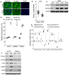
Figure 4. Piezo1 is required for the OSS-induced upregulation of lymphatic valve genes.
(A and B) Primary human LECs exposed to OSS for 24 hours became cuboidal shapes. (A) Immunofluorescence staining for F-actin and (B) a box-and-whisker plot showing the ratio of length/width of the cells. (C and D) OSS upregulated FOXC2, GATA2, CX37, LAMA5, and ITGA9 in LECs, based on (C) Western blot analyses and (D) quantitative PCR (qPCR). The box plots depict the minimum and maximum values (whiskers), the upper and lower quartiles, and the median. The length of the box represents the interquartile range. (E and F) Piezo1 knockdown prevented the OSS-mediated upregulation of lymphatic valve genes. Piezo1 expression in primary LECs was inhibited by Piezo1 siRNA (siPiezo1), or not inhibited by control siRNA (siCTR), for 24 hours. Cells were then exposed to OSS for (E) 4 hours or (F) 24 hours before (E) qPCR data assay or (F) Western blot analysis, respectively. OSS with approximately at 6 dyne/cm2 maximum was applied directly onto the cells by reversing the flow at 0.5 Hz (peak). **P < 0.01; ***P < 0.001; unpaired, 2-tailed t test compared with the static culture.
Figure 5
Figure 5. Ectopic expression of Piezo1 induces lymphatic valve signature genes.
(A and B) Primary LECs were transfected with a Piezo1/EGFP-expressing plasmid and cultured for 48 hours without OSS, before immunostaining for lymphatic valve genes FOXC2, GATA2, CX37, LAMA5, and ITGA9. (A) Arrowheads point to transfected EGFP+ (thus Piezo1-overexpressing) cells, whereas arrows point to EGFP, untransfected cells. O/E, overexpression; LV, lymphatic valve. (B) The intensity of these cells was measured and charted in a box-and-whisker plot. (C and D) Expression of the lymphatic valve genes in LECs that were transfected with a control (CTR) or Piezo1/EGFP-expressing plasmid (Piezo1) was determined by qRT-PCR after (C) 24 hours, or by Western blot assay after (D) 48 hours, in the absence of OSS. The box plots depict the minimum and maximum values (whiskers), the upper and lower quartiles, and the median. The length of the box represents the interquartile range. Scale bars: 50 μm (A). **P < 0.01; ***P < 0.001; unpaired, 2-tailed t test compared with (B) the untransfected cells or (C) the control plasmid.
Figure 6
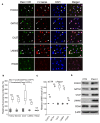
Figure 6. Activation of Piezo1 accelerates in vivo lymphatic valve formation.
(A) Experimental design. Pregnant Prox1-EGFP females were i.p. injected with vehicle or Yoda1 (70 μg/kg) at E18.5 and then allowed to give birth to pups. Individual pups at P0 were injected once again with vehicle or Yoda1 (70 μg/kg) and then euthanized at P1 for lymphatic valve analyses. The effect of Yoda1 on developing mesenteric lymphatic valves was evaluated by selecting comparable anatomic locations in the (B) jejunum, (C) colon, and (D) tail skin of the vehicle or Yoda1-treated pups. (E and F) Images of the lymphatics were captured using EGFP signal, and lymphatic valve formation was quantified. The box plots depict the minimum and maximum values (whiskers), the upper and lower quartiles, and the median. The length of the box represents the interquartile range. More than 6 pups (mixed sex) were analyzed per group. Scale bars: 500 μm (B–D). ***P < 0.001, unpaired, 2-tailed t test compared with the vehicle-treated group (E and F).
Figure 7
Figure 7. Yoda1 activates the expression of some lymphatic valve genes in a Piezo1-dependent manner.
(A and B) Regulation of (A) mRNA and (B) protein of lymphatic valve genes in LECs by Yoda1 was determined by qPCR and Western blot analyses, respectively. Primary human LECs were treated by Yoda1 at the indicated concentrations for 24 hours. Yoda1 upregulated the expression of GATA2, CX37, LAMA5, and ITGA9 but not FOXC2 and PROX1. (C and D) Piezo1 is required for the Yoda1-induced upregulation of GATA2, CX37, and LAMA5 but not ITGA9. LECs were transfected with control siRNA (siCTR) or Piezo1 siRNA (siPiezo1) for 24 hours, followed by Yoda1 treatment (250 nM) for 24 hours, before isolation of (C) RNA or (D) whole-cell lysates for qPCR and Western blot analyses, respectively. The box plots depict the minimum and maximum values (whiskers), the upper and lower quartiles, and the median. The length of the box represents the interquartile range. *P < 0.05; **P < 0.01; ***P < 0.001; unpaired, 2-tailed t test.

Similar articles

Cited by

References

    1. Breslin JW. Mechanical forces and lymphatic transport. Microvasc Res. 2014;96:46–54. doi: 10.1016/j.mvr.2014.07.013. - DOI - PMC - PubMed
    1. Bazigou E, Wilson JT, Moore JE. Primary and secondary lymphatic valve development: molecular, functional and mechanical insights. Microvasc Res. 2014;96:38–45. doi: 10.1016/j.mvr.2014.07.008. - DOI - PMC - PubMed
    1. Bazigou E, Makinen T. Flow control in our vessels: vascular valves make sure there is no way back. Cell Mol Life Sci. 2013;70(6):1055–1066. doi: 10.1007/s00018-012-1110-6. - DOI - PMC - PubMed
    1. Schmid-Schonbein GW. The second valve system in lymphatics. Lymphat Res Biol. 2003;1(1):25–29. doi: 10.1089/15396850360495664. - DOI - PubMed
    1. Chiu JJ, Chien S. Effects of disturbed flow on vascular endothelium: pathophysiological basis and clinical perspectives. Physiol Rev. 2011;91(1):327–387. doi: 10.1152/physrev.00047.2009. - DOI - PMC - PubMed

Publication types