Review
. 2019 Sep 1;27(17):642-651.
doi: 10.5435/JAAOS-D-18-00347.

Flexion Instability After Total Knee Arthroplasty

Affiliations
Review

Flexion Instability After Total Knee Arthroplasty

Jeffrey B Stambough et al. J Am Acad Orthop Surg. .

Abstract

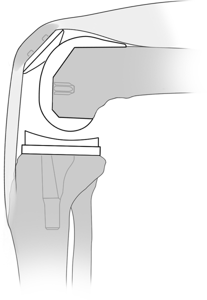
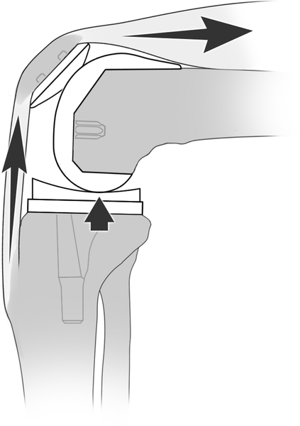
Flexion instability after total knee arthroplasty (TKA) is caused by an increased flexion gap compared with extension gap. Patients present with recurrent effusions, subjective instability (especially going downstairs), quadriceps weakness, and diffuse periretinacular pain. Manual testing for laxity in flexion is commonly done to confirm a diagnosis, although testing positions and laxity grades are inconsistent. Nonsurgical treatment includes quadriceps strengthening and bracing treatment. The mainstays to surgical management of femoral instability involve increasing the posterior condylar offset, decreasing the tibial slope, raising the joint line in combination with a thicker polyethylene insert, and ensuring appropriate rotation of implants. Patient outcomes after revision TKA for flexion instability show the least amount of improvement when compared with revisions for other TKA failure etiologies. Future work is needed to unify reproducible diagnostic criteria. Advancements in biomechanical analysis with motion detection, isokinetic quadriceps strength testing, and computational modeling are needed to advance the collective understanding of this underappreciated failure mechanism.


Figures

Figure 1.
Figure 1.
In patients with pre-existing flexion contracture, it is possible to create a situation of flexion instability when the surgeon does not resect enough distal femur (a). The shaded area signifies the bone that should have been cut to properly balance the gaps. This scenario with under resection of the distal femur translates to a knee remaining tight in extension (b) necessitating the surgeon use a thin polyethylene insert, thereby increasing the flexion space (c) thereby creating laxity in flexion.
Figure 1.
Figure 1.
In patients with pre-existing flexion contracture, it is possible to create a situation of flexion instability when the surgeon does not resect enough distal femur (a). The shaded area signifies the bone that should have been cut to properly balance the gaps. This scenario with under resection of the distal femur translates to a knee remaining tight in extension (b) necessitating the surgeon use a thin polyethylene insert, thereby increasing the flexion space (c) thereby creating laxity in flexion.
Figure 2.
Figure 2.
An overly aggressive posterior condylar resection can lead to flexion instability. These bone cuts often lead to undersizing of the femoral component, thereby not recreating the appropriate posterior femoral offset, as indicated by the dashed line.
Figure 3.
Figure 3.
By cutting too much posterior slope into the tibia, the gap posteriorly increases disproportionately to the extension gap. This allows the knee to roll posterior on the tibia in flexion, thereby translating the tibia anterior, leading to flexion instability.
Figure 4.
Figure 4.
When a patient with flexion instability rest with their knee at a 90 degree flexed position without their foot contacting the floor, the femoral component does not contact the polyethylene (a). In order to initiate knee extension, the quadriceps and extensor mechanism must first contract to pull the tibia up to contact the femur (b) before any extension can occur. The quadriceps must generate additional increased force to subsequently extend the knee once femoral-polyethylene contact is established (c).
Figure 4.
Figure 4.
When a patient with flexion instability rest with their knee at a 90 degree flexed position without their foot contacting the floor, the femoral component does not contact the polyethylene (a). In order to initiate knee extension, the quadriceps and extensor mechanism must first contract to pull the tibia up to contact the femur (b) before any extension can occur. The quadriceps must generate additional increased force to subsequently extend the knee once femoral-polyethylene contact is established (c).
Figure 5.
Figure 5.
Flow diagram highlighting operative steps during revision knee surgery to address flexion instability.
Figure 6.
Figure 6.
Lateral radiographs demonstrating flexion instability as a result of over-resection of the posterior femoral condyle (a) that was surgically corrected by increasing the posterior femoral offset by 4mm using a revision femoral component and posterior augment (b).
Figure 6.
Figure 6.
Lateral radiographs demonstrating flexion instability as a result of over-resection of the posterior femoral condyle (a) that was surgically corrected by increasing the posterior femoral offset by 4mm using a revision femoral component and posterior augment (b).

Similar articles

Cited by

References

    1. Sharkey PF, Lichstein PM, Shen C, Tokarski AT, Parvizi J. Why Are Total Knee Arthroplasties Failing Today—Has Anything Changed After 10 Years? J Arthroplasty. 2014;29(9):1774–1778. - PubMed
    1. Dalury DF, Pomeroy DL, Gorab RS, Adams MJ. Why are Total Knee Arthroplasties Being Revised? J Arthroplasty. 2013;28(8):120–121. - PubMed
    1. Le DH, Goodman SB, Maloney WJ, Huddleston JI. Current modes of failure in TKA: infection, instability, and stiffness predominate. Clin Orthop Relat Res. 2014;472(7):2197–2200. - PMC - PubMed
    1. Cottino U, Sculco PK, Sierra RJ, Abdel MP. Instability After Total Knee Arthroplasty. Orthop Clin North Am. 2016;47(2):311–316. - PubMed
    1. Rodriguez-Merchan EC. Instability Following Total Knee Arthroplasty. HSS J. 2011. - PMC - PubMed