. 2019 Apr;33(4):5468-5481.
doi: 10.1096/fj.201801860RR. Epub 2019 Jan 24.

Lysyl oxidase-like 2 (LOXL2)-mediated cross-linking of tropoelastin

Affiliations

Lysyl oxidase-like 2 (LOXL2)-mediated cross-linking of tropoelastin

Christian E H Schmelzer et al. FASEB J. 2019 Apr.

Abstract

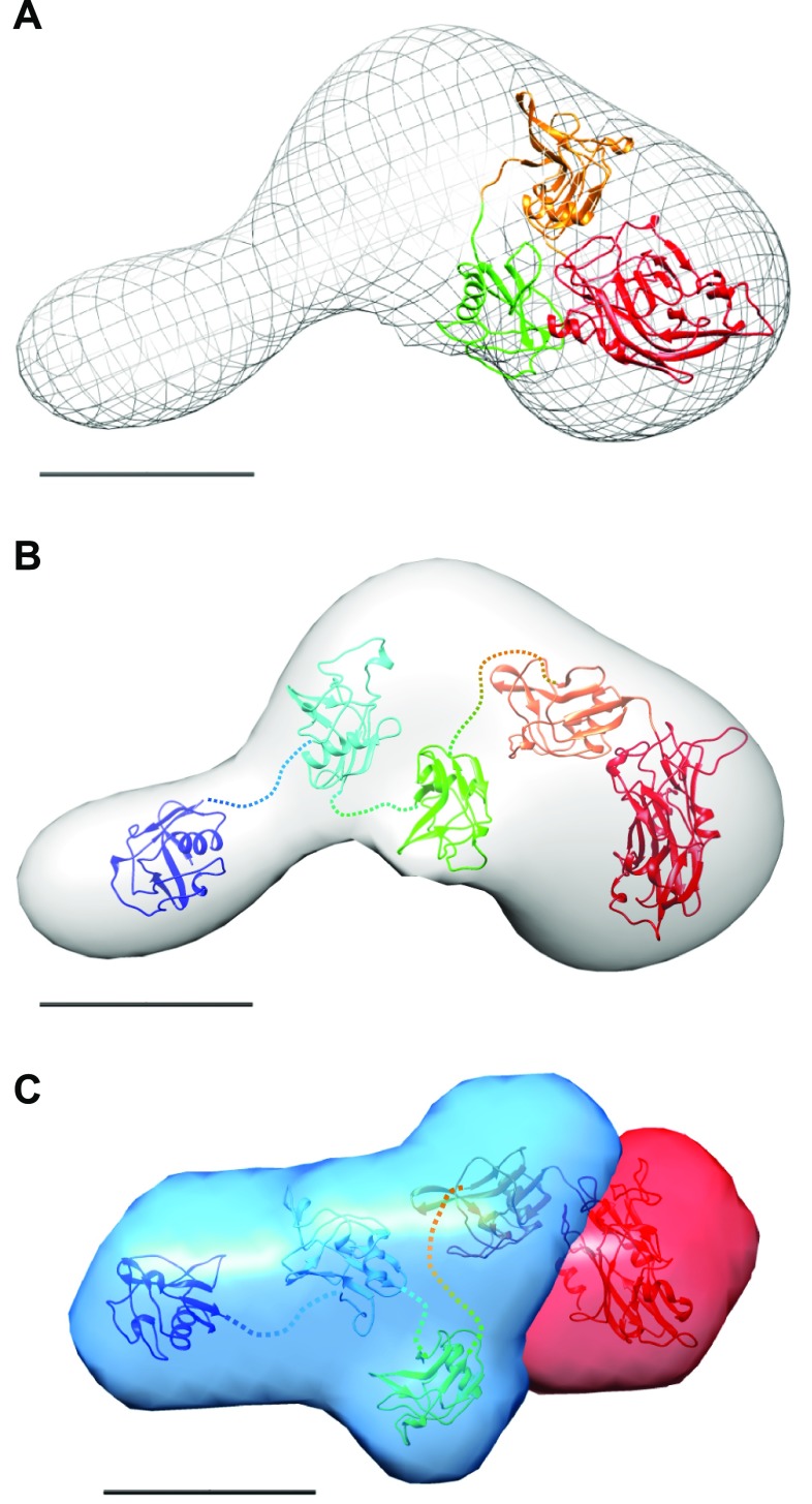
Lysyl oxidases (LOXs) play a central role in extracellular matrix remodeling during development and tumor growth and fibrosis through cross-linking of collagens and elastin. We have limited knowledge of the structure and substrate specificity of these secreted enzymes. LOXs share a conserved C-terminal catalytic domain but differ in their N-terminal region, which is composed of 4 repeats of scavenger receptor cysteine-rich (SRCR) domains in LOX-like (LOXL) 2. We investigated by X-ray scattering and electron microscopy the low-resolution structure of the full-length enzyme and the structure of a shorter form lacking the catalytic domain. Our data demonstrate that LOXL2 has a rod-like structure with a stalk composed of the SRCR domains and the catalytic domain at its tip. We detected direct interaction between LOXL2 and tropoelastin (TE) and also LOXL2-mediated deamination of TE. Using proteomics, we identified several allysines together with cross-linked TE peptides. The elastin-like material generated was resistant to trypsin proteolysis and displayed mechanical properties similar to mature elastin. Finally, we detected the codistribution of LOXL2 and elastin in the vascular wall. Altogether, these data suggest that LOXL2 could participate in elastogenesis in vivo and could be used as a means of cross-linking TE in vitro for biomimetic and cell-compatible tissue engineering purposes.-Schmelzer, C. E. H., Heinz, A., Troilo, H., Lockhart-Cairns, M.-P., Jowitt, T. A., Marchand, M. F., Bidault, L., Bignon, M., Hedtke, T., Barret, A., McConnell, J. C., Sherratt, M. J., Germain, S., Hulmes, D. J. S., Baldock, C., Muller, L. Lysyl oxidase-like 2 (LOXL2)-mediated cross-linking of tropoelastin.

Keywords: SAXS; elastin; matrix remodeling; protein structure; proteomics.


Conflict of interest statement

The authors thank the Structure Fédérative de Recherche Biosciences (UMS3444/CNRS), US8/INSERM, École Normale Supérieure de Lyon, Université Claude Bernard Lyon 1 Protein Science Facility for access to the circular dichroism instrument. The authors thank Christian Ihling (Martin Luther University Halle-Wittenberg, Germany) for assistance with mass spectrometry measurements, Prof. Tony Weiss (University of Sydney, Sydney, NSW, Australia) for providing the recombinant tropoelastin used in surface plasmon resonance experiments, Markus Pietzsch (Martin Luther University Halle-Wittenberg) for providing the recombinant tropoelastin used in other experiments, and Jürgen Brinckmann (University of Lübeck, Germany) for the amino acid analysis, and Dr. Catherine Moali (Université de Lyon, France) for helpful discussions and reading the manuscript, as well as providing BMP-1 and PCPE-1. The authors thank staff in the University of Manchester Biomolecular Analysis Core Facility and Electron Microscopy Centre for assistance and Diamond Light Source and European Synchrotron Radiation Facility (ESRF) (BioStruct-X FP7-Infractrustures Project 283570 Midland BAG MX1783) for beamtime. The work was supported by the Fraunhofer Internal Programs (Attract Grant 069–608203 to C.E.H.S.), the European Regional Development Fund in Saxony-Anhalt (project ElastoHEAL; to CEHS), the Biotechnology and Biological Sciences Research Council (BBSRC) (BB/L00612X/1 to C.B.), the LEO Foundation (Grant LF17063 to A.H.), and Prevent Breast Cancer (to M.J.S.). M.F.M. was supported by Fédération pour la Recherche Médicale (FRM-ECO2016-0736055). The authors declare no conflicts of interest.

Figures

Figure 1
Figure 1
Characterization of recombinant human FL-LOXL2 and the SRCR14 region. A) Schematic representation of recombinant FL-LOXL2 and of the catalytic domain truncated form (SRCR14). Black box corresponds to the signal peptide. Dark gray box corresponds to the catalytic domain, containing the copper binding site (Cu), the lysine tyrosylquinone cofactor (LTQ), and the cytokine receptor–like domain (CRL, in black box). The C-terminal myc-histidine tag (MH) is in red. B, C) Purified recombinant proteins were separated by SDS-PAGE and stained with Coomassie Blue (B) and analyzed by CD (C). D, E) Purity and MWs were also assessed using multiangle laser light scattering AUC. Multiangle laser light scattering was performed with a Superdex 200 column for LOXL2 and a Superdex 75 for SRCR14 (D). Ultracentrifugation was analyzed using Sedfit2 (E). dRI, differential refractive index; C(s), sedimentation coefficient distribution; Sapp, apparent sedimentation; deg, degrees; MRW = mean residue weight.
Figure 2
Figure 2
Low-resolution structures and modeling of LOXL2 and SRCR by SAXS analysis. A) DAMMIF ab initio models of LOXL2. B) The SRCR DAMMIF ab initio model is superimposed with the rigid-body model of SRCR14 colored from blue to red. C) The ab initio model of the SRCR region (blue) inside the ab initio model of LOXL2 (mesh). D) MONSA model of LOXL2 with the catalytic domain shown as a solid red density and the SRCR domains shown in blue, into which the rigid-body model of the SRCR domains is docked (shown colored from blue to red). Scale bar, 5 nm.
Figure 3
Figure 3
Structural analysis of LOXL2 by single-particle EM and image reconstruction. A) Raw image of LOXL2 collected at ×23,000, 2.8 Å/pixel. B) Selected class averages of LOXL2. A total of 32 classes were generated from ∼10,000 particles selected from 150 micrographs. C) Corresponding projections from the 3D reconstruction generated using EMAN2. Box size is 36 nm (B, C). D) LOXL2 3D reconstruction shown in 2 orientations. E) MONSA-SAXS model (catalytic domain shown in red and SRCR region in blue) fitted within the EM model (mesh). Two orientations are shown. Scale bar, 5 nm (D, E).
Figure 4
Figure 4
Comparison of SAXS and EM data for FL-LOXL2 with the crystal structure of LOXL2 N455Q lacking SRCR domains 1 and 2. A) Crystal structure directly superimposed on the EM model (mesh). B, C) The 2 rigid-body models that best fit the FL-LOXL2 EM reconstruction (B) and MONSA-SAXS model (C). SRCR domains are numbered and colored from blue at the N terminus to red at the C terminus. The catalytic domain and SRCR domain 4 from the crystal structure were treated as a rigid body, whereas SRCR domains 1–3 had flexible linkers (represented as dashed lines). The catalytic domain is shown in red and SRCR region in blue (C). Scale bars, 5 nm.
Figure 5
Figure 5
Direct interaction between LOXL2 and TE. A) Binding between immobilized LOXL2 was analyzed with TE as analyte using surface plasmon resonance. B) Binding constants were determined using steady-state binding analysis, which gives a dissociation constant of 146 ± 16 nM (n = 4).
Figure 6
Figure 6
LOXL2-mediated modification of lysines results in cross-linking of TE. A) Recombinant LOXL2 activity was measured using the Amplex UltraRed assay using either cadaverine (blue) or TE (yellow) as a substrate. No release of H2O2 was detected in the presence of 500 µM β-APN (gray) or when using laminin (green) as a substrate. B, C) Annotated product ion spectra of an allysine (Lya)-containing peptide with m/z 849.9803 (z = 2) (B) as well as an intramolecularly cross-linked peptide (m/z 509.7868; z = 2) possessing a dehydrolysinonorleucine (ΔLNL) (C). Both peptides were identified in a digest of cTE. The amino acid sequences are displayed in the upper-right corners of the spectra. Corresponding b-ions and a-ions are highlighted in blue, y-ions are shown in red, and internal fragments are shown in green; product ions denoted with ° indicate loss of water. D) Tandem mass spectrum of free desmosine (M+ 526.29) detected in a TE sample cross-linked by LOXL2 after total hydrolysis (bottom). The fragment pattern shows clear conformity with a desmosine standard (top) and verifies the presence of desmosine in the sample. E) Domain structure of human TE (isoform 2) indicating the identified allysines and cross-linking sites within the sequence. Lys residues are shown as filled circles within Lys-Ala (KA) and Lys-Pro (KP) domains highlighted in dark and light blue, respectively. The domain numbering shown is based on exon assignment, and all domains are displayed in appropriate size corresponding to their relative length. In total, we identified 3 intramolecular bifunctional cross-links and 6 allysine residues.
Figure 7
Figure 7
LOXL2-mediated cross-linking of TE results in formation of an insoluble elastin-like material. A, B) Scanning electron micrographs of isolated aortic elastin (AE) (A) and cTE (B). C) Local micromechanical stiffness was measured by AFM indentation of aortic elastin and cTE. The distribution of reduced modulus values (main panel) was significantly shifted to the right for cTE (red) when compared with aortic elastin (blue). Analysis of mean reduced modulus values from 2 replicates (inset panel) showed that cTE (red; 193.1 ± 13.9 kPa) exhibited a significantly increased reduced modulus when compared with aortic elastin (blue; 144.2 ± 10.8 kPa). ***P < 0.0005. D) Total ion current intensity chromatograms obtained by nano–HPLC-MS runs of the 24-h tryptic digests of cTE (red) vs. unmodified TE in solution (black).
Figure 8
Figure 8
LOXL2 is expressed in the vascular wall. A) LOXL2 was detected in cryostat sections of mouse tibialis anterior. Whereas type IV collagen (Col IV) was detected in the basement membrane of both blood vessels and muscle fibers, LOXL2 was restricted to blood vessels (arrows), with a very high expression in arterioles (arrowheads). Scale bars, 50 µm. B) LOX and LOXL2 were detected in the intima (arrows) and media of mouse aorta cryosection by immunofluorescence, together with elastin autofluorescence (green). Scale bars, 50 µm. C) Aorta were frozen and extracted with Triton X-100 followed by SDS extraction of the insoluble fraction before separation by SDS-PAGE. LOX and LOXL2 were both detected in the Triton X-100–resistant ECM fraction together with fibronectin.

References

    1. Lucero H. A., Kagan H. M. (2006) Lysyl oxidase: an oxidative enzyme and effector of cell function. Cell. Mol. Life Sci. 63, 2304–2316 - PMC - PubMed
    1. Moon H. J., Finney J., Ronnebaum T., Mure M. (2014) Human lysyl oxidase-like 2. Bioorg. Chem. 57, 231–241 - PMC - PubMed
    1. Trackman P. C., Bais M. V. (2018) Measurement of lysyl oxidase activity from small tissue samples and cell cultures. Methods Cell Biol. 143, 147–156 - PubMed
    1. López-Jiménez A. J., Basak T., Vanacore R. M. (2017) Proteolytic processing of lysyl oxidase-like-2 in the extracellular matrix is required for crosslinking of basement membrane collagen IV. J. Biol. Chem. 292, 16970–16982 - PMC - PubMed
    1. Mäki J. M., Räsänen J., Tikkanen H., Sormunen R., Mäkikallio K., Kivirikko K. I., Soininen R. (2002) Inactivation of the lysyl oxidase gene Lox leads to aortic aneurysms, cardiovascular dysfunction, and perinatal death in mice. Circulation 106, 2503–2509 - PubMed

Publication types

LinkOut - more resources