

Comparison of prostate delineation on multimodality imaging for MR-guided radiotherapy
- PMID: 30676772
- PMCID: PMC6540870
- DOI: 10.1259/bjr.20180948
Comparison of prostate delineation on multimodality imaging for MR-guided radiotherapy
Abstract
Objective:: With increasing incorporation of MRI in radiotherapy, we investigate two MRI sequences for prostate delineation in radiographer-led image guidance.
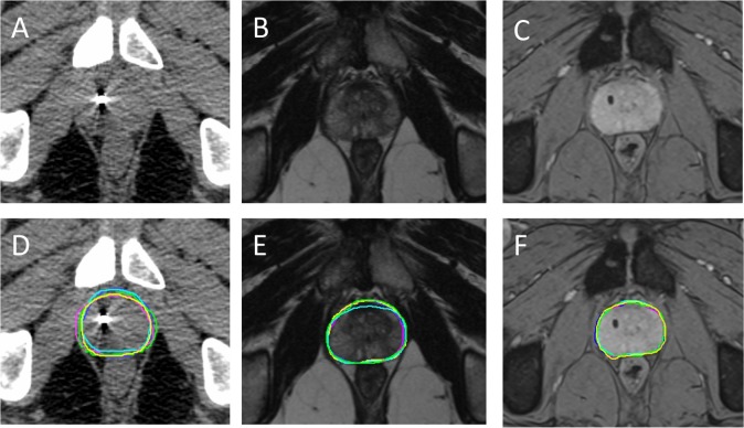
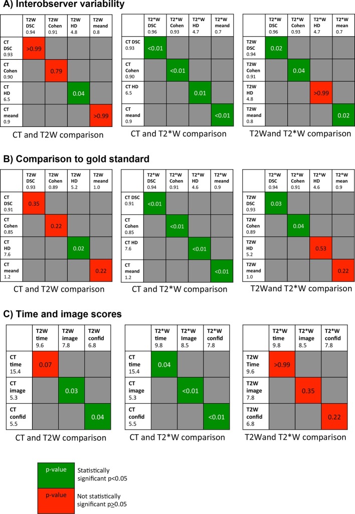
Methods:: Five therapeutic radiographers contoured the prostate individually on CT, T2 weighted (T2W) and T2* weighted (T2*W) imaging for 10 patients. Contours were analysed with Monaco ADMIRE (research v. 2.0) to assess interobserver variability and accuracy by comparison with a gold standard clinician contour. Observers recorded time taken for contouring and scored image quality and confidence in contouring.
Results:: There is good agreement when comparing radiographer contours to the gold-standard for all three imaging types with Dice similarity co-efficient 0.91-0.94, Cohen's κ 0.85-0.91, Hausdorff distance 4.6-7.6 mm and mean distance between contours 0.9-1.2 mm. In addition, there is good concordance between radiographers across all imaging modalities. Both T2W and T2*W MRI show reduced interobserver variability and improved accuracy compared to CT, this was statistically significant for T2*W imaging compared to CT across all four comparison metrics. Comparing MRI sequences reveals significantly reduced interobserver variability and significantly improved accuracy on T2*W compared to T2W MRI for DSC and Cohen's κ. Both MRI sequences scored significantly higher compared to CT for image quality and confidence in contouring, particularly T2*W. This was also reflected in the shorter time for contouring, measuring 15.4, 9.6 and 9.8 min for CT, T2W and T2*W MRI respectively. Conclusion: Therapeutic radiographer prostate contours are more accurate, show less interobserver variability and are more confidently and quickly outlined on MRI compared to CT, particularly using T2*W MRI. Advances in knowledge: Our work is relevant for MRI sequence choice and development of the roles of the interprofessional team in the advancement of MRI-guided radiotherapy.
Figures
References
-
- Pathmanathan AU, van As NJ, Kerkmeijer LGW, Christodouleas J, Lawton CAF, Vesprini D, et al. Magnetic Resonance Imaging-Guided Adaptive Radiation Therapy: A “Game Changer” for Prostate Treatment? International Journal of Radiation Oncology*Biology*Physics 2018; 100: 361–73. doi: 10.1016/j.ijrobp.2017.10.020 - DOI - PubMed
-
- Schieda N, Avruch L, Shabana WM, Malone SC. Multi-echo gradient recalled echo imaging of the pelvis for improved depiction of brachytherapy seeds and fiducial markers facilitating radiotherapy planning and treatment of prostatic carcinoma. J Magn Reson Imaging 2015; 41: 715–20. doi: 10.1002/jmri.24590 - DOI - PubMed
Publication types
MeSH terms
Grants and funding
LinkOut - more resources
Full Text Sources
Medical
