. 2019 Jan 24;15(1):e1007896.
doi: 10.1371/journal.pgen.1007896. eCollection 2019 Jan.

Serotonin and neuropeptides are both released by the HSN command neuron to initiate Caenorhabditis elegans egg laying

Affiliations

Serotonin and neuropeptides are both released by the HSN command neuron to initiate Caenorhabditis elegans egg laying

Jacob C Brewer et al. PLoS Genet. .

Abstract

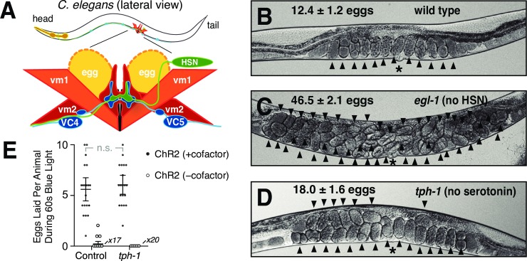
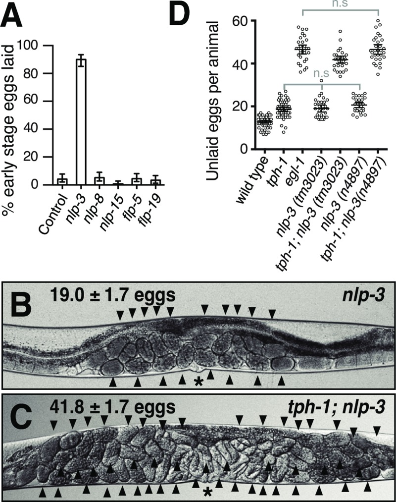
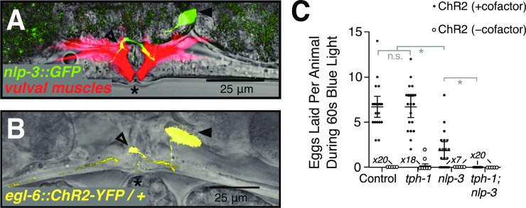
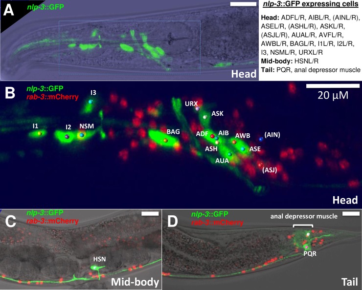
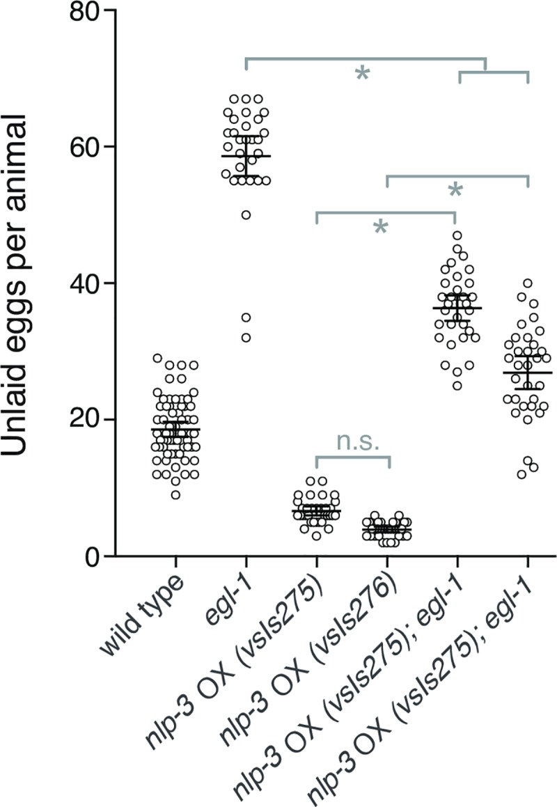
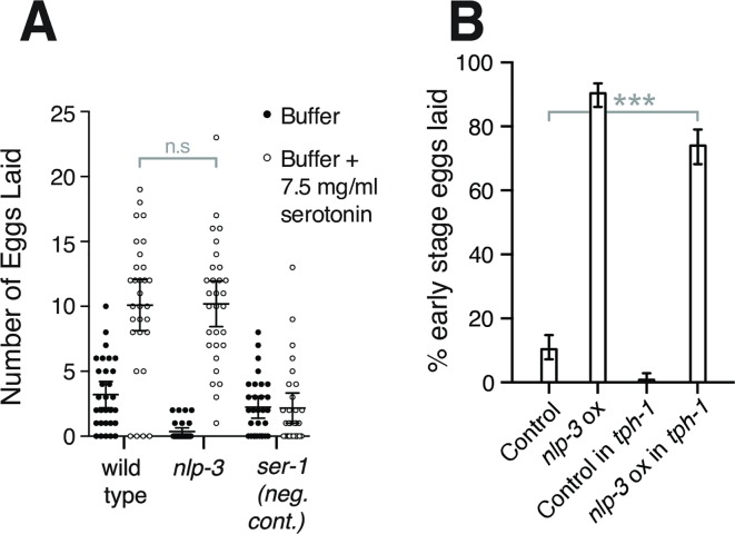
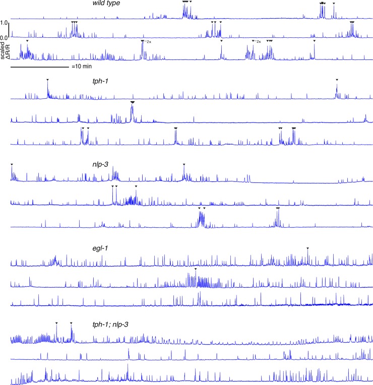
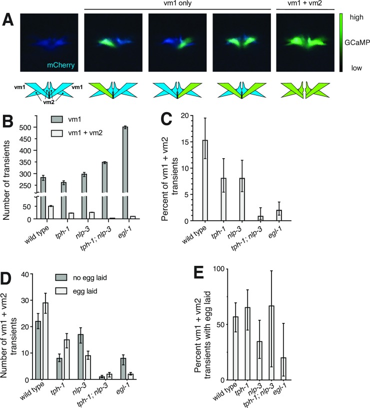
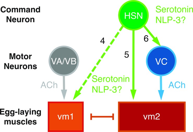
Neurons typically release both a small-molecule neurotransmitter and one or more neuropeptides, but how these two types of signal from the same neuron might act together remains largely obscure. For example, serotonergic neurons in mammalian brain express the neuropeptide Substance P, but it is unclear how this co-released neuropeptide might modulate serotonin signaling. We studied this issue in C. elegans, in which all serotonergic neurons express the neuropeptide NLP-3. The serotonergic Hermaphrodite Specific Neurons (HSNs) are command motor neurons within the egg-laying circuit which have been shown to release serotonin to initiate egg-laying behavior. We found that egg-laying defects in animals lacking serotonin were far milder than in animals lacking HSNs, suggesting that HSNs must release other signal(s) in addition to serotonin to stimulate egg laying. While null mutants for nlp-3 had only mild egg-laying defects, animals lacking both serotonin and NLP-3 had severe defects, similar to those of animals lacking HSNs. Optogenetic activation of HSNs induced egg laying in wild-type animals, and in mutant animals lacking either serotonin or NLP-3, but failed to induce egg laying in animals lacking both. We recorded calcium activity in the egg-laying muscles of animals lacking either serotonin, NLP-3, or both. The single mutants, and to a greater extent the double mutant, showed muscle activity that was uncoordinated and unable to expel eggs. Specifically, the vm2 muscles cells, which are direct postsynaptic targets of the HSN, failed to contract simultaneously with other egg-laying muscle cells. Our results show that the HSN neurons use serotonin and the neuropeptide NLP-3 as partially redundant co-transmitters that together stimulate and coordinate activity of the target cells onto which they are released.


Conflict of interest statement

The authors have declared that no competing interests exist.

Figures

Fig 1
Fig 1. Serotonin is not required for the HSN to stimulate egg laying.
A) Schematic of the C. elegans egg-laying circuit, adapted from [23]. HSN and VC motorneurons synapse onto vm2 vulval muscle cells, which along with vm1 muscle cells contract to open the vulva and release eggs. Only the left HSN and vulval muscle cells are shown–equivalent cells are also found on the right side of the animal. The uv1 neuroendocrine cells that inhibit the circuit are not shown. B-D) Images of representative animals of the indicated genotypes, showing the average number of unlaid eggs +/- 95% confidence intervals, n = 30. Arrowheads indicate individual unlaid eggs. Asterisks indicate the location of the vulva. E) Number of eggs laid during 60 seconds of blue light exposure by animals expressing ChR2 in the HSNs. Both control and tph-1 animals also carry lite-1(ce314) mutations that eliminate a locomotion response to blue light [69]. Black filled data points, animals that were grown for a generation in the presence of ChR2’s required cofactor all-trans retinal (ATR). Open circle data points, negative control animals grown in the absence of ATR. For this and all subsequent graphs: error bars represent 95% confidence intervals for the mean unless otherwise indicated; n.s. indicates no statistically significant difference (p>0.05); the number measurements of zero eggs for some genotypes is indicated at the horizontal axis.
Fig 2
Fig 2. The neuropeptide gene nlp-3, together with serotonin, stimulates egg laying.
A) Overexpression of nlp-3, but not of four other neuropeptide genes, increased the rate of egg-laying behavior. Genomic clones for each neuropeptide gene, or the coinjection marker alone (control), were injected into C. elegans to generate high-copy extrachromosomal transgenes. For each gene, 250 freshly laid eggs (50 from each of five independent transgenic lines) were examined and the percent laid at early stages of development (eight cells or fewer) was determined. Only nlp-3 overexpression gave results significantly different from the control (p<0.05). B-C) Representative images of nlp-3 and tph-1; nlp-3 animals showing the average number of unlaid eggs, n = 30. D) Graph of the number of unlaid eggs for the strains indicated. Two independent deletion alleles of nlp-3 were used. n≥30 for each strain. Comparisons labeled n.s. showed no significant differences (p>0.05). All other pairwise comparisons were significantly different with p<0.033.
Fig 3
Fig 3. The HSNs require the nlp-3 neuropeptide gene and tph-1 to stimulate egg laying.
A) nlp-3 is expressed specifically in the HSNs. Vulval region of an adult animal carrying an nlp-3::GFP transgene and a second transgene that expresses mCherry in the vulval muscles from the unc-103e promoter [47]. B) The egl-6p::ChR2::YFP transgene is expressed specifically in the HSN. In A and B, asterisks indicate the vulva, filled arrowheads the HSN cell body and open arrowheads the HSN synapse onto the vulval muscles. C) Average number of eggs laid during 60 seconds of blue light exposure by animals heterozygous for the egl-6p::ChR2::YFP transgene and homozygous for the indicated null mutations in tph-1 and/or nlp-3. Control, animals wild-type for tph-1 and nlp-3. Black filled data points, animals grown in the presence of ChR2’s required cofactor all-trans retinal (ATR). Open circle data points, animals grown in the absence of ATR. All animals in this experiment were homozygous for a lite-1 mutation that abolished an endogenous C. elegans response to blue light [64]. Error bars, 95% confidence intervals of the mean. The number of measurements of zero eggs for some genotypes is indicated at the horizontal axis. Key pairwise comparisons are indicated (n.s., not significantly different, p>0.05; or *, significantly different, p<0.001).
Fig 4
Fig 4. Expression pattern of nlp-3.
The nlp-3 promoter was used to drive expression of GFP in transgenic animals. Animals also express nuclear-localized tagRFP in all neurons from the rab-3 promoter. A) nlp-3::GFP fluorescence superimposed on a bright-field image of the head. B) The area outlined by the rectangle in A is enlarged, and both GFP and tagRFP fluorescence are shown. Colored balls indicate cell bodies of GFP-expressing neurons, and are labeled with their cell designations. C) and D) Show midbody and tail regions, respectively, with GFP and mCherry fluorescence superimposed on bright-field images, with GFP-expressing cells identified. All images show the left side of the animal only and all scale bars are 20 μm. A list of all cells that express nlp-3 is shown at upper right. AIN and ASJ are not visibly GFP labeled in the image shown, and along with ASH are more weakly labeled than other nlp-3-expressing cells. The AVFL/R neurons are just outside of the area displayed in B but are shown in S4 Video.
Fig 5
Fig 5. Overexpression of nlp-3 in cells other than HSNs has some ability to stimulate egg laying.
Two different chromosomally-integrated transgenes that carry multiple copies of nlp-3 genomic DNA were used to overexpress ("OX") nlp-3. Egg-laying behavior was assessed by counting unlaid eggs in adult animals either carrying the overexpressor transgenes by themselves or in combination with an egl-1 mutation that results in absence of HSN neurons. nlp-3 overexpression significantly reduced the accumulation of eggs caused by the egl-1 mutation. n.s., no statistically significant difference (p>0.05); *, significant difference with p<0.0001.
Fig 6
Fig 6. Serotonin and NLP-3 neuropeptides can stimulate egg laying in the absence of each other.
A) Exogenous serotonin stimulates egg laying in wild-type and nlp-3 animals. The number of eggs laid by 10 animals over 30 minutes in M9 buffer or M9 buffer plus 7.5 mg/ml serotonin was measured, and this assay was repeated >10 times per genotype. The ser-1 serotonin receptor null mutant is the negative control. B) nlp-3 overexpression stimulates egg laying even in the absence of serotonin. Animals wild-type for tph-1 or tph-1 null mutants were injected with marker DNA alone (control) or nlp-3 genomic DNA plus marker DNA to overexpress nlp-3 (nlp-3 ox). In each case, five independent transgenic lines were produced, and 50 freshly laid eggs per line (250 eggs total per condition) were examined to determine their developmental stages. Error bars, 95% confidence intervals.
Fig 7
Fig 7. Vulval muscle activity, but not egg release, occurs frequently in mutants lacking serotonin, NLP-3, or both.
Graphs of Ca2+ transients showing ΔR/R of GCaMP5/mCherry signal recorded over 1 hour for each of three different animals per genotype. Arrows indicate a calcium transient associated with an egg-laying event. “2x” indicates that two eggs were laid nearly simultaneously during the same calcium transient. Scale bar, 10 minutes. Vertical scales have been normalized to depict comparable peak heights in all animals shown.
Fig 8
Fig 8. Egg-laying events are associated with vm1 + vm2 Ca2+ transients.
A) Still frames from ratiometric recordings illustrating different patterns of activity observed in the vulval muscles. The mCherry channel is rendered in blue. The GCaMP channel is superimposed in green, with intensity rendered by ranging from transparent (low) to bright green (high). Schematics are shown below the images to indicate the individual muscle cells where activity appears to occur. B-C) A graph of the number (B) and percent (C) of calcium transients occurring in the vm1 only compared to those appearing to occur in vm1 + vm2 for each genotype. p-values for decreases in percentage of vm1 + vm2 transients for mutants compared to the wild type in (C) were = 0.05 (tph-1), = 0.04 (nlp-3), <0.001 (tph-1; nlp-3); and <0.001 (egl-1). These values indicate that tph-1; nlp-3 and egl-1 have significantly fewer vm1 + vm2 transients after correcting for multiple comparisons. D-E) The number (D) and proportion (E) of either vm1 or vm1 + vm2 transients that are associated with egg-laying events. All 57 egg-laying events occurred during vm1 + vm2 events. In (B-E) the data analyzed are from the three hours of recordings for each genotype depicted in Fig 7, and error bars are 95% confidence intervals for the results expected if an infinite sample size was used.
Fig 9
Fig 9. Model depicting signaling events that activate the egg-laying circuit.
Solid arrows, synaptic signaling; dashed arrow, extrasynaptic signaling; and bar, gap junctions. vm1 and vm2 cells are known to express multiple serotonin receptor isoforms that each mediate activation of egg laying. The signaling of serotonin onto VC neurons (arrow 3) remains hypothetical since no serotonin receptors have yet been described as expressed on these neurons. The possible direct signaling of NLP-3 depicted onto vm1 (arrow 2), vm2 (arrow 2), or and/or VC (arrow 3) is also hypothetical: NLP-3 receptors have yet to be identified and it thus remains unknown which cell(s) of the circuit express them.

References

    1. Nautiyal KM, Hen R. Serotonin receptors in depression: from A to B. F1000Res. 2017:6 Available from: https://www.ncbi.nlm.nih.gov/pmc/articles/PMC5302148/ - PMC - PubMed
    1. Baker KG, Halliday GM, Hornung J-P, Geffen LB, Cotton RGH, Törk I. Distribution, morphology and number of monoamine-synthesizing and substance P-containing neurons in the human dorsal raphe nucleus. Neuroscience. 1991;42: 757–775. - PubMed
    1. Appel NM, Wessendorf MW, Elde RP. Thyrotropin-releasing hormone in spinal cord: coexistence with serotonin and with substance P in fibers and terminals apposing identified preganglionic sympathetic neurons. Brain Res. 1987;415: 137–143. - PubMed
    1. Henry JN, Manaker S. Colocalization of substance P or enkephalin in serotonergic neuronal afferents to the hypoglossal nucleus in the rat. J Comp Neurol. 1998;391: 491–505. - PubMed
    1. Hökfelt T, Pernow B, Wahren J. Substance P: a pioneer amongst neuropeptides. J Intern Med. 2001;249: 27–40. - PubMed

Publication types