. 2019 Jan 24;15(1):e1007474.
doi: 10.1371/journal.ppat.1007474. eCollection 2019 Jan.

CD4+ T cells promote humoral immunity and viral control during Zika virus infection

Affiliations

CD4+ T cells promote humoral immunity and viral control during Zika virus infection

Annie Elong Ngono et al. PLoS Pathog. .

Erratum in

Abstract

Several Zika virus (ZIKV) vaccines designed to elicit protective antibody (Ab) responses are currently under rapid development, but the underlying mechanisms that control the magnitude and quality of the Ab response remain unclear. Here, we investigated the CD4+ T cell response to primary intravenous and intravaginal infection with ZIKV. Using the LysMCre+Ifnar1fl/fl (myeloid type I IFN receptor-deficient) C57BL/6 mouse models, we identified six I-Ab-restricted ZIKV epitopes that stimulated CD4+ T cells with a predominantly cytotoxic Th1 phenotype in mice primed with ZIKV. Intravenous and intravaginal infection with ZIKV effectively induced follicular helper and regulatory CD4+ T cells. Treatment of mice with a CD4+ T cell-depleting Ab reduced the plasma cell, germinal center B cell, and IgG responses to ZIKV without affecting the CD8+ T cell response. CD4+ T cells were required to protect mice from a lethal dose of ZIKV after infection intravaginally, but not intravenously. However, adoptive transfer and peptide immunization experiments showed a role for memory CD4+ T cells in ZIKV clearance in mice challenged intravenously. These results demonstrate that CD4+ T cells are required mainly for the generation of a ZIKV-specific humoral response but not for an efficient CD8+ T cell response. Thus, CD4+ T cells could be important mediators of protection against ZIKV, depending on the infection or vaccination context.


Conflict of interest statement

The authors have declared that no competing interests exist.

Figures
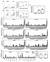
Fig 1
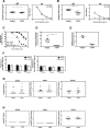
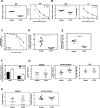
Fig 1. Mapping of the CD4+ T cell response in the LysMCre+Ifnar1fl/fl mouse model of primary ZIKV infection.
Five-week-old LysMCre+Ifnar1fl/fl C57BL/6 mice were infected retro-orbitally with 104 FFU of ZIKV strain MR766 or FSS13025 in 10% FBS/PBS or were mock-infected (10% FBS/PBS alone). Data are the mean ± SEM of n = 4–6 mice/group. (A–C) Splenocytes were removed on day 7 post-infection and analyzed for the percentage of (A) CD44+CD4+ T cells, (B) CD49d+CD11a+ T cells, and (C) granzyme B+CD4+ T cells. (D–F) Splenocytes were removed on day 7 post-infection and stimulated with the indicated ZIKV-derived peptides and brefeldin A. The percentage CD44+CD4+ T cells producing (D) IFNγ, (E) IFNγ and TNF, and (F) IL-2 was measured by ICS. Cells stimulated with DMSO or PMA/ionomycin served as negative and positive controls, respectively. (G) Summary of the data shown in (D–F). (H) In vivo killing of ZIKV peptide-pulsed target cells. LysMCre+Ifnar1fl/fl mice were retro-orbitally mock-infected (n = 4) or infected with 104 FFU ZIKV MR766 (n = 5) and FSS13025 (n = 4) for 7 days, and then injected retro-orbitally with naïve C57BL/6 splenocytes (n = 4) pulsed with a pool of ZIKV peptides (E346-360, E644-658, NS31740-1754, NS4B2480-2494, NS52604-2618, NS52738-2752) or treated with DMSO. After 12 h, the splenocytes were harvested from recipient mice, analyzed by flow cytometry, and the percentage ZIKV-specific cytotoxicity was calculated. *P < 0.05, **P < 0.01 by the Mann–Whitney U test. The production of cytokines after stimulation with each peptide was compared to the negative control (DMSO) using one-way ANOVA t-test. **** P < 0.0001. Data are representative of two independent experiments.
Fig 2
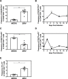
Fig 2. Kinetics of the follicular helper and regulatory CD4+ T cell responses in the LysMCre+Ifnar1fl/fl mouse model of primary ZIKV infection.
Five-week-old LysMCre+Ifnar1fl/fl C57BL/6 mice were infected retro-orbitally with 104 FFU of ZIKV strain FSS13025 or were mock-infected. (A and C) Splenocytes were removed on day 7 post-infection and the percentage (A) CXCR5+PD1+CD44+CD4+ TFH cells or (C) CD25+FoxP3+CD44+CD4+ Treg cells was assessed by flow cytometry. (B and D) Splenocytes were analyzed on the indicated days post-infection for the percentage (B) CXCR5+PD1+CD44+CD4+ TFH cells or (D) CD25+FoxP3+CD44+CD4+ Treg cells. (E) Splenocytes were removed on day 7 post-infection and analyzed for the frequency of IL-10-producing CD44+CD4+ cells. Data are the mean ± SEM of n = 7 (A), n = 4 (B), n = 5 (C), n = 4 (D), n = 5 (E) mock-infected and n = 9 (A), n = 4–6 (B), n = 8 (C), n = 4–6 (D), n = 6 (E) ZIKV-infected mice. **P < 0.01, ***P < 0.001, by the Mann–Whitney U test. For the kinetic analysis, each time point was compared to day 0 using the Mann–Whitney U test, *P < 0.05, ***P < 0.001. Data are representative of two independent experiments.
Fig 3
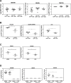
Fig 3. Contribution of CD4+ T cells to Ab and CD8+ T cell responses and to viral control during primary ZIKV infection in LysMCre+Ifnar1fl/fl mice. LysMCre+Ifnar1fl/fl C57BL/6 mice were treated with a depleting anti-CD4 Ab (n = 8) or isotype control Ab (n = 8) on days −3 and −1 prior to and every 2 days after retro-orbital infection with 105 FFU of ZIKV FSS13025.
(A–C) Sera were collected on day 7 post-infection to measure anti-ZIKV IgM (A) and IgG (B) titers by ZIKV E-specific ELISA and (C) ZIKV neutralizing activity using a U937 DC-SIGN cell-based assay with sera from both groups and sera from the anti-CD4 depleted mice group after inactivation of IgM. Data are the mean ± SEM. (D and E) Splenocytes were collected on day 7 post-infection and analyzed by flow cytometry for the percentage CD138+IgD plasma cells (D) or GL7+Fas+ germinal center B cells (E). Data are the mean ± SEM of n = 6 (D) or n = 3 (E) isotype control mice and n = 7 (D) or n = 3 (E) anti-CD4-treated mice. (F) Splenocytes were collected on day 7 post-infection, stimulated with the class I-binding ZIKV peptides PrM169-177, E297-305, or NS52783-2792 and analyzed by flow cytometry for the percentage IFNγ-producing (left) or IFNγ + TNF-producing (right) CD8+ T cells. Data are the mean ± SEM of n = 8 for both isotype control and anti-CD4-treated mice. (G) Serum, brain, and testes were harvested on day 7 post-infection from mice treated with isotype control or depleting anti-CD4 Ab and inoculated with 105 FFU of ZIKV FSS13025, and infectious ZIKV titers were determined using a focus-forming assay. Data are the mean ± SEM of n = 8 (serum and brain), n = 4 (testes) for both isotype control and anti-CD4-treated mice. (H) Serum, brain, and testes were harvested on day 7 post-infection from mice treated with isotype control or depleting anti-CD4 Ab and inoculated with 103 FFU, and infectious ZIKV levels were measured using a focus-forming assay; n = 7 (serum, brain, testes) for both isotype control and anti-CD4-treated mice. *P < 0.05, **P < 0.01, ***P < 0.001 by the Mann–Whitney U test. Data were pooled from two and three independent experiments for high and low viral challenge dose challenge, respectively.
Fig 4
Fig 4. Contribution of memory CD4+ T cells to ZIKV clearance in LysMCre+Ifnar1fl/fl mice.
LysMCre+Ifnar1fl/fl C57BL/6 mice were retro-orbitally infected with 104 FFU of ZIKV strain FSS13025. After 30 days, CD4+ T cells were purified from the spleens and 107 or 1.5 × 10⁷ cells were transferred into naïve LysMCre+Ifnar1fl/fl C57BL/6 mice. One day later, mice were infected retro-orbitally with (A) 105 FFU (n = 9 for serum and brain and n = 4 for testes) or (B) 103 FFU of ZIKV strain FSS13025 (n = 4 for mice receiving CD4+ T cells and n = 5 for control mice receiving no T cells). The indicated organs were isolated 3 days later and infectious ZIKV titers were determined using a focus-forming assay. (C) LysMCre+Ifnar1fl/fl C57BL/6 mice were immunized subcutaneously with a mixture of six immunodominant peptides (n = 5) or DMSO (n = 5) on days −28 and −14, and infected with 105 FFU of ZIKV FSS13025 on day 0. Three days later, the brain and testes were removed and infectious ZIKV titers were determined using a focus-forming assay. (D) LysMCre+Ifnar1fl/fl C57BL/6 mice were infected with 104 FFU of ZIKV FSS13025 on day 0 and treated with CD4-depleting antibody or isotype control Ab on day-3 and day-1 prior to challenge with 103 FFU of ZIKV FSS13025 on day 28 post-priming. Three days later, infectious ZIKV titers in serum, brain, and testes were determined using a focus-forming assay. Data are the mean ± SEM. *P < 0.05, by the Mann–Whitney U test.
Fig 5
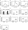
Fig 5. Characterization of the CD4+ T cell response after primary intravaginal ZIKV infection in LysMCre+Ifnar1fl/fl mice.
LysMCre+Ifnar1fl/fl C57BL/6 female mice (8- to 9-week-old) were treated with progesterone and infected via IVag route with 105 FFU of ZIKV FSS13025 3 days later. (A–C) On the indicated days, splenocytes (n = 4–8 mice) were stimulated in vitro with E644-658 FSS13025 peptide and analyzed by flow cytometry for the frequency of CD44+CD4+ T cells producing (A) IFNγ, (B) IFNγ + TNF, and (C) IL-2. (D) As described for (A–C) except cells were harvested from the spleen and iliac lymph nodes (n = 8) on day 10 post-infection, stimulated in vitro, and analyzed for the frequency of CD44+CD4+ T cells producing IFNγ (white bars), IFNγ + TNF (black bars), or IL-2 (gray bars). (E–J) On day 10 post-infection, splenocytes were analyzed for the frequency of (E) CD44+CD4+ T cells, (F) CD49d+CD11a+ cells, and (G) granzyme B+ cells, (H) TFH (CXCR5+PD1+CD44+CD4+) cells, (I) Treg cells (FoxP3+CD25+CD4+CD44+) cells, and (J) IL-10-producing CD44+CD4+ T cells. All experiments were performed twice. Data are the mean ± SEM of n = 3 (E), n = 4 (F), n = 4 (G), n = 4 (H), n = 4 (I), n = 4 (J) mock-infected and n = 4 (E), n = 7 (F), n = 4 (G), n = 7 (H), n = 5 (I), n = 6 (J) ZIKV-infected mice. *P < 0.05, **P < 0.01 by the Mann–Whitney U test.
Fig 6
Fig 6. Contribution of CD4+ T cells to Ab production, CD8+ T cell response, and local viral control during primary intravaginal ZIKV infection of LysMCre+Ifnar1fl/fl mice.
Female LysMCre+Ifnar1fl/fl C57BL/6 mice were treated with a depleting anti-CD4 Ab (n = 7) or isotype control Ab (n = 6) on days −3 and −1 prior to intravaginal infection with 105 FFU of ZIKV FSS13025. Mice were also treated with progesterone on day −3. (A–C) Sera were collected on day 10 post-infection to measure anti-ZIKV IgM (A) and IgG (B) titers by ZIKV E-specific ELISA and neutralizing activity (C) using a U937 DC-SIGN cell-based flow cytometric assay. (D and E) Splenocytes were collected on day 10 post-infection and analyzed by flow cytometry for the percentage plasma cells (CD138+IgD) (D) or germinal center B cells (GL7+Fas+) (E). Data are the mean± SEM of n = 7 (D) or 4 (E) for isotype control mice and n = 6 (D) or 3 (E) for anti-CD4-treated mice. (F) Splenocytes were collected on day 10 post-infection, stimulated with the immunodominant CD8+ T cell ZIKV epitope E297-305, and analyzed by flow cytometry for the percentage IFNγ- or IFNγ + TNF-producing CD8+ T cells. Data are the mean ± SEM of n = 8 mice/group. (G) Infectious ZIKV particles were measured in serum (n = 7, n = 9), vaginal washes (n = 9, n = 9), and the female reproductive tract (FRT) (n = 4, n = 6) on day 10 post-infection for the isotype control or anti-CD4 treated groups. (H) As described for (G) except mice were treated with an anti-CD8 Ab (n = 6) or isotype control Ab (n = 8) before infection, only serum and vaginal washes are represented. *P < 0.05, **P < 0.01 by the Mann–Whitney U test.

Similar articles

Cited by

References

    1. D'Ortenzio E, Matheron S, de Lamballerie X, Hubert B, Piorkowski G, Maquart M, et al. Evidence of Sexual Transmission of Zika Virus. N Engl J Med. 2016. Epub 2016/04/14. 10.1056/NEJMc1604449 . - DOI - PubMed
    1. Freour T, Mirallie S, Hubert B, Splingart C, Barriere P, Maquart M, et al. Sexual transmission of Zika virus in an entirely asymptomatic couple returning from a Zika epidemic area, France, April 2016. Euro Surveill. 2016;21(23). Epub 2016/06/18. 10.2807/1560-7917.ES.2016.21.23.30254 . - DOI - PubMed
    1. Ma W, Li S, Ma S, Jia L, Zhang F, Zhang Y, et al. Zika Virus Causes Testis Damage and Leads to Male Infertility in Mice. Cell. 2016;167(6):1511–24 e10. Epub 2016/11/26. 10.1016/j.cell.2016.11.016 . - DOI - PubMed
    1. Govero J, Esakky P, Scheaffer SM, Fernandez E, Drury A, Platt DJ, et al. Zika virus infection damages the testes in mice. Nature. 2016;540(7633):438–42. Epub 2016/11/01. 10.1038/nature20556 - DOI - PMC - PubMed
    1. Tang WW, Young MP, Mamidi A, Regla-Nava JA, Kim K, Shresta S. A Mouse Model of Zika Virus Sexual Transmission and Vaginal Viral Replication. Cell Rep. 2016;17(12):3091–8. Epub 2016/12/24. 10.1016/j.celrep.2016.11.070 - DOI - PMC - PubMed

Publication types