Review
. 2019 Jan 24:14:293-318.
doi: 10.1146/annurev-pathmechdis-012418-012927.

Malformations of Cerebral Cortex Development: Molecules and Mechanisms

Affiliations
Review

Malformations of Cerebral Cortex Development: Molecules and Mechanisms

Gordana Juric-Sekhar et al. Annu Rev Pathol. .

Abstract

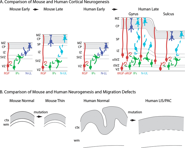
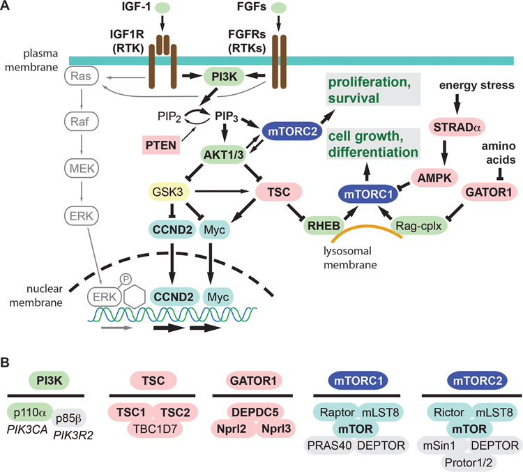
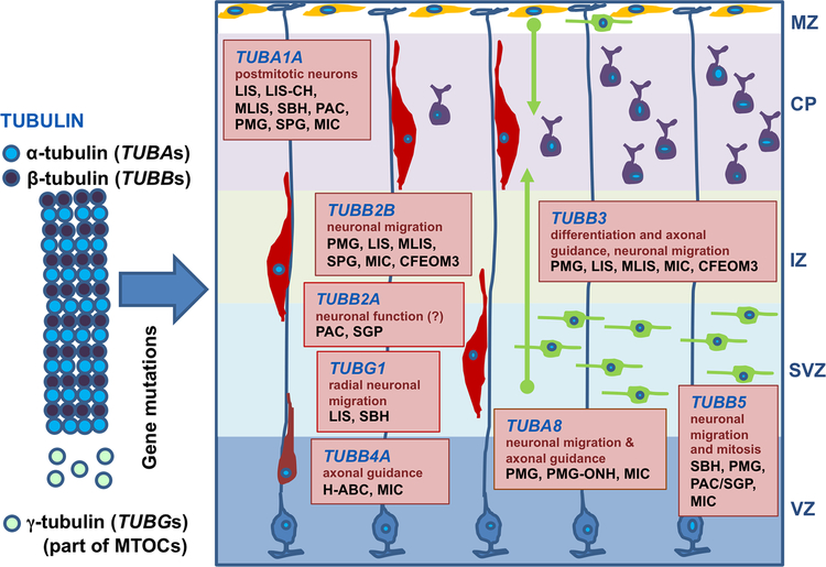
Malformations of cortical development encompass heterogeneous groups of structural brain anomalies associated with complex neurodevelopmental disorders and diverse genetic and nongenetic etiologies. Recent progress in understanding the genetic basis of brain malformations has been driven by extraordinary advances in DNA sequencing technologies. For example, somatic mosaic mutations that activate mammalian target of rapamycin signaling in cortical progenitor cells during development are now recognized as the cause of hemimegalencephaly and some types of focal cortical dysplasia. In addition, research on brain development has begun to reveal the cellular and molecular bases of cortical gyrification and axon pathway formation, providing better understanding of disorders involving these processes. New neuroimaging techniques with improved resolution have enhanced our ability to characterize subtle malformations, such as those associated with intellectual disability and autism. In this review, we broadly discuss cortical malformations and focus on several for which genetic etiologies have elucidated pathogenesis.

Keywords: focal cortical dysplasia; hemimegalencephaly; hippocampal dysgenesis; lissencephaly; microcephaly; polymicrogyria.


Figures

Figure 1.
Figure 1.
Comparison of cortical development and defective neurogenesis in mice and humans. (A) Normal development. In mice, classic RGPs predominate during early (deep-layer) and late (superficial-layer) neurogenesis, and the cortex lacks gyri and sulci. In humans, a transition from classic RGPs to mainly oRGPs occurs between early and late neurogenesis, when gyri and sulci begin to form. The abundance of oRGPs and outer IPs is much higher beneath prospective gyri. (B) Reduced neurogenesis in mice leads to cortical thinning. In humans, reduced neurogenesis is usually associated with impaired gyrification (lissencephaly or pachygyria), and leads to increased cortical thickness. Abbreviations: CP: cortical plate; ctx: cortex; IP: intermediate progenitor; IZ: intermediate zone; LIS: lissencephaly; MZ: marginal zone; N-LL: neuron destined for lower layers; N-UL: neuron destined for upper layers; oRGP: outer radial glial progenitor; PAC: pachygyria; RGP: radial glial progenitor; SVZ: subventricular zone; tRGP: truncated radial glial progenitor; VZ: ventricular zone; wm: white matter.
Figure 2.
Figure 2.
The RTK-PI3K-AKT-mTOR signaling pathway. (A) The pathways is driven by growth factors interacting with their receptors (RTKs), as well as by intracellular metabolic factors including energy status and amino acids. Activators are colored green, and inhibitors red. Molecules known to be linked to brain overgrowth or FCD are indicated by bold type. (B) Subunit composition of key protein complexes linked to brain overgrowth or FCD. The genes encoding p110α (PIK3CA) and p85β (PIK3R2) are also indicated.
Figure 3.
Figure 3.
Patchy mTORC1 activation in a case of FCD-IIa reflects somatic mosaicism. (A) Phospho-S6 immunoreactivity in cortical resection specimen at low magnification. Note local variations in the abundance of immunoreactive cells. (B) Higher magnification shows an area of relatively abundant dysplastic neurons with high phospho-S6 immunoreactivity. (C) An area of sparse dysplastic neurons. (D, E) Two-color immunofluorescence to detect phospho-S6 (red) and NeuN (green) demonstrates that a single dysplastic neuron with high phospho-S6 expression. (F, G) High expression of phospho-S6 in subpial (F) and deep-layer (G) astrocytes is not diagnostically significance, as this may be seen in all forms of epilepsy (Jansen et al., 2015). Scale bar (A): 1 mm. Scale bar (B): 50 µm for B, C; 25 µm for C–F.
Figure 4.
Figure 4.
Tubulinopathies affect multiple processes in cortical development, and cause heterogeneous MCDs. Boxes represent the expression, functions, and MCDs associated with TUBA1A, TUBA8, TUBB2A, TUBB2B, TUBB3, TUBB4A, TUBB5, and TUBG1. Abbreviations: CFEOM3: congenital fibrosis of extraocular muscles type 3; CP: cortical plate; H-ABC: hypomyelination with atrophy of the basal ganglia and cerebellum; IZ: intermediate zone; LIS: lissencephaly; LIS-CH: lissencephaly with cerebellar hypoplasia; MIC: congenital microcephaly; MLIS: microlissencephaly; MTOCs: microtubule organizing centers; MZ: marginal zone (Cajal-Retzius neurons in yellow); PAC: pachygyria; PMG: polymicrogyria; PMG-ONH: polymicrogyria with optic nerve hypoplasia; SBH: subcortical band heterotopia; SGP: simplified gyral pattern; SVZ: subventricular zone; VZ: ventricular zone.

Similar articles

Cited by

References

    1. Bystron I, Blakemore C, Rakic P. 2008. Development of the human cerebral cortex: Boulder Committee revisited. Nat Rev Neurosci 9:110–132 - PubMed
    1. Barkovich AJ, Guerrini R, Kuzniecky RI, Jackson GD, Dobyns WB. 2012. A developmental and genetic classification for malformations of cortical development: update 2012. Brain 135:1348–1369 - PMC - PubMed
    1. Guerrini R, Dobyns W. 2014. Malformations of cortical development: clinical features and genetic causes. Lancet Neurology 13:710–726 - PMC - PubMed
    1. Kuzniecky RI. 1994. Magnetic resonance imaging in developmental disorders of the cerebral cortex. Epilepsia 35 Suppl 6:S44–56 - PubMed
    1. Leventer RJ, Phelan EM, Coleman LT, et al. 1999. Clinical and imaging features of cortical malformations in childhood. Neurology 53:715–722 - PubMed

Publication types

MeSH terms

LinkOut - more resources