. 2019 Apr:69:153-165.
doi: 10.1016/j.meegid.2019.01.022. Epub 2019 Jan 21.

Genetic evolution analysis and pathogenicity assessment of porcine epidemic diarrhea virus strains circulating in part of China during 2011-2017

Affiliations

Genetic evolution analysis and pathogenicity assessment of porcine epidemic diarrhea virus strains circulating in part of China during 2011-2017

Pengfei Chen et al. Infect Genet Evol. 2019 Apr.

Abstract

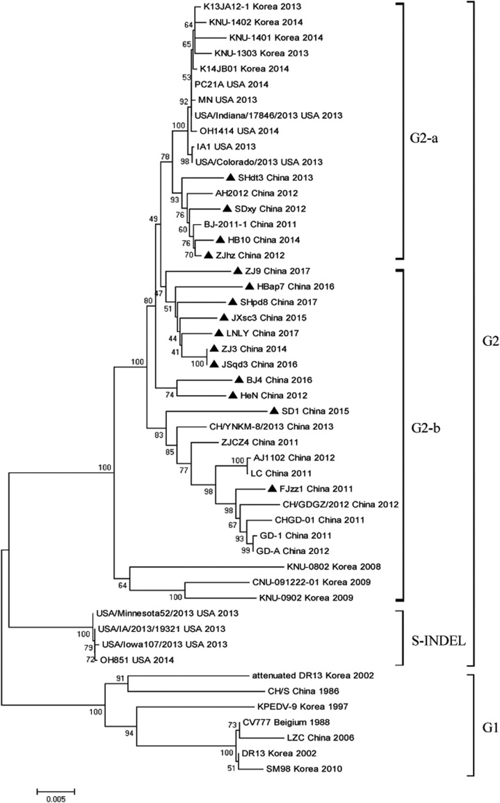
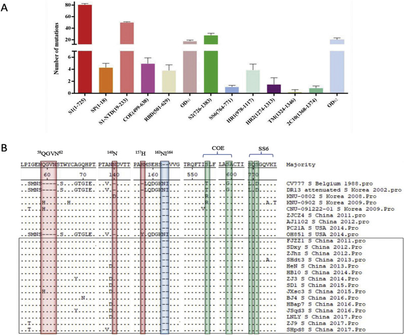
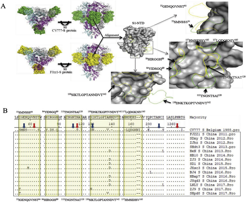
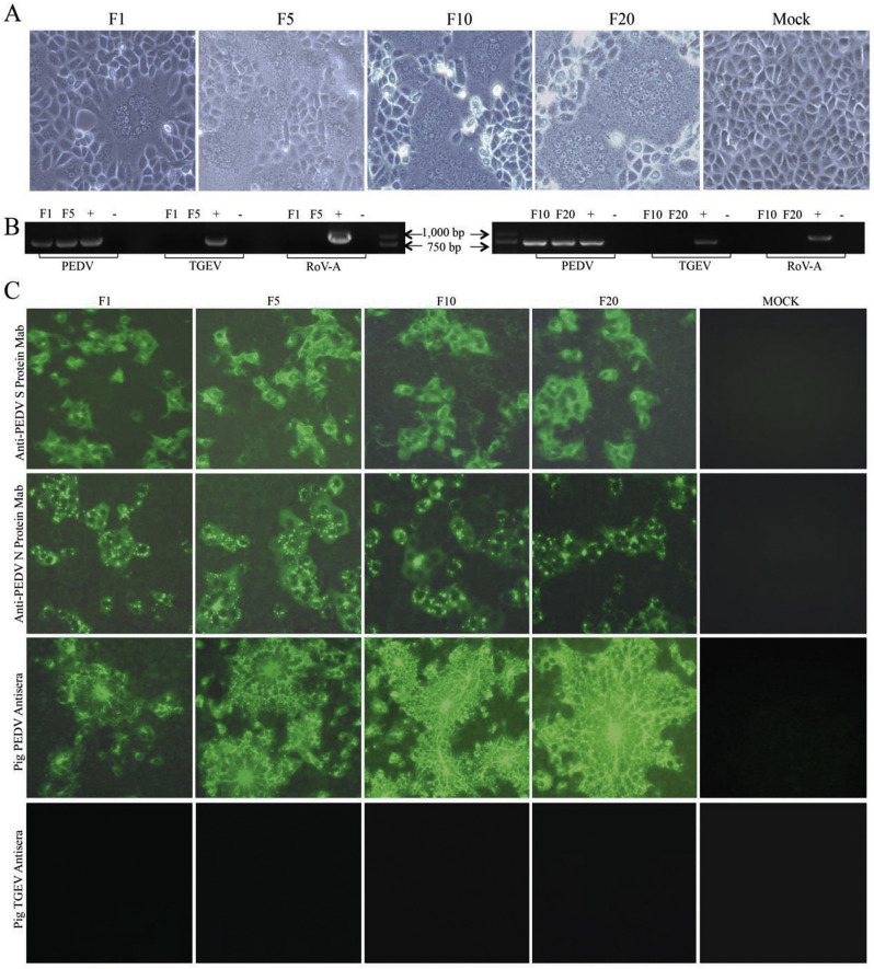
In recent years, the outbreaks of porcine epidemic diarrhea (PED) caused by the highly virulent porcine epidemic diarrhea virus (PEDV) variants occurred frequently in China, resulting in severe economic impacts to the pork industry. In this study, we selected and analyzed the genetic evolution of 15 PEDV representative strains that were identified in fecal samples of diarrheic piglets in 10 provinces and cities during 2011-2017. The phylogenetic analysis indicated that all the 15 PEDV isolates clustered into G2 genotype associated with the current circulating strains. Compared with the genome of the prototype strain CV777, these strains had 103-120 amino acid mutations in their S proteins, most of which were in the N terminal domain of S1 (S1-NTD). We also found 37 common mutations in all these 15 strains, although these strains shared 96.9-99.7% nucleotide homology and 96.3-99.8% amino acid homology in the S protein compared with the other original pandemic strains. Computational analysis showed that these mutations may lead to remarkable changes in the conformational structure and asparagine (N)-linked glycosylation sites of S1-NTD, which may be associated with the altered pathogenicity of these variant PEDV strains. We evaluated the pathogenicity of the PEDV strain FJzz1 in piglets through oral and intramuscular infection routes. Compared with oral infection, intramuscular infection could also cause typical clinical signs but with a slightly delayed onset, confirming that the variant PEDV isolate FJzz1 was highly pathogenic to suckling piglets. In conclusion, we analyzed the genetic variation and pathogenicity of the emerging PEDV isolates of China, indicating that G2 variant PEDV strains as the main prevalent strains that may mutate continually. This study shows the necessity of monitoring the molecular epidemiology and the etiological characteristics of the epidemic PEDV isolates, which may help better control the PED outbreaks.

Keywords: FJzz1; PEDV; Pathogenicity; S gene; Variation.


Figures

Fig. 1
Fig. 1
Phylogenetic analysis. The phylogenetic tree was generated based on the complete S gene of 60 PEDV reference strains and 15 isolates in our study by neighbor-joining method of MEGA 6.0 with 1000 bootstrap replicates. The PEDV strains studied in this research were marked with black triangle.
Fig. 2
Fig. 2
Analysis of amino acid mutations in the S protein. (A) The number of amino acid mutations in the S protein of different domains including the S1 subunit (residue 1–725), the S2 subunit (residue 726–1383), the signal peptide (SP, residues1–18), the N-terminal domain of S1 (S1-NTD, residue 19–233), the neutralizing epitopes (COE, residues 499–638; SS6, residues 764–771; 2C10, residues 1368–1374), two heptad repeat regions (HR1, residues 978–1117 and HR2, residues 1274–1313), the transmembrane domain (TM, residues 1324–1346) and the other domains of both S1 and S2 (ODS1, ODS2)were counted. (B)The S protein amino acid sequences of 24 PEDV strains were aligned using Clustal W method. The insertion and deletion areas were highlighted in red and blue, respectively. The substitutions in neutralizing epitopes were highlighted in green. The PEDV strains studied in this research were marked with black box. (For interpretation of the references to colour in this figure legend, the reader is referred to the web version of this article.)
Fig. 3
Fig. 3
Analysis of the 3D structural models and the predicted N-linked glycosylation sites of CV777 and FJzz1 in the S protein. (A) The predicted S protein 3D structural models of CV777 and FJzz1 were generated and observed on the top and front sides. The three monomers that form the glycoprotein trimer were colored in grey, magenta and cyan. The S1-NTDs of CV777 and FJzz1 were shown as ‘surface’ in green and yellow, respectively. Also, the structural alignment of S1-NTDs of these two proteins was carried out. The residues with no obvious structural variations were displayed as ‘surface’ in grey, while the residues with most obvious structural variations were shown as ‘ribbon’ in green for CV777 and yellow for FJzz1. (B) The S protein amino acid sequences of 15 PEDV strains in this study and the classical CV777 strain were aligned as described above. The residues with most obvious structural variations described in Fig. 3A were highlighted in yellow, while the predicted N-linked glycosylation sites of CV777 and our 15 strains were marked by blue and red arrows, respectively. The PEDV strains studied in this research were marked with black box. (For interpretation of the references to colour in this figure legend, the reader is referred to the web version of this article.)
Fig. 4
Fig. 4
Detection of FJzz1 in Vero cells. (A) The CPE of FJzz1 F1, F5, F10 and F20 at 24hpi was observed. (B) The cell culture supernatants of FJzz1 F1, F5, F10 and F20 were examined by RT-PCR using the primers specific for PEDV, TGEV and RoV-A. (C) Also, the monolayers of Vero cells inoculated with FJzz1 F1, F5, F10 and F20 were tested by IFA using MAb against PEDV S protein, MAb against PEDV N protein, pig PEDV antisera and pig TGEV antisera.
Fig. 5
Fig. 5
In vitro characterization of FJzz1. (A) The negative-staining EM image of FJzz1 F5 virus particles. (B) Multi-step growth curve of FJzz1 from different passages (F5, F10 and F20) on Vero cells at an MOI of 0.01. (C) The crystal violet stained plaques formed in the monolayers of Vero cells inoculated with FJzz1 from different passages (F5, F10 and F20) at 3 days post infection. (For interpretation of the references to colour in this figure legend, the reader is referred to the web version of this article.)
Fig. 6
Fig. 6
Pathogenicity analysis of FJzz1. (A) The average body weight changes in each group. (B) Fecal scores of piglets in different groups. Evaluation standard: 0 = normal; 1 = soft; 2 = semi-fluid; 3 = watery diarrhea. (C) The survival rate of piglets in each group. The survival curves of piglets infected with FJzz1 by oral inoculation or muscular injection are shown respectively. (D) The changes in average body temperature of each group within the first 14 dpi. Asterisk (*) indicates a significant difference between Group A and Group C (* p < .05; ** p < .01; *** p < .001). Pound (#) indicates a significant difference between Group B and Group C (# p < .05; ## p < .01; ### p < .001).
Fig. 7
Fig. 7
Virus shedding in feces and quantification of viral load in different intestine segments. (A) Virus shedding in feces. Daily virus shedding in feces of different groups as measured by real-time RT-PCR detection of virus genome in fecal swabs. (B) Quantification of viral load in different parts of intestine by TaqMan real-time RT-PCR targeting PEDV N gene. Labels without the same letters indicate significant differences. .
Fig. 8
Fig. 8
Gross lesions of piglets and histopathologic examination of intestines. (A) Gross lesions of piglets of each group. The intestinal lesions were examined at the day of death or at the final time points. (B) Histopathologic examination of jejunum. Jejunums collected from each group were processed for HE staining. (C) Immunohistochemical detection. Jejunum of each groups was stained with PEDV monoclonal antibody against spike protein (1:100 dilution).

Similar articles

Cited by

References

    1. Arnold K., Bordoli L., Kopp J., Schwede T. The SWISS-MODEL workspace: a web-based environment for protein structure homology modelling. Bioinformatics. 2006;22:195–201. - PubMed
    1. Beall A., Yount B., Lin C.M., Hou Y., Wang Q., Saif L., Baric R. Characterization of a pathogenic full-length cDNA clone and transmission model for porcine epidemic diarrhea virus strain PC22A. MBio. 2016;7 (e01451–01415) - PMC - PubMed
    1. Ben Salem A.N., Chupin Sergei A., Bjadovskaya Olga P., Andreeva Olga G., Mahjoub A., Prokhvatilova Larissa B. Multiplex nested RT-PCR for the detection of porcine enteric viruses. J. Virol. Methods. 2010;165:283–293. - PMC - PubMed
    1. Benkert P., Biasini M., Schwede T. Toward the estimation of the absolute quality of individual protein structure models. Bioinformatics. 2011;27:343–350. - PMC - PubMed
    1. Biasini M., Bienert S., Waterhouse A., Arnold K., Studer G., Schmidt T., Kiefer F., Gallo Cassarino T., Bertoni M., Bordoli L., Schwede T. SWISS-MODEL: modelling protein tertiary and quaternary structure using evolutionary information. Nucleic Acids Res. 2014;42:W252–W258. - PMC - PubMed

Publication types

MeSH terms

LinkOut - more resources