. 2019 Jan 23;20(3):478.
doi: 10.3390/ijms20030478.

Ganoderma lucidum Polysaccharides Prevent Palmitic Acid-Evoked Apoptosis and Autophagy in Intestinal Porcine Epithelial Cell Line via Restoration of Mitochondrial Function and Regulation of MAPK and AMPK/Akt/mTOR Signaling Pathway

Affiliations

Ganoderma lucidum Polysaccharides Prevent Palmitic Acid-Evoked Apoptosis and Autophagy in Intestinal Porcine Epithelial Cell Line via Restoration of Mitochondrial Function and Regulation of MAPK and AMPK/Akt/mTOR Signaling Pathway

Zengenni Liang et al. Int J Mol Sci. .

Abstract

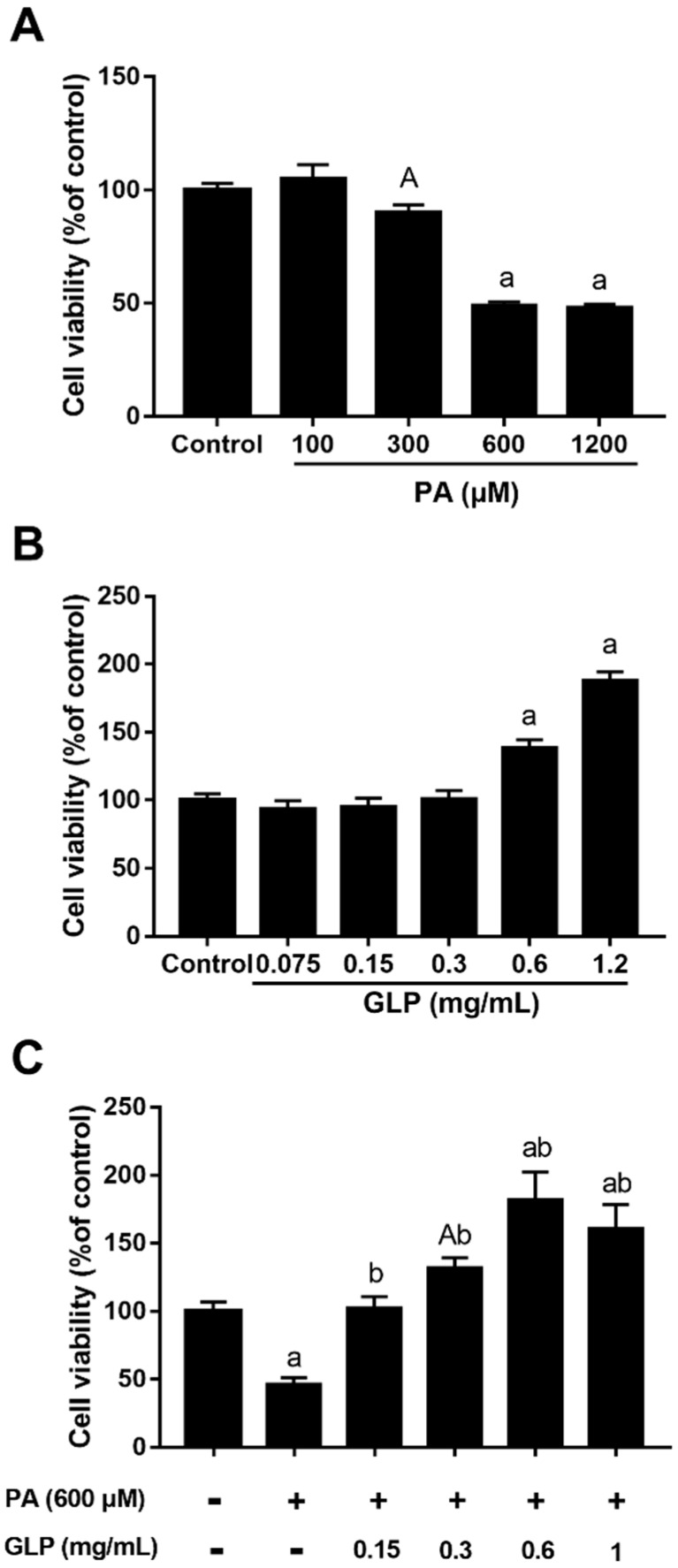
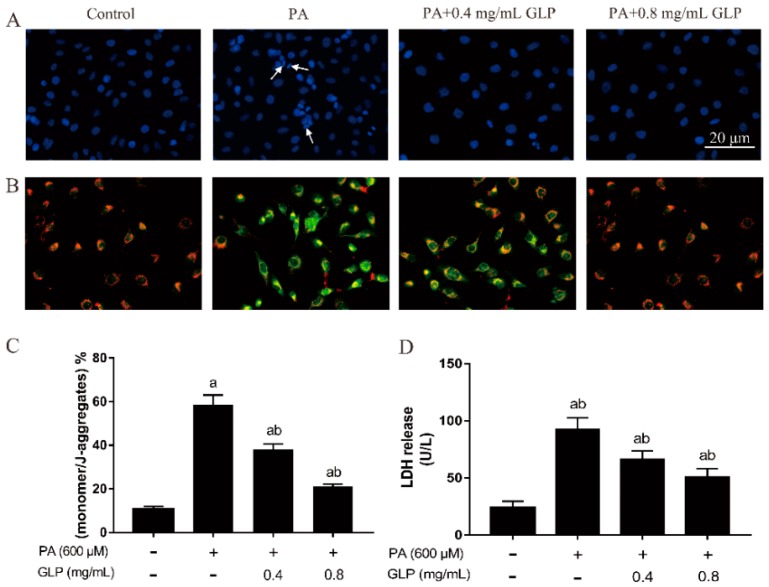
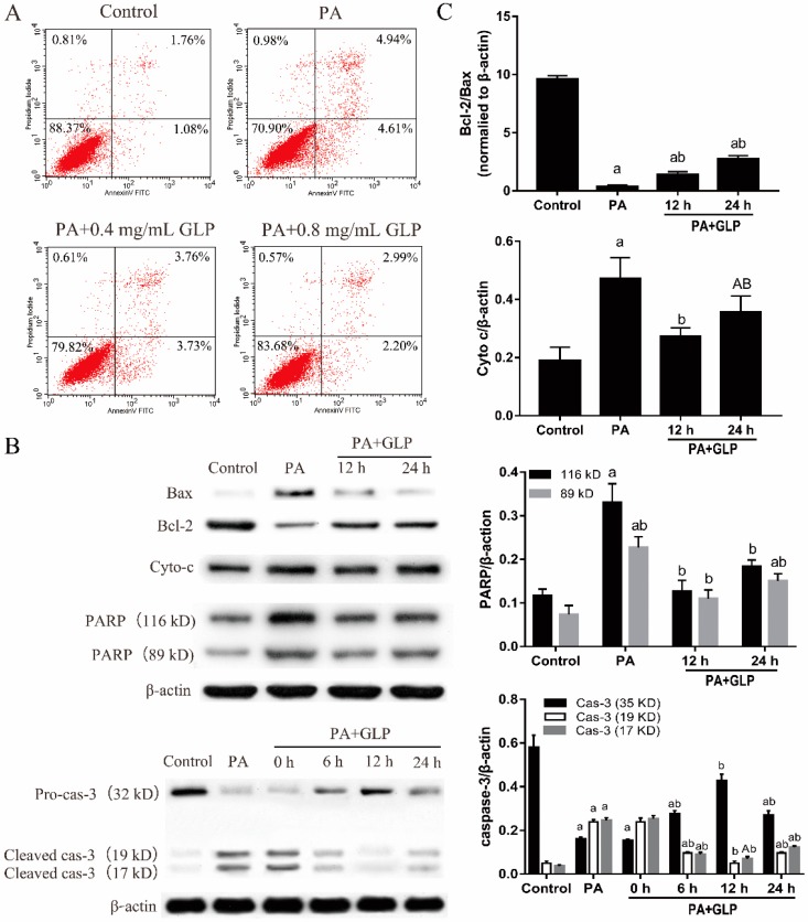
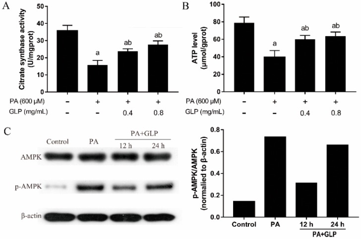
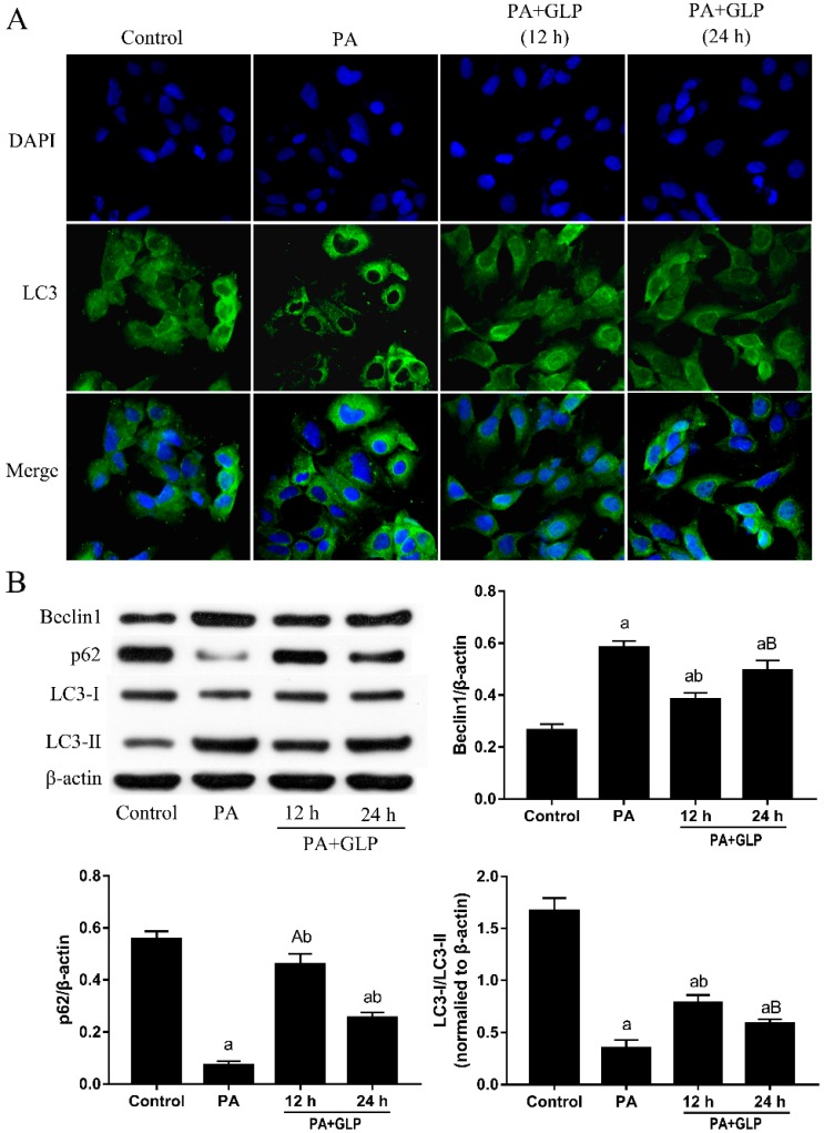
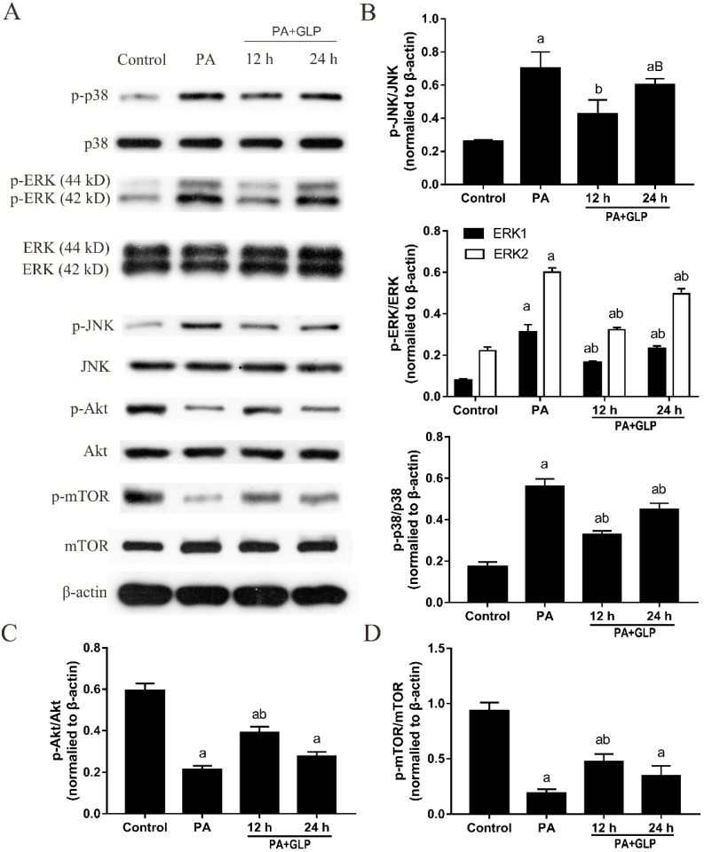
Ganoderma lucidum polysaccharide (GLP) extracted from Ganoderma lucidum (Leyss. ex Fr.) Karst, a traditional Chinese medicine, is a biologically active substance reported to possess anti-oxidative, anti-apoptotic, and neurological protection. However, it is unknown whether GLP have any protective effect against high-fat constituents-induced epithelial cell injury. The aim of this study was to investigate the protection and molecular mechanism of GLP on injury induced by palmitic acid (PA) in the intestinal porcine epithelial cell line (IPEC-J2). First, we tested whether the treatment of GLP attenuate PA-induced IPEC-J2 cell death. GLP markedly blocked PA-caused cytotoxicity and apoptosis in IPEC-J2 cells. Moreover, GLP recovered the decreased mitochondrial function and inhibited activation of caspase-dependent apoptotic pathway. Interestingly, PA promoted cell apoptosis and autophagy through stimulation of phosphorylation of mitogen-activated protein kinases (MAPKs), AMP-activated protein kinase (AMPK), and inhibition of phosphorylation of Akt and mammalian target of rapamycin (mTOR), which was reversed by GLP. Taken together, this study revealed a protective effect of GLP against PA-evoked IPEC-J2 cell death through anti-apoptotic and anti-autophagic properties.

Keywords: Ganoderma lucidum; apoptosis; autophagy; mitochondria; polysaccharides.


Conflict of interest statement

The authors declare no conflict of interest. The funders had no role in the design of the study; in the collection, analyses, or interpretation of data; in the writing of the manuscript, or in the decision to publish the results.

Figures

Figure 1
Figure 1
MTT assay determined the effects of palmitic acid (PA) and Ganoderma lucidum polysaccharide (GLP) on IPEC-J2 cell viability. Cells were treated with a 1640 medium containing 10% FBS (control), various concentrations of PA or/and GLP for 24 h. (A) Dose-dependent inhibitory effect of PA on IPEC-J2 cell viability. (B) The effect of various concentrations GLP (0.075–1.2 mg/mL) on IPEC-J2 cell viability. (C) The protective effect of GLP on PA-induced cell viability loss. Values are expressed as percentages of control and are as mean ± SE for three independent experiments (n = 5). A < 0.05 and a < 0.01 vs. control, b < 0.01 vs. PA alone.
Figure 2
Figure 2
The effects of GLP on apoptotic characteristics in PA-induced IPEC-J2 cells. Cells were exposed to 600 μM PA with or without 0.4 and 0.8 mg/mL of GLP for 24 h. (A) Representative images of 4’,6-diamidi-no-2-phenylindole (DAPI) staining (blue). Arrows denote chromatin condensation and fragmentation. Original magnification 400×. (B) The mitochondrial membrane potential was examined by JC-1 staining. JC-1 aggregate and JC-1 monomer exhibited red and green fluorescence, respectively (400×). (C) JC-1 staining quantification results are presented as the mean of monomer/J-aggregates ± SE. (D) The release of LDH was analyzed using a commercially available kit and calculated as U/L. Data were presented as mean ± SE for three independent experiments (n = 3), a < 0.01 vs. control, b < 0.01 vs. PA alone.
Figure 3
Figure 3
GLP prevented PA-induced apoptosis in IPEC-J2 cells. Cells were exposed to 0.4 and 0.8 mg/mL GLP in presence of 600 μM PA for 24 h. (A) Quantification of IPEC-J2 apoptotic cells by flow cytometry. Four parts were observed: necrotic cells (upper left quadrants), late apoptotic and necrotic cells (upper right quadrants), viable cells (lower left quadrants), and early apoptotic cells (lower right quadrant). (B) The expressions of Bax, Bcl-2, Cyto c, caspase-3 (cas-3), and PARP were analyzed by Western blot. (C) The data were quantified and normalized to β-actin. Data shown are representative of three independent experiments (n = 3). A < 0.05 and a < 0.01 vs. control, B < 0.05 and b < 0.01 vs. PA alone.
Figure 4
Figure 4
The effects GLP and PA on mitochondrial energy metabolism in IPEC-J2 cells. Cells were treated with 600 μM PA or/and GLP (0.4 and 0.8 mg/mL) for 24 h. (A) Citrate synthase activity was assayed using a citrate synthase assay kit and expressed as U/mg protein (n = 5). (B) Mitochondrial proteins were extracted using a mitochondria isolation kit after cells were lysed. The ATP level was determined by bioluminescence measurement and expressed as μmol/g protein (n = 5). (C) Representative Western blot of AMPK and p-AMPK. The data were quantified and normalized to β-actin. Data were presented as mean ± SE for three independent experiments, a < 0.01 vs. control, b < 0.01 vs. PA alone.
Figure 5
Figure 5
GLP ameliorated PA-induced autophagy in IPEC-J2 cells. Cells were treated with 600 μM PA and GLP (0.4 and 0.8 mg/mL) for 24 h. (A) Representative LC3/DAPI images. Cells were stained for LC3 (green) and DAPI (blue), and the fluorescence was visualized in a fluorescence microscope. Original magnification 400×. (B) The protein expressions of Beclin1, p62, LC3-I and LC3-II were subjected to Western blot analysis using indicating antibodies. The data were quantified and normalized to β-actin. Data shown are representative of three independent experiments (n = 3). A < 0.05 and a < 0.01 vs. control, B < 0.05 and b < 0.01 vs. PA alone.
Figure 6
Figure 6
GLP activated Akt/mTOR signaling pathway and suppressed MAPK pathway in PA-induced IPEC-J2 cells. (A) Western blotting was used to analyze the protein expressions. (B) p-JNK/JNK, p-ERK/ERK, p-p38/p38, (C) p-Akt/Akt, and (D) p-mTOR/mTOR were calculated and presented. The data were quantified and normalized to β-actin. Data were presented as mean ± SE for three independent experiments (n = 3), a < 0.01 vs. control, B < 0.05 and b < 0.01 vs. PA alone.

Similar articles

Cited by

References

    1. Moreira A.P.B., Texeira T.F.S., Ferreira A.B., Peluzio M.C.G., Alfenas R.C.G. Influence of a high-fat diet on gut microbiota, intestinal permeability and metabolic endotoxaemia. Br. J. Nutr. 2012;108:801–809. doi: 10.1017/S0007114512001213. - DOI - PubMed
    1. Akbari P., Fink-Gremmels J., Willems R.H.A.M., Difilippo E., Schols M.H.C., Garssen J., Braber S. Characterizing microbiota-independent effects of oligosaccharides on intestinal epithelial cells: Insight into the role of structure and size. Eur. J. Nutr. 2017;56:1919–1930. doi: 10.1007/s00394-016-1234-9. - DOI - PMC - PubMed
    1. Nguyen H.T.T., Dalmasso G., Müller S., Carrière J., Seibold F., Darfeuille-Michaud A. Crohn’s disease-associated adherent invasive escherichia coli modulate levels of microRNAs in intestinal epithelial cells to reduce autophagy. Gastroenterology. 2014;146:508–519. doi: 10.1053/j.gastro.2013.10.021. - DOI - PubMed
    1. Peterson L.W., Artis D. Intestinal epithelial cells: Regulators of barrier function and immune homeostasis. Nat. Rev. Immunol. 2014;14:141–153. doi: 10.1038/nri3608. - DOI - PubMed
    1. Elkahoui S., Bartley G.E., Yokoyama W.H., Friedman M. Dietary supplementation of potato peel powders prepared from conventional and organic russet and non-organic gold and red potatoes reduces weight gain in mice on a high-fat diet. J. Agric. Food Chem. 2018;66:6064–6072. doi: 10.1021/acs.jafc.8b01987. - DOI - PubMed

MeSH terms

LinkOut - more resources