. 2019 Jan 23;8(2):135.
doi: 10.3390/jcm8020135.

Elevated Serum Melatonin under Constant Darkness Enhances Neural Repair in Spinal Cord Injury through Regulation of Circadian Clock Proteins Expression

Affiliations

Elevated Serum Melatonin under Constant Darkness Enhances Neural Repair in Spinal Cord Injury through Regulation of Circadian Clock Proteins Expression

Yunkyung Hong et al. J Clin Med. .

Abstract

We investigated the effects of environmental lighting conditions regulating endogenous melatonin production on neural repair, following experimental spinal cord injury (SCI). Rats were divided into three groups randomly: the SCI + L/D (12/12-h light/dark), SCI + LL (24-h constant light), and SCI + DD (24-h constant dark) groups. Controlled light/dark cycle was pre-applied 2 weeks before induction of spinal cord injury. There was a significant increase in motor recovery as well as body weight from postoperative day (POD) 7 under constant darkness. However, spontaneous elevation of endogenous melatonin in cerebrospinal fluid was seen at POD 3 in all of the SCI rats, which was enhanced in SCI + DD group. Augmented melatonin concentration under constant dark condition resulted in facilitation of neuronal differentiation as well as inhibition of primary cell death. In the rostrocaudal region, elevated endogenous melatonin concentration promoted neural remodeling in acute phase including oligodendrogenesis, excitatory synaptic formation, and axonal outgrowth. The changes were mediated via NAS-TrkB-AKT/ERK signal transduction co-regulated by the circadian clock mechanism, leading to rapid motor recovery. In contrast, exposure to constant light exacerbated the inflammatory responses and neuroglial loss. These results suggest that light/dark control in the acute phase might be a considerable environmental factor for a favorable prognosis after SCI.

Keywords: circadian clock; endogenous melatonin; environmental lighting condition; neural remodeling; spinal cord injury.


Conflict of interest statement

The authors declare no conflict of interest.

Figures

Figure 1
Figure 1
Experimental scheme of the study. Experimental animals were randomly divided into 3 groups: 12/12-h light/dark (L/D), 24-h constant light (LL), and 24-h constant dark (DD). The animals were sacrificed 3, 7, 14, and 28 days after injury for further analyses.
Figure 2
Figure 2
Spontaneous motor recovery of spinal cord injured animals and time-dependent changes of endogenous cerebrospinal fluid (CSF) melatonin. (A) There were significant differences in the changes of body weight among the spinal cord injury (SCI) groups were observed. Animals caged in the constant dark condition showed greater body weight from postoperative days (POD) 7 compared to other groups; (B) Spontaneous behavioral recovery was suppressed under LL condition, but DD condition enhanced motor function after the seven postoperative days (POD) compared to natural light/dark cycle; (C) Cerebrospinal fluid (CSF) was collected between ZT13 and ZT15 under the light/dark cycle. Endogenous CSF melatonin was more concentrated at POD 3 under DD condition, but the mean value did not differ thereafter. L/D, 12/12-h light/dark; LL, 24-h constant light; DD, 24-h constant dark. a p < 0.05, vs. L/D; b p < 0.05, vs. LL.
Figure 3
Figure 3
The neuroprotective effects of elevated melatonin on the primary injury region through regulation of anti-inflammatory and anti-oxidant genes. The damaged segments of spinal cord (T9-11) were analyzed at POD 3. (A) LL condition exacerbated the histological deterioration of the primary lesion, where Iba1 expression (microglial marker) was upregulated. DD condition not only protected injured tissue but also increased the levels of endogenous pluripotency markers (Nestin, Vimentin, Oct4), neuron (NeuN), and oligodendrocytes (Olig2). Magnification = ×200; Scale bar = 40 μm; (B) Immunoblots demonstrate that activation of apoptotic machinery was reduced under DD condition, which was mediated through regulation of inflammatory (Tnfα) and anti-oxidant molecules (Catalase, Sod2). L/D, 12/12-h light/dark; LL, 24-h constant light; DD, 24-h constant dark. a p < 0.05, vs. L/D; b p < 0.05, vs. LL.
Figure 3
Figure 3
The neuroprotective effects of elevated melatonin on the primary injury region through regulation of anti-inflammatory and anti-oxidant genes. The damaged segments of spinal cord (T9-11) were analyzed at POD 3. (A) LL condition exacerbated the histological deterioration of the primary lesion, where Iba1 expression (microglial marker) was upregulated. DD condition not only protected injured tissue but also increased the levels of endogenous pluripotency markers (Nestin, Vimentin, Oct4), neuron (NeuN), and oligodendrocytes (Olig2). Magnification = ×200; Scale bar = 40 μm; (B) Immunoblots demonstrate that activation of apoptotic machinery was reduced under DD condition, which was mediated through regulation of inflammatory (Tnfα) and anti-oxidant molecules (Catalase, Sod2). L/D, 12/12-h light/dark; LL, 24-h constant light; DD, 24-h constant dark. a p < 0.05, vs. L/D; b p < 0.05, vs. LL.
Figure 4
Figure 4
Constant dark condition accelerates excitatory synapse formation and axonal outgrowth in the rostral region via TrkB-ERK signaling pathway. The mid-thoracic segments (T6-8) were analyzed at POD 3. (A) The animals under LL condition showed hemorrhage in dorsal white column, interrupted neurite outgrowth, and massive loss of immature (DCX) and mature neurons (NeuN) after spinal cord injury. The DD condition promoted not only excitatory synaptic formation (PSD-95, nNOS, Nr2a) but also axonal outgrowth (Tau) compared with the L/D group. VC, ventral commissure; Magnification = ×200; Scale bar = 40 μm; (B) TrkB-mediated ERK signal transduction was enhanced under DD condition, while LL condition decreased their activation in spite of the higher level of BDNF. Transcriptional regulation of Aanat gene was affected by the light/dark condition; (C) DD condition increased the core clock proteins (CKOCK, BMAL1, nuclear PER1) as well as mRNA (Per1) in the rostral region. The level of melatonin receptor 1A (MT1) and 1B (MT2) was greater in the SCI + DD group than those of other groups. L/D, 12/12-h light/dark; LL, 24-h constant light; DD, 24-h constant dark. a p < 0.05 vs. L/D; b p < 0.05 vs. LL.
Figure 5
Figure 5
Constant dark condition preserves excitatory circuits at the acute phase in the lumbar enlargement. The spinal segments caudal to the injury epicenter (L1-2) were analyzed at POD 3. (A) LL condition led to the reduction of the neuroglial markers (GFAP, NeuN) as well as extensive tissue damage in the secondary lesion. Both PSD-95 and Olig2 proteins were augmented when DD condition was applied. Neuronal degeneration found in histological examination could be an evidence supporting these results. VC, ventral commissure; Magnification = ×200; Scale bar = 40 μm; (B) Increased Aanat expression under DD condition activated TrkB-ERK signal transduction, but its repression under LL condition inhibited AKT-dependent intracellular signal; (C) LL condition decreased Per1 expression, nuclear distribution of PER1 protein, and glycosylated MT1 level, which was preserved under DD condition. L/D, 12/12-h light/dark; LL, 24-h constant light; DD, 24-h constant dark. a p < 0.05 vs. L/D; b p < 0.05 vs. LL.
Figure 5
Figure 5
Constant dark condition preserves excitatory circuits at the acute phase in the lumbar enlargement. The spinal segments caudal to the injury epicenter (L1-2) were analyzed at POD 3. (A) LL condition led to the reduction of the neuroglial markers (GFAP, NeuN) as well as extensive tissue damage in the secondary lesion. Both PSD-95 and Olig2 proteins were augmented when DD condition was applied. Neuronal degeneration found in histological examination could be an evidence supporting these results. VC, ventral commissure; Magnification = ×200; Scale bar = 40 μm; (B) Increased Aanat expression under DD condition activated TrkB-ERK signal transduction, but its repression under LL condition inhibited AKT-dependent intracellular signal; (C) LL condition decreased Per1 expression, nuclear distribution of PER1 protein, and glycosylated MT1 level, which was preserved under DD condition. L/D, 12/12-h light/dark; LL, 24-h constant light; DD, 24-h constant dark. a p < 0.05 vs. L/D; b p < 0.05 vs. LL.
Figure 6
Figure 6
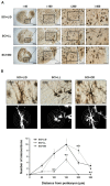
Golgi-cox stained sections of spinal cord from the rats subjected to controlled light/dark cycle. (A) Experimental animals were kept in controlled light/dark cycle; L/D, LL, and DD. A total of 3 days after SCI surgery, histological differences in the injured spinal segment were investigated. Magnification = ×4, ×100, ×200, ×400; Scale bar = 500 μm (×40, ×100), 200 μm (×200, ×400). (B) Neurite outgrowth of the neurons in Golgi-stained spinal sections was quantified using Sholl analysis method. The Sholl rings are separated by 10 μm. The data were expressed as mean ± SD. L/D, 12/12-h light/dark; LL, 24-h constant light; DD, 24-h constant dark. a p < 0.05 vs. L/D; b p < 0.05 vs. LL; Magnification = ×1000; Scale bar = 100 μm.
Figure 7
Figure 7
Schematic diagram summarizing our results. (A) Different light/dark conditions influenced the level of endogenous melatonin distributed in CSF, which might regulate the clock-driven neural remodeling processes at the acute phase. Time required for neural repair might be shortened under DD condition, but LL condition delayed the recovery speed; (B) Early elevated endogenous melatonin might act on the rostral (T6-8) and caudal penumbra (L1-2) as well as the injury epicenter (T9-11). Increased melatonin secretion under DD condition elevated neuronal and glial population, and exerted the anti-inflammatory and anti-oxidative actions, which might have protective role against primary cell death in the epicenter. Early dark condition enhanced clock-controlled Aanat expression in the rostral and caudal penumbra, leading to activation of NAS-TrkB-AKT/ERK signal transduction pathway. These might influence post-SCI neural remodeling including excitatory neurotransmission as well as axonal sprouting, followed by advance in motor recovery, ultimately.

Similar articles

Cited by

References

    1. Silva N.A., Sousa N., Reis R.L., Salgado A.J. From basics to clinical: A comprehensive review on spinal cord injury. Prog. Neurobiol. 2014;114:25–57. doi: 10.1016/j.pneurobio.2013.11.002. - DOI - PubMed
    1. Oyinbo C.A. Secondary injury mechanisms in traumatic spinal cord injury: A nugget of this multiply cascade. Acta Neurobiol. Exp. 2011;71:281–299. - PubMed
    1. Donnelly D.J., Popovich P.G. Inflammation and its role in neuroprotection, axonal regeneration and functional recovery after spinal cord injury. Exp. Neurol. 2008;209:378–388. doi: 10.1016/j.expneurol.2007.06.009. - DOI - PMC - PubMed
    1. Abematsu M., Tsujumura K., Yamano M., Saito M., Kohno K., Kohyama J., Namihira M., Komiya S., Nakashima K. Neurons derived from transplanted neural stem cells restore disrupted neuronal circuitry in a mouse model of spinal cord injury. J. Clin. Investig. 2010;120:3255–3266. doi: 10.1172/JCI42957. - DOI - PMC - PubMed
    1. Coutts M., Keirstead H.S. Stem cells for the treatment of spinal cord injury. Exp. Neurol. 2008;209:368–377. - PubMed

LinkOut - more resources