. 2019 Jan 22;11(2):214.
doi: 10.3390/nu11020214.

Toll-Like Receptor-Dependent Immunomodulatory Activity of Pycnogenol®

Affiliations

Toll-Like Receptor-Dependent Immunomodulatory Activity of Pycnogenol®

Annelies Verlaet et al. Nutrients. .

Abstract

Background: Pycnogenol® (PYC), an extract of French maritime pine bark, is widely used as a dietary supplement. PYC has been shown to exert anti-inflammatory actions via inhibiting the Toll-like receptor 4 (TLR4) pathway. However, the role of the other receptors from the TLR family in the immunomodulatory activity of PYC has not been described so far.

Aim: The aim of this study was to investigate whether PYC might exert its immunomodulatory properties through cell membrane TLRs (TLR1/2, TLR5, and TLR2/6) other than TLR4. Moreover, the effect of gastrointestinal metabolism on the immunomodulatory effects of PYC was investigated.

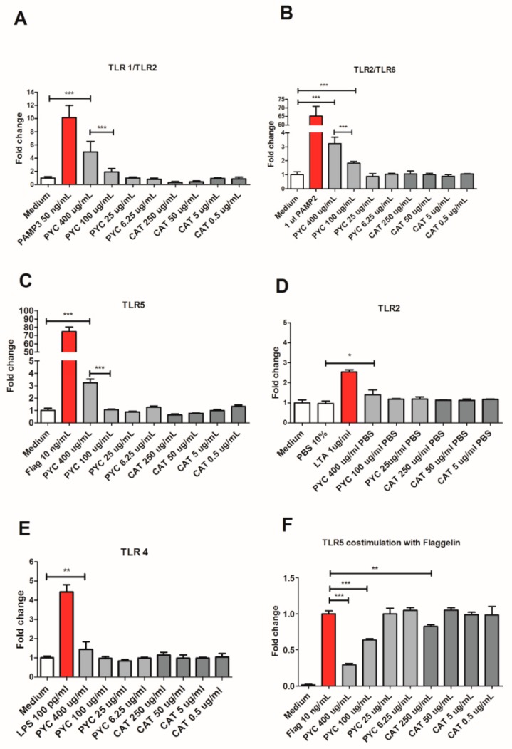
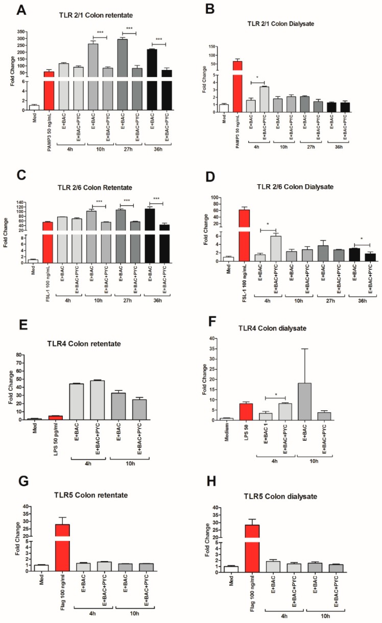
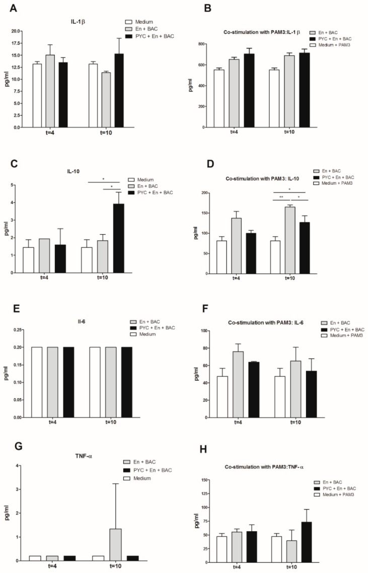
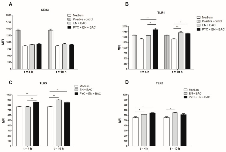
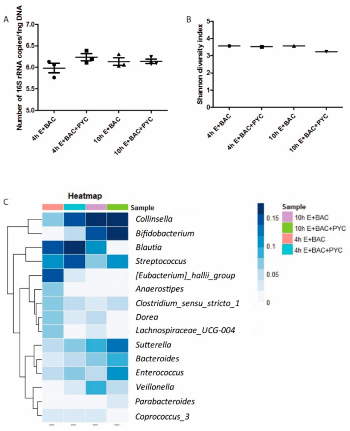
Findings: We showed that intact non-metabolized PYC dose-dependently acts as an agonist of TLR1/2 and TLR2/6 and as a partial agonist of TLR5. PYC on its own does not agonize or antagonize TLR4. However, after the formation of complexes with lipopolysaccharides (LPS), it is a potent activator of TLR4 signaling. Gastrointestinal metabolism of PYC revealed the immunosuppressive potential of the retentate fraction against TLR1/2 and TLR2/6 when compared to the control fraction containing microbiota and enzymes only. The dialyzed fraction containing PYC metabolites revealed the capacity to induce anti-inflammatory IL-10 secretion. Finally, microbially metabolized PYC affected the colonic microbiota composition during in vitro gastrointestinal digestion.

Conclusions: This study showed that gastrointestinal metabolism of PYC reveals its biological activity as a potential inhibitor of TLRs signaling. The results suggest that metabolized PYC acts as a partial agonist of TLR1/2 and TLR2/6 in the presence of the microbiota-derived TLR agonists (retentate fraction) and that it possesses anti-inflammatory potential reflected by the induction of IL-10 from THP-1 macrophages (dialysate fraction).

Keywords: Pycnogenol®; Toll-like receptors; catechin; gastrointestinal metabolism; immunomodulation; metabolites; partial agonist.


Conflict of interest statement

The authors declare no conflict of interest.

Figures

Figure 1
Figure 1
Fingerprint chromatogram. (A) 20 mg/mL PYC (Pycnogenol®) reference in 10% MeOH (methanol). (B) 50 mg/mL PYC in PBS (Phosphate Buffered Saline).
Figure 2
Figure 2
PYC extract contains non-detectable amounts of LPS and does not influence the viability of Human Embryonic Kidney (HEK)-Blue 293 and Human Acute Monocytic Leukaemia (THP-1) cell lines. (A) Lipopolysaccharide (LPS) concentration in Pycnogenol® (PYC) extract without and after spiking with 5 EU/mL of LPS detected with EndoZyme recombinant factor C assay (n = 4 as technical replicates). Spiking recovery between 50% and 200% excludes interference of sample components with the assay. (B) Viability of HEK-293 cells expressed as fold change of absorbance (490 nm) compared to unstimulated cells cultured in medium as control (n = 4 as technical replicates). (C) THP-1 macrophages cultured for 24 h in the presence of PYC. Results are expressed as fold change of absorbance (490 nm) comparedto unstimulated cells cultured in medium as control (n = 4 as technical replicates). Ethanol was used as the positive control that affects cell viability. All data are expressed as the mean ± standard deviation (SD). p-values < 0.05 are considered statistically significant, as analyzed with one-way ANOVA with Tukey post hoc comparison test (GraphPad Prism). Significant differences are indicated by asterisks: * p < 0.05; *** p < 0.001.
Figure 3
Figure 3
PYC extract acts as an agonist of Toll-Like Receptor (TLR)1/2 and TLR2/6 and as a partial agonist of TLR5. HEK 293 cells expressing the receptors (A) TLR1/2, (B) TLR2/6, (C) TLR5, (D) TLR2, and (E) TLR4 were incubated for 24 h with different concentrations of PYC and catechin (CAT) or their known ligands as positive controls (PAM3, PAM2, flagellin, lipoteichoic acid (LTA), and LPS, resp.). All corresponding positive ligands induced TLR activation, confirming functionality of the assays. (F) HEK 293 cells expressing TLR5 were incubated for 24 h with different concentrations of PYC and CAT in combination with its known ligand as positive control (flagellin). Results are expressed as fold change of fluorescence intensity to unstimulated cells cultured in medium as control (n = 4 as technical replicates) All data are expressed as the mean ± SD. p-values < 0.05 are considered statistically significant, as analyzed with one-way ANOVA with Tukey post hoc comparison test (GraphPad Prism). Significant differences are indicated by asterisks: * p < 0.05; ** p < 0.01; *** p < 0.001.
Figure 4
Figure 4
PYC and LPS form complexes, which boost activation of TLR4. (A) PYC was pre-incubated with LPS (10 ng/mL) 30 min before addition to the TLR4-expressing HEK cells, followed by 24 h incubation. (B) TLR4-expressing HEK cells were pre-incubated with PYC, followed by washing and incubation with LPS (10 ng/mL) for 24 h. (C) LPS concentration in pure PYC sample (400 µg/mL) and after 30 min incubation with LPS standard (50 and 550 pg/mL) expressed in LPS units EU/mL. (D) CAT was pre-incubated with LPS (10ng/mL) for 30 min before addition to the TLR4-expressing HEK cells for 24 h. Results of A, B, and D are expressed as fold change of fluorescence intensity to the cells stimulated with 10 ng/mL LPS. Concentration of (E) interleukin (IL)-8, (F) tumor necrosis factor (TNF)-α, and (G) IL-1β in supernatants of THP-1 macrophages after incubation with LPS (1 and 50 pg/mL) and with PYC-LPS complexes formed during 30 min pre-incubation of LPS (50 pg/mL) with different concentrations of PYC. ((H) Concentration of IL-8 in supernatants of THP-1 macrophages after incubation with LPS (1 and 50 pg/mL) and with CAT-LPS complexes formed during 30 min pre-incubation of LPS (50 pg/mL) with different concentrations of CAT. All data (n = 4 technical replicates from two independent experiments) are expressed as the mean ± SD. p-values < 0.05 are considered statistically significant, as analyzed with one-way ANOVA with Tukey post hoc comparison test (GraphPad Prism). Significant differences are indicated by asterisks: * p < 0.05; ** p < 0.01; *** p < 0.001.
Figure 5
Figure 5
Gastrointestinal metabolism modulates the interaction of PYC with TLRs. PYC was subjected to gastrointestinal metabolism in the presence of human faecal suspension. In the intestinal phase, a semi-permeable dialysis membrane mimics one-way absorption. Both retentate and dialysate samples were taken at specific time points (4 h, 10 h, 27 h, and 36 h) and used for stimulation of the HEK 293 cells expressing (A,B) TLR1/2, (C,D) TLR2/6, (E,F) TLR4, and (G,H) TLR5, which were also stimulated with their known ligands as positive controls (PAM3, FSL-1, LPS, and flagellin, resp.). After 24 h of incubation, the luciferase activity in HEK 293 cells was measured and expressed as fold change of fluorescence intensity to unstimulated cells cultured in medium as control. PYC samples were metabolized in the presence of E: enzymes; BAC: intestinal bacteria (human faecal suspension). All data are expressed as the mean ± SD of n = 4 technical replicates from two independent experiments. p-values < 0.05 are considered statistically significant, as analyzed with one-way ANOVA with Tukey post hoc comparison test (GraphPad Prism). Significant differences are indicated by asterisks: * p < 0.05; *** p < 0.001.
Figure 6
Figure 6
Proinflammatory activity of PYC is abolished by metabolism in the presence of human faecal suspension. THP-1 macrophages were incubated 18h with dialysates collected after 4 h and 10 h of metabolism with (A,C,E,G)) and without (B,D,F,H)) co-stimulation with the known ligand PAM3 at a concentration of 0.1 µg/mL. After 24 h of incubation the concentration of (A,B) IL-1β, (C,D) IL-10, (E,F) IL-6 (below detection limit on figure (E)), and (G,H) TNF-α in the supernatants was determined. PYC samples were metabolized in the presence of E: enzymes; BAC: intestinal bacteria (human faecal suspension). All data are expressed as the mean ± SD of n = 4 independent replicates. p-values < 0.05 are considered statistically significant, as analyzed with one-way ANOVA with Tukey post hoc comparison test (GraphPad Prism). Significant differences are indicated by asterisks: * p < 0.05; ** p < 0.01.
Figure 7
Figure 7
Metabolized PYC up-regulates the expression of cluster of differentiation (CD) 83, TLR1, TLR5, and TLR6. THP-1 macrophages were incubated for 18h with dialysates collected after 4 h and 10 h of metabolism or PAM3 as positive control. After 18h of incubation, the cells were harvested and the expression of (A) CD83, (B) TLR1, (C) TLR5, and (D) TLR6 was determined and expressed as differences in mean fluorescence intensity (MFI). PYC samples were metabolized in the presence of E: enzymes; BAC: intestinal bacteria (human faecal suspension). All data are expressed as the mean ± SD of n = 4 independent replicates. p-values < 0.05 are considered statistically significant, as analyzed with one-way ANOVA with Tukey post hoc comparison test (GraphPad Prism). Significant differences are indicated by asterisks: * p < 0.05; ** p < 0.01.
Figure 8
Figure 8
PYC metabolites modulate microbiota composition. (A) Absolute abundance of total bacteria present in the four samples, determined by quantitative polymerase chain reaction (qPCR), with total counts log 10 transformed. (B) Alpha diversity of the four samples calculated using the Shannon diversity index, which accounts for both evenness and richness of a community. (C) Heatmap representing the relative abundances of the fifteen most abundantly present bacterialgenera. E: enzymes; BAC: intestinal bacteria (human faecal suspension). In A, mean and SD are shown of n = 3 independent determinations.

References

    1. Chovanová Z., Muchová J., Sivoňová M., Dvořáková M., Žitňanová I., Waczulíková I., Trebatická J., Škodáček I., Ďuračková Z. Effect of polyphenolic extract, pycnogenol®, on the level of 8-oxoguanine in children suffering from attention deficit/hyperactivity disorder. Free Radic. Res. 2006;40:1003–1010. doi: 10.1080/10715760600824902. - DOI - PubMed
    1. Rohdewald P. Pycnogenol. In: Rice-Evans C.A., Packer L., editors. Flavonoids in Health and Disease. Marcel Dekker, Inc.; New York, NY, USA: 1998.
    1. Mülek M., Seefried L., Genest F., Högger P. Distribution of Constituents and Metabolites of Maritime Pine Bark Extract (Pycnogenol®) into Serum, Blood Cells, and Synovial Fluid of Patients with Severe Osteoarthritis: A Randomized Controlled Trial. Nutrients. 2017;9:443. doi: 10.3390/nu9050443. - DOI - PMC - PubMed
    1. Grimm T., Skrabala R., Chovanová Z., Muchová J., Sumegová K., Liptáková A., Ďuračková Z., Högger P. Single and multiple dose pharmacokinetics of maritime pine bark extract (Pycnogenol) after oral administration to healthy volunteers. BMC Clin. Pharmacol. 2006;6:4. doi: 10.1186/1472-6904-6-4. - DOI - PMC - PubMed
    1. Wei Z.H., Peng Q.L., Lau B.H.S. Pycnogenol enhances endothelial cell antioxidant defenses. Redox. Rep. 1997;3:219–224. doi: 10.1080/13510002.1997.11747113. - DOI - PubMed

MeSH terms

LinkOut - more resources