Review
. 2019 Jan 22;11(2):239.
doi: 10.3390/nu11020239.

Substances of Interest That Support Glaucoma Therapy

Affiliations
Review

Substances of Interest That Support Glaucoma Therapy

Sergio Claudio Saccà et al. Nutrients. .

Abstract

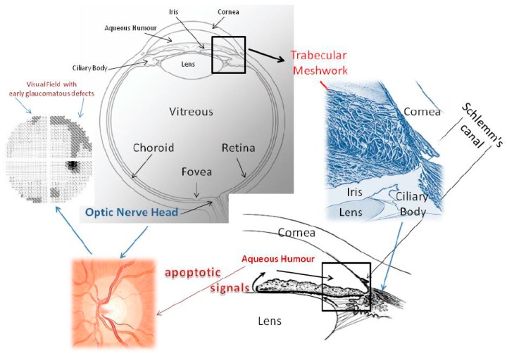
Glaucoma is a multifactorial disease in which pro-apoptotic signals are directed to retinal ganglion cells. During this disease the conventional outflow pathway becomes malfunctioning. Aqueous humour builds up in the anterior chamber, leading to increased intraocular pressure. Both of these events are related to functional impairment. The knowledge of molecular mechanisms allows us to better understand the usefulness of substances that can support anti-glaucoma therapy. The goal of glaucoma therapy is not simply to lower intraocular pressure; it should also be to facilitate the survival of retinal ganglion cells, as these constitute the real target tissue in this disease, in which the visual pathway is progressively compromised. Indeed, an endothelial dysfunction syndrome affecting the endothelial cells of the trabecular meshwork occurs in both normal-tension glaucoma and high-tension glaucoma. Some substances, such as polyunsaturated fatty acids, can counteract the damage due to the molecular mechanisms - whether ischemic, oxidative, inflammatory or other - that underlie the pathogenesis of glaucoma. In this review, we consider some molecules, such as polyphenols, that can contribute, not only theoretically, to neuroprotection but which are also able to counteract the metabolic pathways that lead to glaucomatous damage. Ginkgo biloba extract, for instance, improves the blood supply to peripheral districts, including the optic nerve and retina and exerts a neuro-protective action by inhibiting apoptosis. Polyunsaturated fatty acids can protect the endothelium and polyphenols exert an anti-inflammatory action through the down-regulation of cytokines such as TNF-α and IL-6. All these substances can aid anti-glaucoma therapy by providing metabolic support for the cells involved in glaucomatous injury. Indeed, it is known that the food we eat is able to change our gene expression.

Keywords: cell cultures; oxidative damage; primary open angle glaucoma; trabecular meshwork NFkB.


Conflict of interest statement

The authors declare no conflict of interest.

Figures

Figure 1
Figure 1
This picture shows the whole eye and the iridocorneal angle, seen both two-dimensionally and three-dimensionally, to explain the aqueous humour outflow pathway. Malfunctioning of the trabecular meshwork causes IOP to increase. The conventional outflow pathway is probably the tissue which is responsible for the proapoptotic signals that determine alterations of the optic nerve head and of the visual field. The trabecular meshwork consists of endothelial cells immersed in their fundamental substance. Aqueous humour flows through the intercellular spaces of the TM and crosses the inner wall of Schlemm’s canal. From a functional point of view, the conventional aqueous outflow is endowed with two barriers. The first is formed by the trabecular meshwork cells and the second by the endothelial cells that line the lumen of Schlemm’s canal. Located between the iris and cornea, it has a particular architecture, which considerably increases the filtration surface; its endothelial cells are constantly in contact with free radicals. Oxidative stress is chiefly responsible for molecular damage to its endothelial cells and triggers all those events that lead to glaucoma. Oxidative attack induces a loss of trabecular meshwork cells, impairing TM functionality. It is not known whether oxidative damage is due to reduced antioxidant defences or to primary damage to mitochondria. In addition, free radicals are implicated in the mechanism of senescence. During the course of glaucoma, the TM displays cell loss, subclinical inflammation, increased accumulation of extracellular matrix, endothelial dysregulation and dysfunction, changes in the cytoskeleton, altered motility and outflow impairment. The aqueous humour proteome profile also undergoes dramatic changes, reflecting cellular and molecular damage to the TM. We still do not know the mechanism that links trabecular damage to the apoptosis of ganglion cells. From a clinical standpoint, the death of ganglion cells causes alterations in the visual field. Furthermore, in glaucoma, the aqueous humour proteins, which are an expression of TM failure, among other things, might constitute biological signals for the posterior segment, where the cascade of events leading to the process of degeneration involves ganglion cells [12,13].
Figure 2
Figure 2
Glaucoma stages. Row (a) representation of a normal optic nerve, which presents nerve fibre layers and unaltered visual field. Row (b) early-stage glaucoma with initial optic disc excavation at the fundus and loss of nerve fibres more pronounced in the superior sector that determines a nasal step in the visual field. Row (c) moderate-stage glaucoma with partial optic disc excavation at the fundus and loss of nerve fibres in the inferior sector, determining an arciform defect in the visual field. Row (d) severe glaucoma, characterized by an excavated optic disc, loss of nerve fibres in both superior and inferior sectors and absolute defects in all sectors of the visual field.
Figure 3
Figure 3
Inflammation due to oxidative stress is the basis both of alterations to the conventional outflow pathway, leading to IOP increase [13] and of the alterations that induce apoptosis of RGCs. Indeed, in glaucoma, the expression of inflammatory genes occurs. ROS also activate transcription factor NF-kB, which induces the expression of various agents, including pro-inflammatory cytokines (IL-1/6, TNF-α) [90]. Pro-inflammatory cytokines, such as TNF-α or interleukins, are up-regulated, inducing intracellular and extracellular ROS production in human RPE cells [91]. Between oxidative stress and inflammation, NF-kB plays a strategic role, entering into the nucleus to induce transcription of a myriad of genes that mediate diverse cellular processes, such as immunity, inflammation, proliferation, apoptosis and cellular senescence [92].

References

    1. Izzotti A., Bagnis A., Saccà S.C. The role of oxidative stress in glaucoma. Mutat. Res. 2006;612:105–114. doi: 10.1016/j.mrrev.2005.11.001. - DOI - PubMed
    1. Russo R., Varano G.P., Adornetto A., Nucci C., Corasaniti M.T., Bagetta G., Morrone L.A. Retinal ganglion cell death in glaucoma: Exploring the role of neuroinflammation. Eur. J. Pharmacol. 2016;787:134–142. doi: 10.1016/j.ejphar.2016.03.064. - DOI - PubMed
    1. Izzotti A., Saccà S.C., Di Marco B., Penco S., Bassi A.M. Antioxidant activity of timolol on endothelial cells and its relevance for glaucoma course. Eye (Lond.) 2008;22:445–453. doi: 10.1167/iovs.09-3871. - DOI - PubMed
    1. Flammer J., Konieczka K. Retinal venous pressure: The role of endothelin. Epma J. 2015;6:21. doi: 10.1186/s13167-015-0043-1. - DOI - PMC - PubMed
    1. Gauthier A.C., Liu J. Neurodegeneration and Neuroprotection in Glaucoma. Yale J. Biol. Med. 2016;89:73–79. - PMC - PubMed