Review
. 2019 Oct;25(5):475-490.
doi: 10.1177/1073858418824256. Epub 2019 Jan 24.

The Striatum's Role in Executing Rational and Irrational Economic Behaviors

Affiliations
Review

The Striatum's Role in Executing Rational and Irrational Economic Behaviors

Ian J Bamford et al. Neuroscientist. 2019 Oct.

Abstract

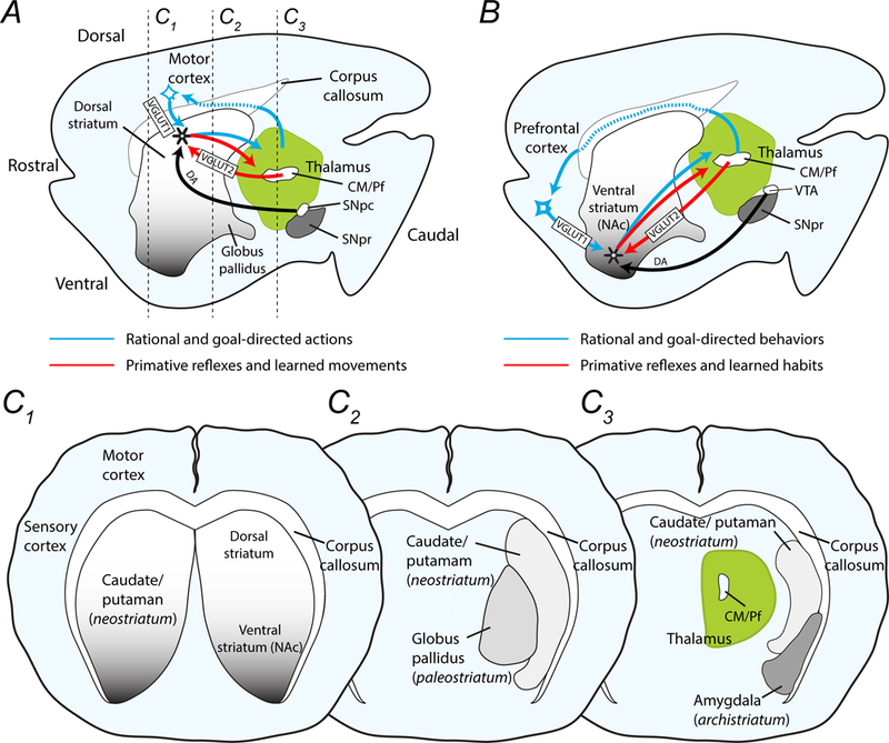
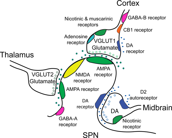
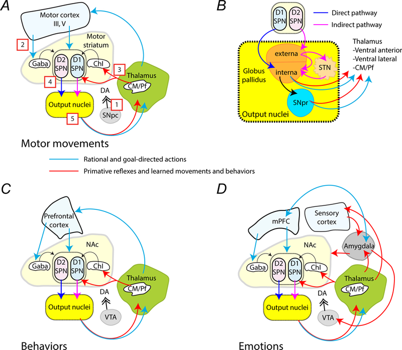
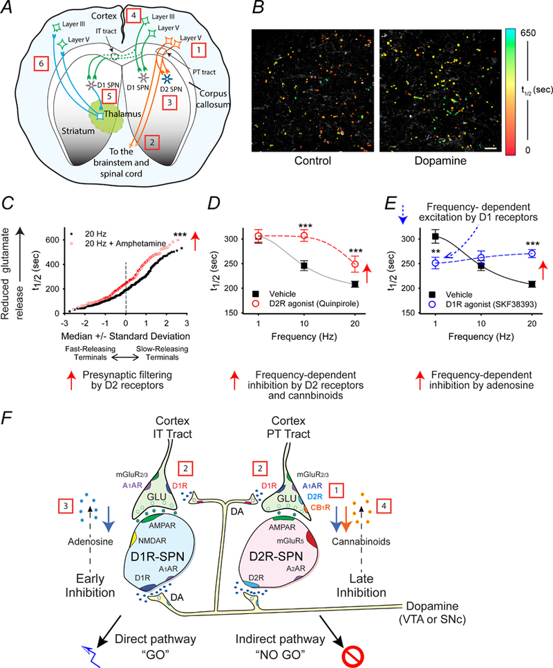
The striatum is a critical component of the brain that controls motor, reward, and executive function. This ancient and phylogenetically conserved structure forms a central hub where rapid instinctive, reflexive movements and behaviors in response to sensory stimulation or the retrieval of emotional memory intersect with slower planned motor movements and rational behaviors. This review emphasizes two distinct pathways that begin in the thalamus and converge in the striatum to differentially affect movements, behaviors, and decision making. The convergence of excitatory glutamatergic activity from the thalamus and cortex, along with dopamine release in response to novel stimulation, provide the basis for motor learning, reward seeking, and habit formation. We outline how the rules derived through research on neural pathways may enhance the predictability of reflexive actions and rational responses studied in behavioral economics.

Keywords: behavioral economics; decision-making; emotion; habits; motor learning; neuroeconomics; neuroscience; reward.


Conflict of interest statement

Declaration of Conflicting Interests

The authors declare that there is no conflict of interest

Figures

Figure 1.
Figure 1.
Drawings demonstrate the major components of the striatal network in mice and the proposed circuits involved in reflexive and rational actions and behaviors. (A) The midline sagittal section shows the dorsal and ventral striatum caudal to the motor and prefrontal cortex. The thalamus, which includes the centromedian and parafascicular nuclei (CM/Pf), is posterior to the striatum and dorsal to the substantia nigra pars compacta (SNpc) and pars reticulata (SNpr). Movements are encoded in the dorsal striatum, while (B) decisions and behaviors are programmed in the nucleus accumbens (NAc) of the ventral striatum. Rational and goal-directed action and behaviors rely on corticostriatal-thalamocortical loops (blue arrows), while reflexive movements and behaviors depend on communication between the thalamus and the striatum (red arrows). These two pathways are modified by dopamine projections from the SNpc to the motor striatum and by dopamine afferents from the VTA to the NAc. (C1-C3) Coronal sections moving from rostral to caudal, as indicated in panel A, show the medial placement of the globus pallidus (palaeostriatum) and the ventrolateral placement of the amygdala (arachistriatum). The caudate and putamen that comprise the neostriatum are not separate structures in mice as they are in humans.
Figure 2.
Figure 2.
The corticostriatal network and the effect of dopamine on corticostriatal terminals. The simplified cartoon shows the tripartite configuration of a glutamatergic corticostriatal input, a thalamostriatal input, and a dopaminergic (DA) midbrain input as they synapse on an SPN. Glutamatergic presynaptic elements generally display an asymmetric synaptic density with docked synaptic vesicles. The corticostriatal density contains VGLUT1 and docks near the spine head, while the thalamostriatal element contains VGLUT2 and docks on the spine shaft. A dopamine terminal is generally associated with the spine neck and shaft and displays a relatively small symmetric synaptic density (Totterdell and Smith 1989). Virtually all striatal synapses are within 2 μm of an apparent dopamine axonal varicosity, and thus are thought to receive dopamine input. SPNs express AMPA, NMDA and GABA-A receptors, in addition to D1 or D2 receptors (Wang and others 2012). Presynaptic afferents from the cortex express acetylcholine nicotinic and muscarinic receptors, adenosine and cannabinoid CB1 receptors and GABA-B receptors (Wang and others 2013a; Wang and others 2012; Wang and others 2013b). In the dorsal and ventral striatum, dopamine D2 receptors are found on presynaptic afferents to D2-SPNs (Bamford and others 2004b; Wang and others 2012). In the ventral striatum, D1-SPNs are expressed on presynaptic afferents to both D1- and D2-SPNs (Wang and others 2012). Presynaptic receptors on excitatory glutamatergic afferents from the thalamus have not been well characterized.
Figure 3.
Figure 3.
Simplified circuits that mediate habitual, rational, and emotional actions and behaviors. (A) This simplified schematic describes the connections between brain regions involved in executing movements. The red arrows are pathways proposed to mediate primitive or learned habitual movements, while blue arrows represent pathways involve in learned rational and goal-directed movements. [1] Signaling by dopamine from the SNpc that is coincident with either [2] excitatory VGLUT1 corticostriatal projections or [3] excitatory VGLUT2 thalamostriatal projections is relayed through [4] striatal SPNs. [5] Information is processed within the striatal output nuclei, which includes the globus pallidus, SNpr and STN, before being sent on to the thalamus. The thalamus then directs information back to the cortex or the striatum. (B) The drawing shows the output nuclei that receive inhibitory GABAergic signals from the SPNs. These nuclei allow signal inversion so that signals from D1R-expressing SPNs excite the thalamus, while signals from D2R-expressing SPNs are inhibitory. (C) The schematic describes the putative connections involved in irrational habits (red arrows) and reward-based, rational behaviors (blue arrows). (D) The simplified schematic shows the interconnections proposed to encode emotional behaviors. The amygdala receives inputs from the thalamus and from the sensory cortex. Emotion-based behaviors (red arrows) extend from the amygdala to the VTA, the mPFC, and the NAc. The mPFC (blue arrows) processes emotional responses using extinction, which allows goal-directed behaviors instead of emotion-directed behaviors to be executed if they conflict.
Figure 4.
Figure 4.
Proposed steps that lead to movements and decision making via the corticostriatal pathway. (A) The schematic shows the origin and destination of IT-type and PT-type projections. [1] Actions generated by the giant Betz cells of cortical layer V are [2] relayed to the midbrain and contralateral spinal cord by PT-type projections. [3] Their axon collaterals to ipsilateral D2-SPNs terminate the movement. [4] IT-type projections from layer III cortico-cortical neurons and from upper layer V neurons promote planning for the next action, [5] which after a processing delay is relayed by ipsilateral and contralateral D1-SPNs to the cortex via the thalamus (ipsilateral circuit is not shown). (B) Examples of optical FM1–43 destaining within the striatum in response to cortical stimulation. Corticostriatal terminals in the NAc were loaded with FM1–43 during a pre-measurement interval, and then the destaining rate during a subsequent stimulation period at individual nerve terminals was monitored as t1/2 values (the time required for florescence to decay to half of it original value). The individual halftimes (t1/2) of FM1–43 release from corticostriatal terminals in an acute slice preparation are shown in false color. Note that the preparation exposed to the dopamine releaser amphetamine (right; n = 112) has slower destaining puncta (higher t1/2) than the control (left; n = 99). Scale bar, 10 μm. (C) The normal probability plot demonstrates presynaptic filtering of glutamatergic terminals in the NAc of mouse brain slices. Comparison of individual half-times of FM1–43 release shows that the dopamine releaser amphetamine filters cortical activity by reducing exocytosis of FM1–43 from terminals with lowest probability of release (i.e. those with the highest t1/2). At 20 Hz, t1/2 = 207 s; n = 330. At 20 Hz with amphetamine, t1/2 = 264 s; n = 193; ***p < 0.001, Mann-Whitney. (D) This plot demonstrates that activation of D2 receptors inhibits glutamate release at higher frequencies. Distribution of mean t1/2 of release for slices containing the NAc as a function of frequency. The release of FM1–43 was reduced at higher frequencies (20 Hz) when the slice was exposed the D2R agonist quinpirole (n = 80 – 96 puncta; ***p < 0.001, Mann-Whitney). (E) This graph demonstrates that activation of D1 receptors excites glutamate release at lower frequencies (1 Hz), while adenosine inhibits glutamate release at higher frequencies (20 Hz). The D1R agonist SKF38393 increased release at low frequencies (1 Hz). At higher frequencies of cortical stimulation (20 Hz), the D1R agonist reduced release by promoting presynaptic inhibition by adenosine (n = 97 – 260 puncta; ***p < 0.001 Mann-Whitney). (F) The schematic shows glutamatergic and dopaminergic inputs to D1- and D2-expressing SPNs in the NAc, including representations of presynaptic filtering. [1] Tonic dopamine selectively inhibits low-release probability synapses of the indirect pathway D2-SPNs through presynaptic D2 receptors (D2Rs). [2] Higher levels of dopamine modulate corticoaccumbal activity through D1Rs that strengthen glutamate release from presynaptic terminals innervating both D1-SPNs and D2R-SPNs. [3] Higher cortical frequencies coincident with dopamine release inhibits presynaptic activity of both direct pathway D1-SPNs and indirect pathway D2-SPNs via stimulation of A1A receptors by adenosine, putatively produced in D1-SPNs following AMPA receptor (AMPAR) and NMDA receptor (NMDAR) activation. [4] Filtering by adenosine is followed by the selective presynaptic inhibition of D2R-expressing SPNs via stimulation of CB1 receptors (CB1R) via endocannabinoids putatively produced by D2-SPNs following activation of their D2 and group 1 metabotropic glutamate (mGluR5) receptors. Data are shown as mean ± SEM. Panel B is reproduced with permission from Bamford et.al. Neuron. 42:653–663 (2004). Panels C - F are reproduced with permission from Wang et.al., J Physiol. 590:3743–69 (2012).
Figure 5.
Figure 5.
Time course of D1-SPN and D2-SPN responses in the rat NAc during an ICSS trial consisting of a cue presentation followed two seconds later by lever extension and then a subsequent lever press (LP), which delivers an electrical stimulation to dopamine cell bodies in the VTA. (A) Time course of D2-SPNs that are cue excitatory and D1-SPNs that are LP-excitatory. The hatched areas indicate the time variation of either cue or LP. (B) Changes in the balance between D2- and D1-SPNs during the cue and the LP. The cue temporarily heightens D2R-SPN activity to create an imbalance in striatal output. The imbalance may alert the animal, perhaps by promoting an urge to move. The lever extension increases D1R-SPN activity, culminating in the lever press (LP). (C) Temporal sequence of chemical activity at SPN synapses. The resting activity of SPNs is low because mature SPNs have prominent potassium currents (Wilson and Kawaguchi 1996) and cortical inputs to D2-SPNs are tonically inhibited by endocannabinoids (Wang and others 2012). [1] The sensory cue triggers excitatory inputs to D2-SPNs, which [2] activate further via presynaptic D1Rs in response to [3] dopamine release. [4] Balance is restored following the release of endocannabinoids from D2-SPNs. [5] In animals that have learned to associate sensory cue to the lever press, another set of glutamatergic inputs subsequently activate with [6] convergent dopamine release to stimulate D1-SPNs. Coactivation of post-synaptic D1Rs, NMDA and AMPA receptors on D1-SPNs release [7] adenosine to inhibit presynaptic activity to both D1- and D2-SPNs and rebalances the circuits.
Figure 6.
Figure 6.
The effect of the thalamostriatal input on motor function was tested by using CAV2 viral manipulations in floxed (CAV2Cre-Slc17a6lox/lox) mice and CAV2Cre-Slc17a6+/+ controls. These VGLUT2 knockout mice specifically reduced excitatory signaling from the thalamus to the striatum. (A) VGLUT2 knockout mice (n = 8) showed no significant difference in spontaneous novelty-induced locomotion compared to control mice (n = 9). (B) VGLUT2 knockout mice (n = 9) had significantly more slips on the balance beam task compared with controls (n = 5). (C) VGLUT2 knockout mice (n = 17) performed significantly worse over time on the rotarod apparatus as compared with controls (n = 11), indicating an impairment in learned motor behaviors and innate balance. Data are shown as mean ± SEM. *p < 0.05, **p < 0.01, ANOVA. Statistical significance of genotype effects (repeated-measures two-way ANOVA) are shown in the headings of panels A and C (not significant, n.s.; **p < 0.01). Reproduced with permission from Melief et.al., npj Parkinson’s Disease. 4:23 (2018). Minor changes were made to the figure legends. http://creativecommons.org/licenses/by/4.0/
Figure 7.
Figure 7.
Hypothesized circuits that mediate reflexive and rational movements and behaviors. [1] Reflexive motor, behavioral and emotional responses or que-based habits are mediated through tonically-inhibited striatal-thalamic-striatal circuits, which are activated by coincident dopamine and glutamate release in response to unexpected sensory input via the [2] spinal cord and thalamus. [3] Motor movements, [4] reward seeking, and [5] action-outcome learning are mediated through the motor, prefrontal and medial prefrontal cortex, respectively. [6] Dopamine release provides motivation to obtain a reward and encodes value.

References

    1. Bamford NS, Joyce JA. 2005. Chronic methamphetamine mediates long-term depression of corticostriatal release in the dorsal striatum. Annals of Neurology 58(S9):S81.
    1. Bamford NS, Robinson S, Palmiter RD, Joyce JA, Moore C, Meshul CK. 2004a. Dopamine modulates release from corticostriatal terminals. J Neurosci 24(43):9541–52. - PMC - PubMed
    1. Bamford NS, Wightman RM, Sulzer D. 2018. Dopamine’s Effects on Corticostriatal Synapses during Reward-Based Behaviors. Neuron 97(3):494–510. - PMC - PubMed
    1. Bamford NS, Zhang H, Joyce JA, Scarlis CA, Hanan W, Wu NP and others. 2008. Repeated exposure to methamphetamine causes long-lasting presynaptic corticostriatal depression that is renormalized with drug readministration. Neuron 58(1):89–103. - PMC - PubMed
    1. Bamford NS, Zhang H, Schmitz Y, Wu NP, Cepeda C, Levine MS and others. 2004b. Heterosynaptic dopamine neurotransmission selects sets of corticostriatal terminals. Neuron 42(4):653–63. - PubMed

Publication types