. 2019 Jan 24;21(1):12.
doi: 10.1186/s13058-018-1079-7.

Intravital imaging reveals systemic ezrin inhibition impedes cancer cell migration and lymph node metastasis in breast cancer

Affiliations

Intravital imaging reveals systemic ezrin inhibition impedes cancer cell migration and lymph node metastasis in breast cancer

Abdi Ghaffari et al. Breast Cancer Res. .

Abstract

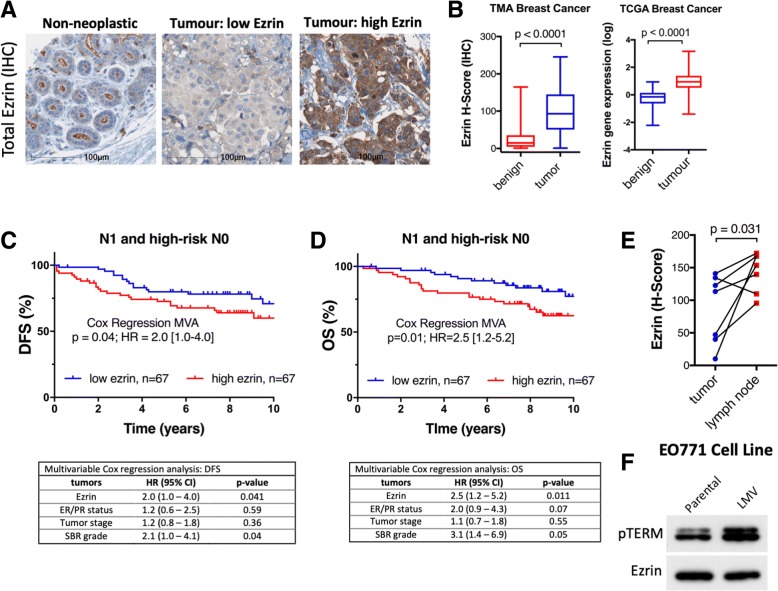
Background: Limited understanding of the cancer biology of metastatic sites is a major factor contributing to poor outcomes in cancer patients. The regional lymph nodes are the most common site of metastasis in most solid cancers and their involvement is a strong predictor of relapse in breast cancer (BC). We have previously shown that ezrin, a cytoskeletal-membrane linker protein, is associated with lymphovascular invasion and promotes metastatic progression in BC. However, the efficacy of pharmacological inhibition of ezrin in blocking cancer cell migration and metastasis remains unexplored in BC.

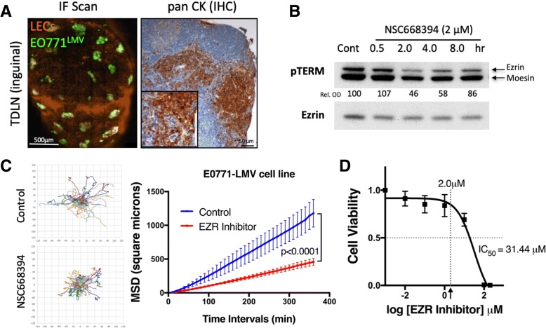
Methods: We quantified ezrin expression in a BC tissue microarray (n = 347) to assess its correlation with risk of relapse. Next, we developed a quantitative intravital microscopy (qIVM) approach, using a syngeneic lymphatic reporter mouse tumor model, to investigate the effect of systemic ezrin inhibition on cancer cell migration and metastasis.

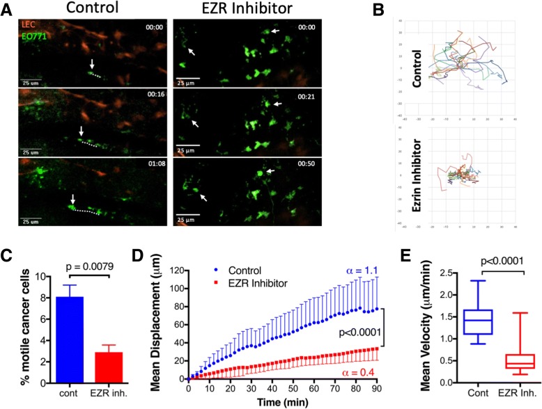
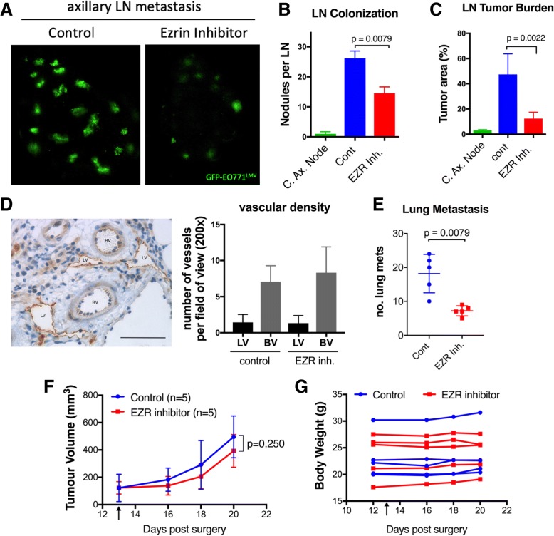
Results: We show that ezrin is expressed at significantly higher levels in lymph node metastases compared to matched primary tumors, and that a high tumor ezrin level is associated with increased risk of relapse in BC patients with regional disease. Using qIVM, we observe a subset of cancer cells that retain their invasive and migratory phenotype at the tumor-draining lymph node. We further show that systemic inhibition of ezrin, using a small molecule compound (NSC668394), impedes the migration of cancer cells in vivo. Furthermore, systemic ezrin inhibition leads to reductions in metastatic burden at the distal axillary lymph node and lungs.

Conclusions: Our findings demonstrate that the tumor ezrin level act as an independent biomarker in predicting relapse and provide a rationale for therapeutic targeting of ezrin to reduce the metastatic capacity of cancer cells in high-risk BC patients with elevated ezrin expression.

Keywords: Biomarker; Cell migration; Ezrin; Lymph node metastasis; Metastatic disease; Quantitative intravital imaging.


Conflict of interest statement

Ethics approval and consent to participate

Written informed consent was obtained from all patients and the studies were conducted in accordance with the Queen’s University Research Ethics Board consent guidelines.

Consent for publication

Not applicable.

Competing interests

The authors declare that they have no competing interests.

Publisher’s Note

Springer Nature remains neutral with regard to jurisdictional claims in published maps and institutional affiliations.

Figures

Fig. 1
Fig. 1
Tumor ezrin levels predict increased risk of relapse in high-risk BC patients. a Immunohistochemistry (IHC) of ezrin expression in non-neoplastic breast tissue and tumours with low and high ezrin are shown. b Box and whisker plots of ezrin histo (H)-score (protein levels, TMA) and relative EZR mRNA expression (TCGA) in benign and tumor tissues (p values from Wilcoxon matched-paired rank test). c, d KM plots showing DFS in node positive (N1, panel C) or node positive plus high-risk node negative (N0, panel D) BC patients stratified by median ezrin score. The corresponding 14 multivariate Cox regression analyses (MVA), adjusted for tumour stage, Scarff-Bloom-Richardson (SBR) grade, and ER/PR status) are shown below each plot. e Ezrin expression (HALO H-score) in paired primary tumour and lymph node metastases is shown (n=7, Wilcoxon matched-pairs signed rank test). f Immunoblot showing elevation of phospho-ezrin (pTERM, activated ezrin) in metastatic variant cell line (LMV) derived from the murine parental cell line EO771 during serial orthotopic injections of lung metastases in C57BL/6 mice. HR, hazard ratio; CI, confidence interval
Fig. 2
Fig. 2
GFP-EO771LMV tumors develop spontaneous LN metastases in subcutaneous and orthotopic models. a Whole tissue confocal scan of inguinal TDLN (left, scale bar 500 μm) and cytokeratin stain (right) confirming presence of GFP-EO771LMV metastases. b Immunoblot analysis of GFP-EO771LMV cells treated with NSC668394 ezrin inhibitor (2 μM) in vitro show reduction in ezrin pT567 (upper pTERM band). The percent ratio of phospho-ezrin to total protein normalized to control (relative optical density (Rel. OD)) shown under each band (mean of n = 2 assays). c Migration of GFP-EO771LMV cells in response to ezrin inhibitor in vitro analyzed by time-lapse microscopy for up to 18 h (see Additional file 3: video 1). Cell trajectories (left, minimum of 30 cells/group, pooled from three independent assays) were used to plot mean square displacement curves (right panel) using DiPer software (p < 0.0001, Wilcoxon matched-paired signed rank test). d Cell viability analysis shows the half maximal inhibitory concentration IC50 in NSC668394 treatment of GFP-EO771LMV cells. Th arrow points to 2.0 μM value on the x axis (mean of three independent assays).
Fig. 3
Fig. 3
Intravital imaging of TDLN metastases reveals reduced migration of cancer cells in response to ezrin inhibition. a Maximum intensity projection (z-stacks of 4–6 μm) of still images from the time-lapse videos (see Additional file 6: video 3 and Additional file 7: video 4) showing migration of cancer cells (dotted line) in the inguinal LN. b Cell trajectory data generated for a minimum of 20 cells per group (3 fields per LN, 3 mice, pool of n=2 IVM studies; motile cells tracked by two observers blinded to the study). c The proportion of motile GFP-EO771LMV cells in TDLNs of control and NSC668394 treated mice (Mann Whitney test). d Mean displacement curves were generated from pooled cell trajectory data in panel B. The slopes of displacement curves (α-value) are shown beside each curve and compared using the Extra-Sum-of-Squares F test. e Overall mean velocities of motile cancer cells in control and treated TDLNs are shown (Mann Whitney test)
Fig. 4
Fig. 4
Ezrin inhibition attenuates metastatic burden in axillary LN and lungs. a On day 20 post injection of GFP-EO771LMV cells into left fourth mammary fat pad of lymphatic reporter mice (five mice per group, n = 2), ipsilateral axillary LNs were harvested and scanned by confocal fluorescent microscope. b The total number of LN tumour nodules and c tumor burden (tumor area/total LN area) in contralateral axillary node (C. Ax. Node) and control and ezrin inhibitor treated ipsilateral axillary nodes are shown (p values calculated by Mann-Whitney test) d An example of LV (high lyve-1) and BV (low lyve-1) in intra-tumour region is shown. Scale bar in IHC image represent 50μm. The graph shows lymphatic (LV) and blood (BV) vascular 15 density assessed by visual count of at least 5 random fields of view (20x objective) in tumours stained with lyve-1 (LV marker) and CD31 (BV marker). e To assess lung metastasis, primary tumours were surgically removed on day 20 and mice allowed to recover for an additional 7 days. Lungs were then harvested and assessed for number of tumour nodules by fluorescence imaging (n=6 per group; p value calculated by Mann-Whitney test). f, g Primary tumour volume and total body weight of mice were measured during the study. Tumour growth curves were compared by Wilcoxon matched-paired signed rank test. Black arrow indicates the start of NSC668394 treatment

References

    1. Li J, Wei K, Yu H, Jin D, Wang G, Yu B. Prognostic Value of Ezrin in Various Cancers: A Systematic Review and Updated Meta-analysis. Sci Rep. 2015;5:17903. - PMC - PubMed
    1. Fehon RG, McClatchey AI, Bretscher A. Organizing the cell cortex: the role of ERM proteins. Nat Rev Mol Cell Biol. 2010;11(4):276–287. - PMC - PubMed
    1. Clucas J, Valderrama F. ERM proteins in cancer progression. J Cell Sci. 2014;127(Pt 2):267–275. - PubMed
    1. Padera TP, Meijer EF, Munn LL. The Lymphatic System in Disease Processes and Cancer Progression. Annu Rev Biomed Eng. 2016;18:125–158. - PMC - PubMed
    1. Ran S, Volk L, Hall K, Flister MJ. Lymphangiogenesis and lymphatic metastasis in breast cancer. Pathophysiology. 2010;17(4):229–251. - PMC - PubMed

Publication types

MeSH terms

Grants and funding