. 2019 Feb;29(2):317-324.
doi: 10.1101/gr.241901.118. Epub 2019 Jan 24.

A chromosome-scale assembly of the axolotl genome

Affiliations

A chromosome-scale assembly of the axolotl genome

Jeramiah J Smith et al. Genome Res. 2019 Feb.

Abstract

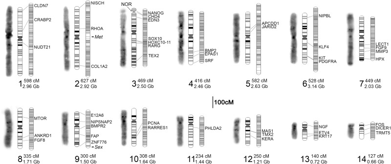
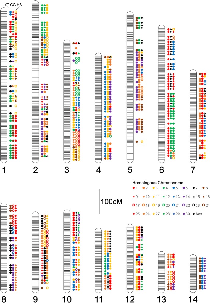
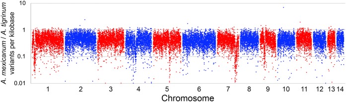
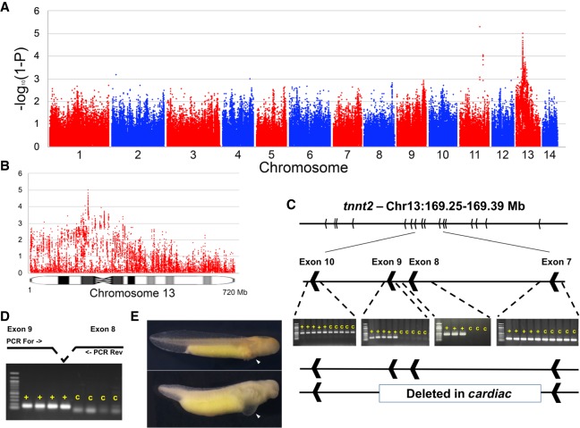
The axolotl (Ambystoma mexicanum) provides critical models for studying regeneration, evolution, and development. However, its large genome (∼32 Gb) presents a formidable barrier to genetic analyses. Recent efforts have yielded genome assemblies consisting of thousands of unordered scaffolds that resolve gene structures, but do not yet permit large-scale analyses of genome structure and function. We adapted an established mapping approach to leverage dense SNP typing information and for the first time assemble the axolotl genome into 14 chromosomes. Moreover, we used fluorescence in situ hybridization to verify the structure of these 14 scaffolds and assign each to its corresponding physical chromosome. This new assembly covers 27.3 Gb and encompasses 94% of annotated gene models on chromosomal scaffolds. We show the assembly's utility by resolving genome-wide orthologies between the axolotl and other vertebrates, identifying the footprints of historical introgression events that occurred during the development of axolotl genetic stocks, and precisely mapping several phenotypes including a large deletion underlying the cardiac mutant. This chromosome-scale assembly will greatly facilitate studies of the axolotl in biological research.


Figures

Figure 1.
Figure 1.
Summary of assembled chromosomes from the A. mexicanum genome. Three images are shown for each chromosome (1–14): (left) microscopic image illustrating DAPI banding; (center) an idiogram summarizing banding patterns and relative sizes of chromosomes from several spreads (Supplemental Fig. S1); and (right) the corresponding linkage group. Groups of linked markers at a given position are represented by horizontal marks on linkage groups. The location of genes and hybridization signals from gene-anchored BACs are labeled with the corresponding gene ID and connecting lines. The length of each linkage group and assembled chromosome is given next to its numerical label. The locations of three additional features are highlighted: (met) a major quantitative trait locus controlling timing and incidence of metamorphosis; (sex) the sex determining locus; and (NOR) the nucleolar organizer, which harbors most ribosomal RNA copies.
Figure 2.
Figure 2.
Patterns of conserved synteny across assembled A. mexicanum chromosomes. Colored shapes represent the consensus homologous chromosomes (see inset legend) for each 10 cM interval. For each chromosome, homologs are shown in a consistent order from left to right: (left) X. tropicalis (XT); (center) chicken (Gallus gallus [GG]); (right) human (Homo sapiens [HS]). A. mexicanum chromosomes are designated by numerical labels corresponding to those in Figure 1.
Figure 3.
Figure 3.
The distribution of segregating variants within the mapping panel used to generate scaffolding information. The density of polymorphisms within 1-Mb intervals is relatively uniform; however, a few intervals show substantially reduced densities, including regions on Chromosomes 1, 5, 7, and 9.
Figure 4.
Figure 4.
Localization and characterization of the cardiac mutation. (A) The genome-wide distribution of P-values for tests of association between transcribed polymorphisms and the c mutant phenotype. (B) Distribution of P-values across Chromosome 13. (C) Identification and PCR validation of a large deletion that is associated with the c phenotype and removes exons 8 and 9 of tnnt2. (D) Verification that these same exons are expressed in wild-type individuals, but not in individuals with the c phenotype. (E) Example of two siblings from a cross segregating for the cardiac mutation: (top) wild-type; (bottom) cardiac homozygote. Note the thoracic edema and lack of erythrocytes in the developing heart (white arrows) of cardiac mutants (Supplemental Fig. S3).

References

    1. Altschul SF, Gish W, Miller W, Myers EW, Lipman DJ. 1990. Basic local alignment search tool. J Mol Biol 215: 403–410. 10.1016/S0022-2836(05)80360-2 - DOI - PubMed
    1. Armstrong JB. 1984. Genetic mapping in the Mexican axolotl, Ambystoma mexicanum. Can J Genet Cytol 26: 1–6. 10.1139/g84-001 - DOI - PubMed
    1. Armstrong JB, Duhon ST, Malacinski GM. 1989. Raising the axolotl in captivity. In Developmental biology of the axolotl (ed. Armstrong JB, Malacinski GM), pp. 220–227. Oxford University Press, New York.
    1. Bryant DM, Johnson K, DiTommaso T, Tickle T, Couger MB, Payzin-Dogru D, Lee TJ, Leigh ND, Kuo TH, Davis FG, et al. 2017. A tissue-mapped axolotl de novo transcriptome enables identification of limb regeneration factors. Cell Rep 18: 762–776. 10.1016/j.celrep.2016.12.063 - DOI - PMC - PubMed
    1. Caballero-Pérez J, Espinal-Centeno A, Falcon F, García-Ortega LF, Curiel-Quesada E, Cruz-Hernández A, Bako L, Chen X, Martinez O, Alberto Arteaga-Vázquez M, et al. 2018. Transcriptional landscapes of Axolotl (Ambystoma mexicanum). Dev Biol 433: 227–239. 10.1016/j.ydbio.2017.08.022 - DOI - PubMed

Publication types

LinkOut - more resources