. 2019 Mar 22;294(12):4511-4519.
doi: 10.1074/jbc.RA118.005428. Epub 2019 Jan 24.

Conformational flexibility and inhibitor binding to unphosphorylated interleukin-1 receptor-associated kinase 4 (IRAK4)

Affiliations

Conformational flexibility and inhibitor binding to unphosphorylated interleukin-1 receptor-associated kinase 4 (IRAK4)

Li Wang et al. J Biol Chem. .

Abstract

Interleukin-1 receptor-associated kinase 4 (IRAK4) is a key player in innate immune and inflammatory responses, performing a critical role in signal transduction downstream of Toll-like receptors and interleukin-1 (IL-1) receptors. Upon ligand binding and via its N-terminal death domain, IRAK4 is recruited to an oligomeric receptor that is proximal to the Myddosome signaling complex, inducing IRAK4 kinase domain dimerization, autophosphorylation, and activation. To date, all known IRAK4 structures are in the active conformation, precluding a good understanding of IRAK4's conformational dynamics. To address this issue, here we first solved three crystal structures of the IRAK4 kinase domain (at ≤2.6 Å resolution), in its unphosphorylated, inactive state bound to either the ATP analog AMP-PNP or to one of the two small-molecule inhibitors JH-I-25 and JH-I-17. The structures disclosed that although the structure in complex with AMP-PNP is in an "αC-out" inactive conformation, those in complex with type I inhibitors assume an active "Asp-Phe-Gly (DFG)-in" and "αC-in" conformation. The ability of unphosphorylated IRAK4 to take on variable conformations prompted us to screen for small-molecule inhibitors that bind preferentially to unphosphorylated IRAK4, leading to the identification of ponatinib and HG-12-6. Solving the structures of unphosphorylated IRAK4 in complex with these two inhibitors, we found that they both bind as type II inhibitors with IRAK4 in a "DFG-out" conformation. Collectively, these structures reveal conformational flexibility of unphosphorylated IRAK4 and provide unexpected insights into the potential use of small molecules to modulate IRAK4 activity in cancer, autoimmunity, and inflammation.

Keywords: IRAK4; Toll-like receptor (TLR); crystal structure; drug development; inactive conformation; inflammation; innate immunity; kinase; structural biology; type II inhibition.


Conflict of interest statement

Although the work was performed solely in academia, Ryan Ferrao and Li Wang are currently employees of Gilead Sciences and SMOC Therapeutics, respectively

Figures
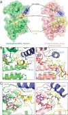
Figure 1.
Figure 1.
Structure of AMP-PNP bound IRAK4 kinase domain. A, cartoon representation of the unphosphorylated IRAK4 kinase domain in an inactive conformation in complex with AMP-PNP (left, green). AMP-PNP–bound phosphorylated IRAK4 in the active conformation (right, pink) (PDB code 2OID) is shown for comparison. The αC-helix and activation loop are colored blue and yellow, respectively. AMP-PNP (magenta) is shown as sticks. B, detailed comparison of the αC-helix and activation loop conformation between unphosphorylated (left, green) and phosphorylated (right, pink) IRAK4. C, detailed interactions between the DFG motif, αC-helix, and AMP-PNP in both the unphosphorylated (left, green) and phosphorylated (right, pink) IRAK4. In both B and C, the αC-helix, activation loop, and AMP-PNP are colored as in A.
Figure 2.
Figure 2.
Regulatory spine of the AMP-PNP bound IRAK4 kinase domain structure. A, detailed view of the residues that form the regulatory spine, shown as red sticks and highlighted with red surface. The αC-helix and activation loop are colored blue and yellow, respectively. AMP-PNP (magenta) is shown in a stick model. Disassembled regulatory spine indicates an inactive conformation of IRAK4. B, detailed interactions between AMP-PNP (magenta), Mg2+, and unphosphorylated IRAK4 (green). IRAK4 DFG motif is colored in yellow.
Figure 3.
Figure 3.
Comparison of the DFG motifs and inhibitory helices of IRAK4, CDK2, EGFR, ABL1, and SRC. A, comparison of the αC-helix and DFG motif conformations of ATP- or AMP-PNP–bound IRAK4 (green), CDK2 (blue, 1HCK), and EGFR (pink, 2GS7). B, comparison of the inhibitory helix conformation of ATP- or AMP-PNP–bound IRAK4 (green), CDK2 (blue, 1HCK), and EGFR (pink, 2GS7). C, sequence alignment of the DFG motif of IRAK4 with other kinases known to adopt the CDK2/c-Src–like inactive conformation.
Figure 4.
Figure 4.
Unphosphorylated IRAK4 in complex with type I inhibitors. A, chemical structure and binding pose of the type I inhibitor JH-I-25 (pink, sticks) bound to unphosphorylated IRAK4 (green). B, chemical structure and binding pose of the type I inhibitor JH-I-17 (blue, sticks) bound to unphosphorylated IRAK4 (cyan). C, 2FoFc electron density map within 1.5 Å of the ligand JH-I-25 contoured at 1.5 σ (colored as in A). D, 2FoFc electron density map within 1.5 Å of the ligand JH-I-17 contoured at 1.5 σ (colored as in B). E, detailed interactions between IRAK4 and JH-I-25 (colored as in A). F, detailed interactions between IRAK4 and JH-I-17 (colored as in B).
Figure 5.
Figure 5.
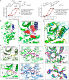
IRAK4 in complex with type II inhibitors. A, chemical structures of the type II inhibitors identified in screen and used in the co-crystallization. The affinities of two type II inhibitors to unphosphorylated and phosphorylated IRAK4 kinase domain were measured by LanthaScreen Eu kinase-binding assay. Both ponatinib and HG-12-6 showed preferred binding to unphosphorylated IRAK4 kinase domain. B, a ribbon diagram showing the overview of IRAK4 in complex with ponatinib. The αC-helix is colored in blue, and the DFG loop is colored in yellow. Ponatinib is shown in a stick model and colored in magenta. C, type II inhibitor binding to a DFG-out conformation of IRAK4, showing disrupted regulatory spine of IRAK4 (colored as in B). Residues forming the regulatory spine are shown as red sticks and highlighted with red surface. D, comparison of the αC-helix position and conserved salt-bridge interaction between Glu233 and Lys213 in active IRAK4 (DFG-in and αC-in, yellow, PDB code 2OID), inactive IRAK4 in complexes with AMP-PNP (αC-out, cyan) and inactive IRAK4 in complexes with ponatinib (DFG-out, green). E, ribbon diagrams comparing DFG-in active conformation in IRAK4 (cartoon, olive green) bound to JH-I-25 (sticks, pink, top panel) and DFG-out inactive conformation in IRAK4 (cartoon, green) bound to ponatinib (sticks, magenta, bottom panel). In these two conformations, Asp329 and Phe330 swap positions with accompanied movement of the αC-helix. F, detailed interaction between IRAK4 (green) with ponatinib (magenta) and between IRAK4 (blue) with HG-12-6 (dark purple). G, superimposition of ponatinib bound IRAK4 structure with reported kinase structures in complex with ponatinib (PDB code of ABL, 3OXZ; PDB code of ABL T315I, 3IK3; PDB code of KIT, 4U0I; and PDB code of RIPK2, 4C8B). These kinases share the conserved DFG-out conformation. The rotation of ring A of ponatinib appears to accommodate the different sizes of gatekeeper residues.

Similar articles

Cited by

References

    1. Kawai T., and Akira S. (2010) The role of pattern-recognition receptors in innate immunity: update on Toll-like receptors. Nat. Immunol. 11, 373–384 10.1038/ni.1863 - DOI - PubMed
    1. Garlanda C., Dinarello C. A., and Mantovani A. (2013) The interleukin-1 family: back to the future. Immunity 39, 1003–1018 10.1016/j.immuni.2013.11.010 - DOI - PMC - PubMed
    1. Lin S. C., Lo Y. C., and Wu H. (2010) Helical assembly in the MyD88-IRAK4-IRAK2 complex in TLR/IL-1R signalling. Nature 465, 885–890 10.1038/nature09121 - DOI - PMC - PubMed
    1. Motshwene P. G., Moncrieffe M. C., Grossmann J. G., Kao C., Ayaluru M., Sandercock A. M., Robinson C. V., Latz E., and Gay N. J. (2009) An oligomeric signaling platform formed by the Toll-like receptor signal transducers MyD88 and IRAK-4. J. Biol. Chem. 284, 25404–25411 10.1074/jbc.M109.022392 - DOI - PMC - PubMed
    1. Ferrao R., Zhou H., Shan Y., Liu Q., Li Q., Shaw D. E., Li X., and Wu H. (2014) IRAK4 dimerization and trans-autophosphorylation are induced by myddosome assembly. Mol. Cell 55, 891–903 10.1016/j.molcel.2014.08.006 - DOI - PMC - PubMed

Publication types

MeSH terms

Substances

LinkOut - more resources