. 2019 Jun;104(6):1176-1188.
doi: 10.3324/haematol.2018.206375. Epub 2019 Jan 24.

Unraveling the cellular origin and clinical prognostic markers of infant B-cell acute lymphoblastic leukemia using genome-wide analysis

Affiliations

Unraveling the cellular origin and clinical prognostic markers of infant B-cell acute lymphoblastic leukemia using genome-wide analysis

Antonio Agraz-Doblas et al. Haematologica. 2019 Jun.

Abstract

B-cell acute lymphoblastic leukemia is the commonest childhood cancer. In infants, B-cell acute lymphoblastic leukemia remains fatal, especially in patients with t(4;11), present in ~80% of cases. The pathogenesis of t(4;11)/KMT2A-AFF1+ (MLL-AF4+) infant B-cell acute lymphoblastic leukemia remains difficult to model, and the pathogenic contribution in cancer of the reciprocal fusions resulting from derivative translocated-chromosomes remains obscure. Here, "multi-layered" genome-wide analyses and validation were performed on a total of 124 de novo cases of infant B-cell acute lymphoblastic leukemia uniformly diagnosed and treated according to the Interfant 99/06 protocol. These patients showed the most silent mutational landscape reported so far for any sequenced pediatric cancer. Recurrent mutations were exclusively found in K-RAS and N-RAS, were subclonal and were frequently lost at relapse, despite a larger number of non-recurrent/non-silent mutations. Unlike non-MLL-rearranged B-cell acute lymphoblastic leukemias, B-cell receptor repertoire analysis revealed minor, non-expanded B-cell clones in t(4;11)+ infant B-cell acute lymphoblastic leukemia, and RNA-sequencing showed transcriptomic similarities between t(4;11)+ infant B-cell acute lymphoblastic leukemias and the most immature human fetal liver hematopoietic stem and progenitor cells, confirming a "pre-VDJ" fetal cellular origin for both t(4;11) and RAS mut The reciprocal fusion AF4-MLL was expressed in only 45% (19/43) of the t(4;11)+ patients, and HOXA cluster genes are exclusively expressed in AF4-MLL-expressing patients. Importantly, AF4-MLL/HOXA-expressing patients had a significantly better 4-year event-free survival (62.4% vs 11.7%, P=0.001), and overall survival (73.7 vs 25.2%, P=0.016). AF4-MLL expression retained its prognostic significance when analyzed in a Cox model adjusting for risk stratification according to the Interfant-06 protocol based on age at diagnosis, white blood cell count and response to prednisone. This study has clinical implications for disease outcome and diagnostic risk-stratification of t(4;11)+ infant B-cell acute lymphoblastic leukemia.


Figures

Figure 1.
Figure 1.
Somatic mutations detected by whole-exome sequencing in the discovery cohort of infant B-cell precursor acute lymphoblastic leukemia. (A) Total number of mutations identified in each individual patient. The total number of non-synonymous mutations (yellow area, right Y axis) and mutant allele frequency (MAF) for each mutation (individual dots, left Y axis) are represented. (B) Oncodrive software identified the PI3K-RAS pathway as the only recurrently mutated pathway in infant B-cell precursor acute lymphoblastic leukemia. The distribution of mutations in genes of the PI3K-RAS pathway is shown for all patients within the three iBCP-ALL subgroups: [total 42 patients: 27 t(4;11)+, 5 t(9;11)+ and 10 MLLwt].
Figure 2.
Figure 2.
Frequent somatic mutations in RAS genes in both the discovery and validation cohorts. (A) Specific KRAS and NRAS mutations recurrently found in each patient by high coverage targeted sequencing. (B) Proportions of patients with mutations in KRAS (brown), NRAS (yellow) or both (gray) within the three infant B-cell precursor acute lymphoblastic leukemia subgroups. (C) Mutant allele frequency of KRAS (brown squares) or NRAS (yellow circles) mutations in each individual patient. Discovery cohort, n=42; validation cohort, n=82.
Figure 3.
Figure 3.
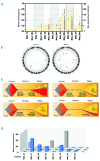
Clonal evolution and genomic instability at relapse. (A) Total number of mutations identified for each patient in paired diagnostic-relapse samples. Total number of non-synonymous mutations (yellow area, right Y axis) and mutant allele frequency (MAF) for each mutation (individual dots, left Y axis) are represented for paired diagnostic and relapsed (R) samples. (B) Circos plot representation of the total number of mutations identified at diagnosis and relapse for a representative patient (MA4_17). Genomic rearrangements are represented by lines connecting both breakpoints. Copy number alterations (blue=gains, red=losses) are represented by the outer gray circle. Somatic mutations (both single nucleotide variants and indels) are depicted in the center of the circle and the affected gene is indicated. (C) Graphic representation of clonal evolution in paired diagnostic (DX)-relapsed (RL) samples. The number of unique somatic mutations called at diagnosis (orange), relapse (yellow) or shared between DX and REL (red) are indicated. Bigger gene names indicate higher MAF for the mutations shared at DX and REL. (D) Dynamics of RAS-mutated clones identified as MAF in matched DX-Remission-REL trios (n=8).
Figure 4.
Figure 4.
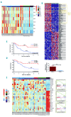
Transcriptional signature of infant B-cell precursor acute lymphoblastic leukemia samples. (A) Heatmap representing FLT3, PROM1, MEIS1 and HOXA gene expression according to the infant B-cell precursor acute lymphoblastic leukemia (iBCP-ALL) cytogenetic group and RAS mutations. (B) Top panel: heatmap showing HOXA cluster gene expression according to the expression of the reciprocal fusion AF4-MLL. Bottom panel: quantitative polymerase chain reaction validating high expression of HOXA cluster genes in t(4;11) iBCP-ALL patients expressing AF4-MLL. (C,D) Four-year event-free survival (C) and overall survival (D) Kaplan-Meier curves for t(4;11) iBCP-ALL patients according to AF4-MLL expression, n=43 t(4;11)+ patients. (E) Heatmap representation of selected genes for the signaling pathways most significantly deregulated. Right panels represent positive pathway enrichment called by gene set enrichment analysis software. Total 42 patients: 27 t(4;11)+, 5 t(9;11)+ and 10 MLLwt.
Figure 5.
Figure 5.
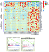
Specific transcriptional differences between MLL-AF4+ and MLL-AF9+ or MLLwt infant B-cell precursor acute lymphoblastic leukemia patients. Here, FL-derived CD34+CD19+ progenitors were not included as normalizers in the analysis in order to avoid potential bias. Gene set enrichment analysis (GSEA) was performed with the genes differentially expressed between MLL-AF4+ patients and MLL-AF9+ or MLLwt patients. MLL-AF4+ infant B-cell precursor acute lymphoblastic leukemia (iBCP-ALL) patients showed a significant overexpression of genes associated with cellular catabolism, coupled to a significant downregulation of negative regulators of the PI3-MAPK pathway, as well as of genes involved in lymphoid differentiation and RNApol II transcriptional regulation as compared to both MLL-AF9+ and MLLwt iBCP-ALL patients. The bottom panels represent positive pathway enrichment called by GSEA software. Total 42 patients: 27 t(4;11)+, 5 t(9;11)+ and 10 MLLwt.
Figure 6.
Figure 6.
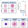
Analysis of B-cell receptor repertoires suggest a hematopoietic stem cell/early pre-VDJ progenitor as the cell-of-origin for t(4;11)/MLL-AF4+ infant B-cell precursor acute lymphoblastic leukemia. (A) Cloud-plots of B-cell receptor (BCR) repertoires from two representative t(4;11)+ infant B-cell precursor acute lymphoblastic leukemia (iBCP-ALL) patients depicting the existence of many minor non-expanded B-cell clones either at diagnosis or relapse. Each vertex represents a unique BCR sequence, and the relative vertex size is proportional to the number of identical reads. (B) Largest BCR clone size in t(4;11)+ iBCP-ALL, healthy individuals and non-t(4;11)+ pediatric BCP-ALL. (C) Cloud-plots of BCR repertoires of representative t(1;19)/E2A-PBX1+, t(12;21)/TEL-AML1+ and t(9;22)/BCR-ABL+ patients showing high clonality of B-cell clones. The samples from the iBCP-ALL patients who were BCR-sequenced were four MLL-AF4+ diagnostic-relapse pairs, three E2A-PBX1+ samples, one TEL-AML1+ sample and one BCR-ABL+ sample.
Figure 7.
Figure 7.
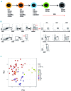
Comparison of the transcriptome of human fetal CD34+ hematopoietic stem and progenitor cell populations to infant B-cell precursor acute lymphoblastic leukemia. (A) Schematic representation of B-cell development in human fetal liver (FL) showing immunophenotypic definitions for hematopoietic stem cells (HSC), multipotent progenitors (MPP), lymphoid-primed multipotent progenitors (LMPP), committed B progenitors (CBP) and B cells. The onset and expected patterns of IgH rearrangements, are depicted as red arrows. (B) Sorting strategy for FL hematopoietic stem and progenitor (HSPC) populations by fluorescence-activated cell sorting. The sorting gates for each population are shown in representative flow plots on the left. The purity of the sorted populations is depicted on the right demonstrating >95% purity. (Lin, Lineage cocktail). (C) Principal component analysis of gene expression of infant B-cell precursor acute lymphoblastic leukemia (iBCP-ALL) samples (n=42) and FL HSPC populations (n=3-7) using the top 1,000 variably expressed genes, as determined by RNA-sequencing. FL HSPC as in (A); MAF4, MLL-AF4+ iBCP-ALL; MA9, MLL-AF9+ iBCP-ALL; MLLwt, MLL wildtype iBCP-ALL.

Comment in

Similar articles

Cited by

References

    1. Pui C-H, Evans WE. A 50-year journey to cure childhood acute lymphoblastic leukemia. Semin Hematol. 2013;50(3):185–196. - PMC - PubMed
    1. Pui CH, Mullighan CG, Evans WE, Relling MV. Pediatric acute lymphoblastic leukemia: where are we going and how do we get there? Blood. 2012;120(6):1165–1174. - PMC - PubMed
    1. Meyer C, Burmeister T, Gröger D, et al. The MLL recombinome of acute leukemias in 2017. Leukemia. 2018;32(2):273–284. - PMC - PubMed
    1. Sanjuan-Pla A, Bueno C, Prieto C, et al. Revisiting the biology of infant t(4;11)/MLL-AF4+ B-cell acute lymphoblastic leukemia. Blood. 2015;126(25):2676–2685. - PMC - PubMed
    1. Marschalek R. Mechanisms of leukemogenesis by MLL fusion proteins. Br J Haematol. 2011;152(2):141–154. - PubMed

Publication types

MeSH terms